Cysts of the jaws

Last updated
Jaw cysts
Specialty Oral and Maxillofacial Surgery, Dentistry

A cyst is a pathological epithelial lined cavity that fills with fluid or soft material and usually grows from internal pressure generated by fluid being drawn into the cavity from osmosis (hydrostatic pressure). The bones of the jaws, the mandible and maxilla, are the bones with the highest prevalence of cysts in the human body. This is due to the abundant amount of epithelial remnants that can be left in the bones of the jaws. The enamel of teeth is formed from ectoderm (the precursor germ layer to skin and mucosa), and so remnants of epithelium can be left in the bone during odontogenesis (tooth development). The bones of the jaws develop from embryologic processes which fuse, and ectodermal tissue may be trapped along the lines of this fusion. [1] This "resting" epithelium (also termed cell rests) is usually dormant or undergoes atrophy, but, when stimulated, may form a cyst. The reasons why resting epithelium may proliferate and undergo cystic transformation are generally unknown, but inflammation is thought to be a major factor. [1] The high prevalence of tooth impactions and dental infections that occur in the bones of the jaws is also significant to explain why cysts are more common at these sites.

Contents

Cysts that arise from tissue(s) that would normally develop into teeth are referred to as odontogenic cysts. Other cysts of the jaws are termed non-odontogenic cysts. [2] Non-odontogenic cysts form from tissues other than those involved in tooth development, and consequently may contain structures such as epithelium from the nose. As the cyst grows from hydraulic pressure it causes the bone around it to resorb, and may cause movement of teeth or other vital structures such as nerves and blood vessels, or resorb the roots of teeth. Most cysts do not cause any symptoms, and are discovered on routine dental radiographs. [1] Some cysts may not require any treatment, but if treatment is required, it usually involves some minor surgery to partially or completely remove the cyst in a one or two-stage procedure.

Classification

Odontogenic cysts

Dental panoramic radiograph showing dentigerous cyst associated with an impacted lower right wisdom tooth in the right mandible (arrowed, appears on the lower left of the image). JawCyst (with arrows).jpg
Dental panoramic radiograph showing dentigerous cyst associated with an impacted lower right wisdom tooth in the right mandible (arrowed, appears on the lower left of the image).
Relative incidence of odontogenic cysts. Relative incidence of odontogenic cysts.jpg
Relative incidence of odontogenic cysts.

Odontogenic cysts have histologic origins in the cells of the dental structures. Some are inflammatory while others are developmental.

Developmental/ Non-odontogenic cysts

There are several development cysts of the head and neck most of which form in the soft tissues rather than the bone. There are also several cysts, previously thought to arise from epithelial remanents trapped in embryonic lines of fusion, most of which are now believed to be odontogenic in origin or have an unknown cause. Their names are included for the sake of completeness.

Developmental cysts of the jaws

Developmental cysts of the soft tissues around the jaws

Developmental cysts of questionable cause

Signs and symptoms

Cysts rarely cause any symptoms, unless they become secondarily infected. [1] The signs depend mostly upon the size and location of the cyst.

If the cyst has not expanded beyond the normal anatomical boundaries of the bone, then there will be no palpable lump outside or inside the mouth. The vast majority of cysts expand slowly, and the surrounding bone has time to increase its density around the lesion, which is the body's attempt to isolate the lesion.

Cysts that have expanded beyond the normal anatomic boundaries of a bone are still often covered with a thin layer of new bone. At this stage, there may be a sign termed "eggshell cracking", where the thinned cortical plate cracks when pressure is applied.

A lump may be felt, which may feel hard if there is still bone covering the cyst, or fluctuant if the cyst has eroded through the bone surrounding it. [5] A cyst may become acutely infected, and discharge into the oral cavity via a sinus. Adjacent teeth may be loosened, tilted or even moved bodily. [6] Rarely, roots of teeth are resorbed, depending upon the type of cyst.

The inferior alveolar nerve runs through the mandible and supplies sensation to the lower lip and chin. As most cysts expand slowly, there will be no altered sensation (anesthesia or paraesthesia), since the inferior alveolar canal is harmlessly enveloped or displaced over time. More aggressive cysts, or acute infection of any cyst may cause altered sensation. Sometimes, they cause higher risk of pathological fracture of lower jaw, especially around angle of mandible. [6]

Diagnosis

Most cysts are discovered as a chance finding on routine dental radiography. [7] They are often asymptomatic unless there has been long-standing with significant enlargement (causing bony expansion or egg-shell cracking feeling [7] ) or secondary infection.

On an x-ray, cysts appear as radiolucent (dark) areas with radiopaque (white) borders. [7] However, cysts in maxillary sinus, also known as antrum, can appear radiopaque as the surrounding air absorbs fewer photons than the cystic fluid content.

Cysts are usually unilocular, but may also be multilocular. Sometimes aspiration (fine needle aspiration) is used to aid diagnosis of a cystic lesion; e.g., fluid aspirated from a radicular cyst may appear straw-colored and display shimmering due to cholesterol content. [5] Almost always, the cyst lining is sent to a pathologist for histopathologic examination after it has been surgically removed. This means that the exact diagnosis of the type of cyst is often made in retrospect, and definitive treatment can be made for the patient.

Treatment

As many cysts of the jaws have similar presentations and treatment options, it is common to perform one of the following treatment options and send the cyst lining to histopathology to provide a retrospective definitive diagnosis.

Cysts treatment is limited to surgical removal for the majority of cysts. There are two techniques used to manage cysts with the deciding factor being the size of the cyst. [8]

The exception to these treatments; is the management of cysts which have a higher rate of recurrence – for example odontogenic keratocysts. Options to reduce the recurrence rate include: curettage post enucleation, Carnoy's solution (treatment of the cavity with a potent fixative) or mandibular resection. These treatments are less conservative than the above options.

Prognosis

The prognosis depends upon the type, size and location of a cyst. Most cysts are entirely benign, and some may require no treatment. Rarely, some cystic lesions represent locally aggressive tumors that may cause destruction of surrounding bone if left untreated. This type of cyst are usually removed with a margin of healthy bone to prevent recurrence of new cysts. If a cyst expands to a very large size, the mandible may be weakened such that a pathologic fracture occurs.

After treatments, the patient should be informed of the risk of recurrence. Some people are more susceptible than others. This can be due to their oral and dental condition or inherited condition. [10] In some cases, there are some cysts remain after the surgery called the residual cysts and most of them arise from a periapical cyst. Glandular odontogenic cysts tend to recur after curettage. [11]

The radicular cyst is the most common type of cyst(65-70%) followed by dentigerous(15-18%). [12] The most common odontogenic cyst is a follicular(dentigerous) cyst. Rarely, the walls of this type of cyst can progress into mucoepidermoid carcinoma, ameloblastoma or squamous carcinoma if the cyst is not properly removed early enough. [11]

Epidemiology

Periapical cysts (also called radicular cysts) are by far the most common cyst occurring in the jaws. [5]

Jaw cysts affect around 3.5% of the population.10 They are more common in males than females at a ratio of 1.6:1 and most people get them between their 40s and 60s. The order of the jaw cysts from most common to least common is; radicular cysts, dentigerous cysts, residual cysts and odontogenic keratocysts. Radicular lesions are most commonly found in the anterior region of the maxilla 12 – usually around the canines.11 The majority of cysts are of inflammatory origin12. They are most commonly found in the posterior mandible 11

Related Research Articles

<span class="mw-page-title-main">Wisdom tooth</span> Large tooth at the back of the human mouth

A third molar, commonly called wisdom tooth, is one of the three molars per quadrant of the human dentition. It is the most posterior of the three. The age at which wisdom teeth come through (erupt) is variable, but this generally occurs between late teens and early twenties. Most adults have four wisdom teeth, one in each of the four quadrants, but it is possible to have none, fewer, or more, in which case the extras are called supernumerary teeth. Wisdom teeth may get stuck (impacted) against other teeth if there is not enough space for them to come through normally. Impacted wisdom teeth are still sometimes removed for orthodontic treatment, believing that they move the other teeth and cause crowding, though this is not held anymore as true. Impacted wisdom teeth may suffer from tooth decay if oral hygiene becomes more difficult. Wisdom teeth which are partially erupted through the gum may also cause inflammation and infection in the surrounding gum tissues, termed pericoronitis. Some more conservative treatments, such as operculectomies, may be fitting for some cases, yet impacted wisdom teeth are commonly extracted as treatment for these problems, many times before these problems, which might never happen, even occur. Some oppose this prophylactic removal of disease-free impacted wisdom teeth, including among them the National Institute for Health and Care Excellence in the UK.

<span class="mw-page-title-main">Ameloblastoma</span> Medical condition

Ameloblastoma is a rare, benign or cancerous tumor of odontogenic epithelium much more commonly appearing in the lower jaw than the upper jaw. It was recognized in 1827 by Cusack. This type of odontogenic neoplasm was designated as an adamantinoma in 1885 by the French physician Louis-Charles Malassez. It was finally renamed to the modern name ameloblastoma in 1930 by Ivey and Churchill.

<span class="mw-page-title-main">Cementoblastoma</span> Medical condition

Cementoblastoma, or benign cementoblastoma, is a relatively rare benign neoplasm of the cementum of the teeth. It is derived from ectomesenchyme of odontogenic origin. Cementoblastomas represent less than 0.69–8% of all odontogenic tumors.

<span class="mw-page-title-main">Dental follicle</span>

The dental follicle, also known as dental sac, is made up of mesenchymal cells and fibres surrounding the enamel organ and dental papilla of a developing tooth. It is a vascular fibrous sac containing the developing tooth and its odontogenic organ. The dental follicle (DF) differentiates into the periodontal ligament. In addition, it may be the precursor of other cells of the periodontium, including osteoblasts, cementoblasts and fibroblasts. They develop into the alveolar bone, the cementum with Sharpey's fibers and the periodontal ligament fibers respectively. Similar to dental papilla, the dental follicle provides nutrition to the enamel organ and dental papilla and also have an extremely rich blood supply.

<span class="mw-page-title-main">Veterinary dentistry</span> Branch of veterinary medicine

Veterinary dentistry is the field of dentistry applied to the care of animals. It is the art and science of prevention, diagnosis, and treatment of conditions, diseases, and disorders of the oral cavity, the maxillofacial region, and its associated structures as it relates to animals.

<span class="mw-page-title-main">Dentigerous cyst</span> Medical condition

Dentigerous cyst, also known as follicular cyst is an epithelial-lined developmental cyst formed by accumulation of fluid between the reduced enamel epithelium and crown of an unerupted tooth. It is formed when there is an alteration in the reduced enamel epithelium and encloses the crown of an unerupted tooth at the cemento-enamel junction. Fluid is accumulated between reduced enamel epithelium and the crown of an unerupted tooth. Dentigerous cyst is the second most common form of benign developmental odontogenic cysts.

<span class="mw-page-title-main">Central giant-cell granuloma</span> Medical condition

Central giant-cell granuloma (CGCG) is a localised benign condition of the jaws. It is twice as common in females and is more likely to occur before age 30. Central giant-cell granulomas are more common in the anterior mandible, often crossing the midline and causing painless swellings.

<span class="mw-page-title-main">Periapical cyst</span> Medical condition

Commonly known as a dental cyst, the periapical cyst is the most common odontogenic cyst. It may develop rapidly from a periapical granuloma, as a consequence of untreated chronic periapical periodontitis.

<span class="mw-page-title-main">Odontogenic keratocyst</span> Medical condition

An odontogenic keratocyst is a rare and benign but locally aggressive developmental cyst. It most often affects the posterior mandible and most commonly presents in the third decade of life. Odontogenic keratocysts make up around 19% of jaw cysts.
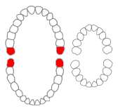
Lateral periodontal cysts (LPCs) are defined as non-keratinised and non-inflammatory developmental cysts located adjacent or lateral to the root of a vital tooth.” LPCs are a rare form of jaw cysts, with the same histopathological characteristics as gingival cysts of adults (GCA). Hence LPCs are regarded as the intraosseous form of the extraosseous GCA. They are commonly found along the lateral periodontium or within the bone between the roots of vital teeth, around mandibular canines and premolars. Standish and Shafer reported the first well-documented case of LPCs in 1958, followed by Holder and Kunkel in the same year although it was called a periodontal cyst. Since then, there has been more than 270 well-documented cases of LPCs in literature.

<span class="mw-page-title-main">Calcifying odontogenic cyst</span> Medical condition

Calcifying odotogenic cyst (COC) is a rare developmental lesion that comes from odontogenic epithelium. It is also known as a calcifying cystic odontogenic tumor, which is a proliferation of odontogenic epithelium and scattered nest of ghost cells and calcifications that may form the lining of a cyst, or present as a solid mass.

<span class="mw-page-title-main">Glandular odontogenic cyst</span> Human jaw cyst

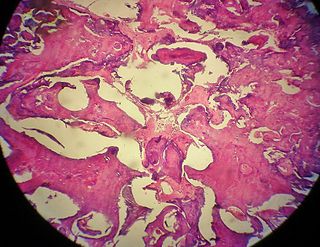
A glandular odontogenic cyst (GOC) is a rare and usually benign odontogenic cyst developed at the odontogenic epithelium of the mandible or maxilla. Originally, the cyst was labeled as "sialo-odontogenic cyst" in 1987. However, the World Health Organization (WHO) decided to adopt the medical expression "glandular odontogenic cyst". Following the initial classification, only 60 medically documented cases were present in the population by 2003. GOC was established as its own biological growth after differentiation from other jaw cysts such as the "central mucoepidermoid carcinoma (MEC)", a popular type of neoplasm at the salivary glands. GOC is usually misdiagnosed with other lesions developed at the glandular and salivary gland due to the shared clinical signs. The presence of osteodentin supports the concept of an odontogenic pathway. This odontogenic cyst is commonly described to be a slow and aggressive development. The inclination of GOC to be large and multilocular is associated with a greater chance of remission. GOC is an infrequent manifestation with a 0.2% diagnosis in jaw lesion cases. Reported cases show that GOC mainly impacts the mandible and male individuals. The presentation of GOC at the maxilla has a very low rate of incidence. The GOC development is more common in adults in their fifth and sixth decades.

<span class="mw-page-title-main">Adenomatoid odontogenic tumor</span> Medical condition

The adenomatoid odontogenic tumor is an odontogenic tumor arising from the enamel organ or dental lamina.

Squamous odontogenic tumors (SOTs) are very rare benign locally infiltrative odontogenic neoplasms of epithelial origin. Only some 50 cases have been documented. They occur mostly from 20-40 and are more common in males. Treatment is by simple enucleation and local curettage, and recurrence is rare.

An ameloblastic fibroma is a fibroma of the ameloblastic tissue, that is, an odontogenic tumor arising from the enamel organ or dental lamina. It may be either truly neoplastic or merely hamartomatous. In neoplastic cases, it may be labeled an ameloblastic fibrosarcoma in accord with the terminological distinction that reserves the word fibroma for benign tumors and assigns the word fibrosarcoma to malignant ones. It is more common in the first and second decades of life, when odontogenesis is ongoing, than in later decades. In 50% of cases an unerupted tooth is involved.

<span class="mw-page-title-main">Odontoma</span> Benign tumour of dental tissue

An odontoma, also known as an odontome, is a benign tumour linked to tooth development. Specifically, it is a dental hamartoma, meaning that it is composed of normal dental tissue that has grown in an irregular way. It includes both odontogenic hard and soft tissues. As with normal tooth development, odontomas stop growing once mature which makes them benign.

The odontogenic myxoma is an uncommon benign odontogenic tumor arising from embryonic connective tissue associated with tooth formation. As a myxoma, this tumor consists mainly of spindle shaped cells and scattered collagen fibers distributed through a loose, mucoid material.

<span class="mw-page-title-main">Bone cyst</span> Medical condition

A bone cyst or geode is a cyst that forms in bone.

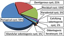
Odontogenic cyst are a group of jaw cysts that are formed from tissues involved in odontogenesis. Odontogenic cysts are closed sacs, and have a distinct membrane derived from rests of odontogenic epithelium. It may contain air, fluids, or semi-solid material. Intra-bony cysts are most common in the jaws, because the mandible and maxilla are the only bones with epithelial components. That odontogenic epithelium is critical in normal tooth development. However, epithelial rests may be the origin for the cyst lining later. Not all oral cysts are odontogenic cyst. For example, mucous cyst of the oral mucosa and nasolabial duct cyst are not of odontogenic origin.
In addition, there are several conditions with so-called (radiographic) 'pseudocystic appearance' in jaws; ranging from anatomic variants such as Stafne static bone cyst, to the aggressive aneurysmal bone cyst.

<span class="mw-page-title-main">Gingival cyst</span> Medical condition

Gingival cyst, also known as Epstein's pearl, is a type of cysts of the jaws that originates from the dental lamina and is found in the mouth parts. It is a superficial cyst in the alveolar mucosa. It can be seen inside the mouth as small and whitish bulge. Depending on the ages in which they develop, the cysts are classified into gingival cyst of newborn and gingival cyst of adult. Structurally, the cyst is lined by thin epithelium and shows a lumen usually filled with desquamated keratin, occasionally containing inflammatory cells. The nodes are formed as a result of cystic degeneration of epithelial rests of the dental lamina.

References

  1. 1 2 3 4 Hupp JR, Ellis E, Tucker MR (2008). Contemporary oral and maxillofacial surgery (5th ed.). St. Louis, Mo.: Mosby Elsevier. pp.  450–456. ISBN   9780323049030.
  2. Neville, Brad W.; Damm, Douglas D.; Allen, Carl M.; Bouquot, Jerry E. (2002). Oral & Maxillofacial Pathology (2nd ed.). Philadelphia, PA: W.B. Saunders Company. pp.  590–609. ISBN   978-0-7216-9003-2.
  3. Leandro Bezerra Borges; Francisco Vagnaldo Fechine; Mário Rogério Lima Mota; Fabrício Bitu Sousa; Ana Paula Negreiros Nunes Alves (2012). "Odontogenic lesions of the jaw: a clinical-pathological study of 461 cases". Revista Gaúcha de Odontologia. 60 (1).
  4. Zadik Y, Yitschaky O, Neuman T, Nitzan DW (May 2011). "On the Self-Resolution Nature of the Buccal Bifurcation Cyst". J Oral Maxillofac Surg. 69 (7): e282–4. doi:10.1016/j.joms.2011.02.124. PMID   21571416.[ dead link ]
  5. 1 2 3 Wray D, Stenhouse D, Lee D, Clark AJ (2003). Textbook of general and oral surgery. Edinburgh [etc.]: Churchill Livingstone. pp. 229–237. ISBN   978-0443070839.
  6. 1 2 Current diagnosis & treatment in otolaryngology : head & neck surgery. Lalwani, Anil K. (3rd ed.). New York: McGraw Hill Medical. 2012. ISBN   978-0-07-162439-8. OCLC   704526362.{{cite book}}: CS1 maint: others (link)
  7. 1 2 3 Whaites, Eric (2013-06-20). Essentials of dental radiography and radiology. Drage, Nicholas (Fifth ed.). Edinburgh. ISBN   978-0-7020-4599-8. OCLC   854310114.
  8. Contemporary oral and maxillofacial surgery. Hupp, James R., Ellis, Edward, DDS., Tucker, Myron R. (5th ed.). St. Louis, Mo.: Mosby Elsevier. 2008. ISBN   978-0-323-04903-0. OCLC   187293319.{{cite book}}: CS1 maint: others (link)
  9. Odell, E. W. (2017-05-02). Cawson's Essentials of Oral Pathology and Oral Medicine. [Place of publication not identified]. ISBN   978-0-7020-7389-2. OCLC   1054910269.
  10. "Dental cysts | Cambridge University Hospitals". www.cuh.nhs.uk. Retrieved 2020-02-23.
  11. 1 2 Dios, Pedro Diz (2016-05-17). Oral medicine and pathology at a glance. Scully, Crispian, Almeida, Oslei Paes de, Bagan, Jose, Taylor, Adalberto Mosqueda, Scully, Crispian, Preceded by (work) (Second ed.). Chichester, West Sussex. ISBN   978-1-119-12135-0. OCLC   942611369.
  12. Evidence-based oral surgery : a clinical guide for the general dental practitioner. Ferneini, Elie M., Goupil, Michael T. Cham, Switzerland. 2019-02-18. ISBN   978-3-319-91361-2. OCLC   1088721095.{{cite book}}: CS1 maint: others (link)