Related Research Articles

Macrocephaly is a condition in which the human head is abnormally large; this includes the scalp, the cranial bone, and the contents of the cranium. It may be pathological or benign, even a familial genetic characteristic. People diagnosed with macrocephaly will have further testing done to determine if the syndrome is accompanied by any other disorders. Those with benign or familial macrocephaly are considered to have megalencephaly, another form of macrocephaly that will not result in the development of neurological disorders in the patient.

Basal-cell carcinoma (BCC), also known as basal-cell cancer, is the most common type of skin cancer. It often appears as a painless raised area of skin, which may be shiny with small blood vessels running over it. It may also present as a raised area with ulceration. Basal-cell cancer grows slowly and can damage the tissue around it, but it is unlikely to spread to distant areas or result in death.

An epidermoid cyst or epidermal inclusion cyst is a benign cyst usually found on the skin. The cyst develops out of ectodermal tissue. Histologically, it is made of a thin layer of squamous epithelium.
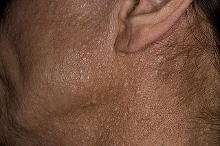
Birt–Hogg–Dubé syndrome (BHD), also Hornstein–Birt–Hogg–Dubé syndrome, Hornstein–Knickenberg syndrome, and fibrofolliculomas with trichodiscomas and acrochordons is a human autosomal dominant genetic disorder that can cause susceptibility to kidney cancer, renal and pulmonary cysts, and noncancerous tumors of the hair follicles, called fibrofolliculomas. The symptoms seen in each family are unique, and can include any combination of the three symptoms. Fibrofolliculomas are the most common manifestation, found on the face and upper trunk in over 80% of people with BHD over the age of 40. Pulmonary cysts are equally common (84%), but only 24% of people with BHD eventually experience a collapsed lung. Kidney tumors, both cancerous and benign, occur in 14–34% of people with BHD; the associated kidney cancers are often rare hybrid tumors.

A bifid rib is a congenital abnormality of the rib cage and associated muscles and nerves which occurs in about 1.2% of humans. Bifid ribs occur in up to 8.4% of Samoans. The sternal end of the rib is cleaved into two. It is usually unilateral.

Nevoid basal-cell carcinoma syndrome (NBCCS), is an inherited medical condition involving defects within multiple body systems such as the skin, nervous system, eyes, endocrine system, and bones. People with this syndrome are particularly prone to developing a common and usually non-life-threatening form of non-melanoma skin cancer. About 10% of people with the condition do not develop basal-cell carcinomas (BCCs).

Dentigerous cyst, also known as follicular cyst is an epithelial-lined developmental cyst formed by accumulation of fluid between the reduced enamel epithelium and crown of an unerupted tooth. It is formed when there is an alteration in the reduced enamel epithelium and encloses the crown of an unerupted tooth at the cemento-enamel junction. Fluid is accumulated between reduced enamel epithelium and the crown of an unerupted tooth. Dentigerous cyst is the second most common form of benign developmental odontogenic cysts.

An odontogenic keratocyst is a rare and benign but locally aggressive developmental cyst. It most often affects the posterior mandible and most commonly presents in the third decade of life. Odontogenic keratocysts make up around 19% of jaw cysts.

Protein patched homolog 1 is a protein that is the member of the patched family and in humans is encoded by the PTCH1 gene.
Schöpf–Schulz–Passarge syndrome is an autosomal recessive condition with punctate symmetric palmoplantar keratoderma, with the keratoderma and fragility of the nails beginning around age 12. In addition to palmoplantar keratoderma, other symptoms include hypodontia, hypotrichosis, nail dystrophies, and eyelid cysts. Patients may also develop syringofibroadenoma and squamous cell carcinomas.
An odontogenic tumor is a neoplasm of the cells or tissues that initiate odontogenic processes.

Pilomatricoma, is a benign skin tumor derived from the hair matrix. These neoplasms are relatively uncommon and typically occur on the scalp, face, and upper extremities. Clinically, pilomatricomas present as a subcutaneous nodule or cyst with unremarkable overlying epidermis that can range in size from 0.5-3.0 cm, but the largest reported case was 24 cm.
A variant of eccrine spiradenoma which can be multiple on the scalp and can coalesce to form a 'Turban' tumour. In pathology, a cylindroma is a tumour with nests of cells that resemble a cylinder in cross section.
Odontogenic cyst are a group of jaw cysts that are formed from tissues involved in odontogenesis. Odontogenic cysts are closed sacs, and have a distinct membrane derived from rests of odontogenic epithelium. It may contain air, fluids, or semi-solid material. Intra-bony cysts are most common in the jaws, because the mandible and maxilla are the only bones with epithelial components. That odontogenic epithelium is critical in normal tooth development. However, epithelial rests may be the origin for the cyst lining later. Not all oral cysts are odontogenic cyst. For example, mucous cyst of the oral mucosa and nasolabial duct cyst are not of odontogenic origin.
In addition, there are several conditions with so-called (radiographic) 'pseudocystic appearance' in jaws; ranging from anatomic variants such as Stafne static bone cyst, to the aggressive aneurysmal bone cyst.
A cyst is a pathological epithelial lined cavity that fills with fluid or soft material and usually grows from internal pressure generated by fluid being drawn into the cavity from osmosis. The bones of the jaws, the mandible and maxilla, are the bones with the highest prevalence of cysts in the human body. This is due to the abundant amount of epithelial remnants that can be left in the bones of the jaws. The enamel of teeth is formed from ectoderm, and so remnants of epithelium can be left in the bone during odontogenesis. The bones of the jaws develop from embryologic processes which fuse together, and ectodermal tissue may be trapped along the lines of this fusion. This "resting" epithelium is usually dormant or undergoes atrophy, but, when stimulated, may form a cyst. The reasons why resting epithelium may proliferate and undergo cystic transformation are generally unknown, but inflammation is thought to be a major factor. The high prevalence of tooth impactions and dental infections that occur in the bones of the jaws is also significant to explain why cysts are more common at these sites.

The ovarian fibroma, also fibroma, is a benign sex cord-stromal tumour.
A cancer syndrome, or family cancer syndrome, is a genetic disorder in which inherited genetic mutations in one or more genes predispose the affected individuals to the development of cancers and may also cause the early onset of these cancers. Cancer syndromes often show not only a high lifetime risk of developing cancer, but also the development of multiple independent primary tumors.

Patched 2 is a protein that in humans is encoded by the PTCH2 gene.
References
- ↑ Rapini, Ronald P.; Bolognia, Jean L.; Jorizzo, Joseph L. (2007). Dermatology: 2-Volume Set. St. Louis: Mosby. p. 1686. ISBN 978-1-4160-2999-1.
- ↑ Cassarino DS; Linden KG; Barr RJ (2005). Cutaneous keratocyst arising independently of the nevoid basal cell carcinoma syndrome. Am J Dermatopathol. pp. 27:177–178.
| This cutaneous condition article is a stub. You can help Wikipedia by expanding it. |