Caroli disease

Last updated
Caroli disease
Other namesCongenital cystic dilatation of the intrahepatic biliar tree
MRI of Caroli disease (b).jpg
Turbo spin echo T2-weighted axial MRI of Caroli disease, showing cystic dilatations of bile ducts (shown as white). [1] [ predatory publisher ]
Specialty Gastroenterology, medical genetics   OOjs UI icon edit-ltr-progressive.svg

Caroli disease (communicating cavernous ectasia, or congenital cystic dilatation of the intrahepatic biliary tree) is a rare inherited disorder characterized by cystic dilatation (or ectasia) of the bile ducts within the liver. There are two patterns of Caroli disease: focal or simple Caroli disease consists of abnormally widened bile ducts affecting an isolated portion of liver. The second form is more diffuse, and when associated with portal hypertension and congenital hepatic fibrosis, is often referred to as "Caroli syndrome". [2] The underlying differences between the two types are not well understood. Caroli disease is also associated with liver failure and polycystic kidney disease. The disease affects about one in 1,000,000 people, with more reported cases of Caroli syndrome than of Caroli disease. [3]

Contents

Caroli disease is distinct from other diseases that cause ductal dilatation caused by obstruction, in that it is not one of the many choledochal cyst derivatives. [2]

Signs and symptoms

The first symptoms typically include fever, intermittent abdominal pain, and an enlarged liver. Occasionally, yellow discoloration of the skin occurs. [4] Caroli disease usually occurs in the presence of other diseases, such as autosomal recessive polycystic kidney disease, cholangitis, gallstones, biliary abscess, sepsis, liver cirrhosis, kidney failure, and cholangiocarcinoma (7% affected). [2] People with Caroli disease are 100 times more at risk for cholangiocarcinoma than the general population. [4] After recognizing symptoms of related diseases, Caroli disease can be diagnosed.[ citation needed ]

Morbidity is common and is caused by complications of cholangitis, sepsis, choledocholithiasis, and cholangiocarcinoma. [5] These morbid conditions often prompt the diagnosis. Portal hypertension may be present, resulting in other conditions including enlarged spleen, hematemesis, and melena. [6] These problems can severely affect the patient's quality of life. In a 10-year period between 1995 and 2005, only 10 patients were surgically treated for Caroli disease, with an average patient age of 45.8 years. [5]

After reviewing 46 cases of Caroli disease before 1990, 21.7% of the cases were the result of an intrahepatic cyst or nonobstructive biliary tree dilation, 34.7% were linked with congenital hepatic fibrosis, 13% were isolated choledochal cystic dilation, and the remaining 24.6% had a combination of all three. [7]

Causes

Figure 2 Location of the PKHD1 gene on chromosome 6, short (p) arm Pkhd1 gene.jpeg
Figure 2 Location of the PKHD1 gene on chromosome 6, short (p) arm

The cause appears to be genetic; the simple form is an autosomal dominant trait, while the complex form is an autosomal recessive trait. [2] Females are more prone to Caroli disease than males. [8] Family history may include kidney and liver disease due to the link between Caroli disease and ARPKD. [6] PKHD1 , the gene linked to ARPKD, has been found mutated in patients with Caroli syndrome. PKHD1 is expressed primarily in the kidneys with lower levels in the liver, pancreas, and lungs, a pattern consistent with phenotype of the disease, which primarily affects the liver and kidneys. [2] [6] The genetic basis for the difference between Caroli disease and Caroli syndrome has not been defined.[ citation needed ]

Diagnosis

Magnetic resonance cholangiopancreatography (MRCP) of Caroli disease, showing cystic dilatations of bile ducts. MRCP of Caroli disease.jpg
Magnetic resonance cholangiopancreatography (MRCP) of Caroli disease, showing cystic dilatations of bile ducts.

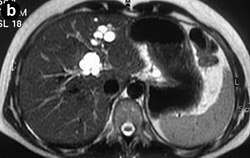
Modern imaging techniques allow the diagnosis to be made more easily and without invasive imaging of the biliary tree. [9] Commonly, the disease is limited to the left lobe of the liver. Images taken by CT scan, X-ray, or MRI show enlarged intrahepatic (in the liver) bile ducts due to ectasia. Using an ultrasound, tubular dilation of the bile ducts can be seen. On a CT scan, Caroli disease can be observed by noting the many fluid-filled, tubular structures extending to the liver. [4] A high-contrast CT must be used to distinguish the difference between stones and widened ducts. Bowel gas and digestive habits make it difficult to obtain a clear sonogram, so a CT scan is a good substitution. When the intrahepatic bile duct wall has protrusions, it is clearly seen as central dots or a linear streak. [10] Caroli disease is commonly diagnosed after this “central dot sign” is detected on a CT scan or ultrasound. [10] However, cholangiography is the best, and final, approach to show the enlarged bile ducts as a result of Caroli disease.[ citation needed ]

Treatment

The treatment depends on clinical features and the location of the biliary abnormality. When the disease is localized to one hepatic lobe, hepatectomy relieves symptoms and appears to remove the risk of malignancy. [11] Good evidence suggests that malignancy complicates Caroli disease in roughly 7% of cases. [11]

Antibiotics are used to treat the inflammation of the bile duct, and ursodeoxycholic acid is used for hepatolithiasis. [9] Ursodiol is given to treat cholelithiasis. In diffuse cases of Caroli disease, treatment options include conservative or endoscopic therapy, internal biliary bypass procedures, and liver transplantation in carefully selected cases. [11] Surgical resection has been used successfully in patients with monolobar disease. [9] An orthotopic liver transplant is another option, used only when antibiotics have no effect, in combination with recurring cholangitis. With a liver transplant, cholangiocarcinoma is usually avoided in the long run. [12]

Family studies are necessary to determine if Caroli disease is due to inheritable causes. Regular follow-ups, including ultrasounds and liver biopsies, are performed.[ citation needed ]

Prognosis

Mortality is indirect and caused by complications. After cholangitis occurs, patients typically die within 5–10 years. [3]

Epidemiology

Caroli disease is typically found in Asia, and diagnosed in persons under the age of 22. Cases have also been found in infants and adults. As medical imaging technology improves, diagnostic age decreases.[ citation needed ]

History

Jacques Caroli, a gastroenterologist, first described a rare congenital condition in 1958 in Paris, France. [8] [13] He described it as "nonobstructive saccular or fusiform multifocal segmental dilatation of the intrahepatic bile ducts"; basically, he observed cavernous ectasia in the biliary tree causing a chronic, often life-threatening hepatobiliary disease. [14] Caroli, born in France in 1902, learned and practiced medicine in Angers. After World War II, he was chief of service for 30 years at Saint-Antoine in Paris. Before dying in 1979, he was honored with the rank of commander in the Legion of Honour in 1976. [13]

Related Research Articles

<span class="mw-page-title-main">Bile duct</span> Type of organ

A bile duct is any of a number of long tube-like structures that carry bile, and is present in most vertebrates.

<span class="mw-page-title-main">Biliary atresia</span> Medical condition

Biliary atresia, also known as extrahepatic ductopenia and progressive obliterative cholangiopathy, is a childhood disease of the liver in which one or more bile ducts are abnormally narrow, blocked, or absent. It can be congenital or acquired. It has an incidence of one in 10,000–15,000 live births in the United States, and a prevalence of one in 16,700 in the British Isles. Biliary atresia is most common in East Asia, with a frequency of one in 5,000.

<span class="mw-page-title-main">Klatskin tumor</span> Medical condition

A Klatskin tumor is a cholangiocarcinoma occurring at the confluence of the right and left hepatic bile ducts. The disease was named after Gerald Klatskin, who in 1965 described 15 cases and found some characteristics for this type of cholangiocarcinoma

<span class="mw-page-title-main">Primary sclerosing cholangitis</span> Medical condition

Primary sclerosing cholangitis (PSC) is a long-term progressive disease of the liver and gallbladder characterized by inflammation and scarring of the bile ducts, which normally allow bile to drain from the gallbladder. Affected individuals may have no symptoms or may experience signs and symptoms of liver disease, such as yellow discoloration of the skin and eyes, itching, and abdominal pain.

<span class="mw-page-title-main">Cholangiocarcinoma</span> Bile duct adenocarcinoma

Cholangiocarcinoma, also known as bile duct cancer, is a type of cancer that forms in the bile ducts. Symptoms of cholangiocarcinoma may include abdominal pain, yellowish skin, weight loss, generalized itching, and fever. Light colored stool or dark urine may also occur. Other biliary tract cancers include gallbladder cancer and cancer of the ampulla of Vater.

Courvoisier's law states that a painless palpably enlarged gallbladder accompanied with mild jaundice is unlikely to be caused by gallstones. Usually, the term is used to describe the physical examination finding of the right-upper quadrant of the abdomen. This sign implicates possible malignancy of the gallbladder or pancreas and the swelling is unlikely due to gallstones.

<span class="mw-page-title-main">Gastrointestinal disease</span> Medical condition

Gastrointestinal diseases refer to diseases involving the gastrointestinal tract, namely the esophagus, stomach, small intestine, large intestine and rectum, and the accessory organs of digestion, the liver, gallbladder, and pancreas.

<span class="mw-page-title-main">Alagille syndrome</span> Medical condition

Alagille syndrome (ALGS) is a genetic disorder that affects primarily the liver and the heart. Problems associated with the disorder generally become evident in infancy or early childhood. The disorder is inherited in an autosomal dominant pattern, and the estimated prevalence of Alagille syndrome is 1 in every 30,000 to 1 in every 40,000 live births. It is named after the French pediatrician Daniel Alagille, who first described the condition in 1969.

<span class="mw-page-title-main">Cholestasis</span> Medical condition

Cholestasis is a condition where bile cannot flow from the liver to the duodenum. The two basic distinctions are an obstructive type of cholestasis where there is a mechanical blockage in the duct system that can occur from a gallstone or malignancy, and metabolic types of cholestasis which are disturbances in bile formation that can occur because of genetic defects or acquired as a side effect of many medications. Classification is further divided into acute or chronic and extrahepatic or intrahepatic.

Progressive familial intrahepatic cholestasis (PFIC) is a group of familial cholestatic conditions caused by defects in biliary epithelial transporters. The clinical presentation usually occurs first in childhood with progressive cholestasis. This usually leads to failure to thrive, cirrhosis, and the need for liver transplantation.

<span class="mw-page-title-main">Ascending cholangitis</span> Medical condition

Ascending cholangitis, also known as acute cholangitis or simply cholangitis, is inflammation of the bile duct, usually caused by bacteria ascending from its junction with the duodenum. It tends to occur if the bile duct is already partially obstructed by gallstones.

<span class="mw-page-title-main">Biliary tract</span> Organ system

The biliary tract refers to the liver, gallbladder and bile ducts, and how they work together to make, store and secrete bile. Bile consists of water, electrolytes, bile acids, cholesterol, phospholipids and conjugated bilirubin. Some components are synthesized by hepatocytes ; the rest are extracted from the blood by the liver.

<span class="mw-page-title-main">Percutaneous transhepatic cholangiography</span>

Percutaneous transhepatic cholangiography, percutaneous hepatic cholangiogram (PTHC) is a radiological technique used to visualize the anatomy of the biliary tract. A contrast medium is injected into a bile duct in the liver, after which X-rays are taken. It allows access to the biliary tree in cases where endoscopic retrograde cholangiopancreatography has been unsuccessful. Initially reported in 1937, the procedure became popular in 1952.

<span class="mw-page-title-main">Choledochal cysts</span> Medical condition

Choledochal cysts are congenital conditions involving cystic dilatation of bile ducts. They are uncommon in western countries but not as rare in East Asian nations like Japan and China.

<span class="mw-page-title-main">Congenital hepatic fibrosis</span> Medical condition

Congenital hepatic fibrosis is an inherited fibrocystic liver disease associated with proliferation of interlobular bile ducts within the portal areas and fibrosis that do not alter hepatic lobular architecture. The fibrosis would affect resistance in portal veins leading to portal hypertension.

<span class="mw-page-title-main">Liver cancer</span> Medical condition

Liver cancer is cancer that starts in the liver. Liver cancer can be primary or secondary. Liver metastasis is more common than that which starts in the liver. Liver cancer is increasing globally.

<span class="mw-page-title-main">Recurrent pyogenic cholangitis</span> Medical condition

Recurrent pyogenic cholangitis (RPC), also known as Hong Kong disease, Oriental cholangitis, and Oriental infestational cholangitis, is a chronic infection characterized by recurrent bouts of bacterial cholangitis with primary hepatolithiasis. It is exclusive to people who live or have lived in southeast Asia.

<span class="mw-page-title-main">Autosomal recessive polycystic kidney disease</span> Medical condition

Autosomal recessive polycystic kidney disease (ARPKD) is the recessive form of polycystic kidney disease. It is associated with a group of congenital fibrocystic syndromes. Mutations in the PKHD1 cause ARPKD.

Vanishing bile duct syndrome is a loose collection of diseases which leads to the injury to hepatic bile ducts and eventual ductopenia.

References

  1. 1 2 Maurea S, Mollica C, Imbriaco M, Fusari M, Camera L, Salvatore M (2010). "Magnetic Resonance Cholangiography with Mangafodipir Trisodium in Caroli's Disease with Pancreas Involvement". Journal of the Pancreas. 11 (5): 460–3. PMID   20818116.
  2. 1 2 3 4 5 Karim B (August 2007). "Caroli's Disease Case Reports" (PDF). Indian Pediatrics. 41 (8): 848–50. PMID   15347876.
  3. 1 2 Romano WJ: Caroli Disease at eMedicine
  4. 1 2 3 "Carolis disease". Medcyclopaedia. GE. Archived from the original on 2012-02-07.
  5. 1 2 Lendoire J, Schelotto PB, Rodríguez JA, et al. (2007). "Bile duct cyst type V (Caroli's disease): surgical strategy and results". HPB (Oxford). 9 (4): 281–4. doi:10.1080/13651820701329258. PMC   2215397 . PMID   18345305.
  6. 1 2 3 Friedman JR: Caroli Disease at eMedicine
  7. Choi BI, Yeon KM, Kim SH, Han MC (January 1990). "Caroli disease: central dot sign in CT". Radiology. 174 (1): 161–3. doi:10.1148/radiology.174.1.2294544. PMID   2294544.
  8. 1 2 Kahn, Charles E, Jr. January 2003. Collaborative Hypertext of Radiology. Medical College of Wisconsin. Archived September 29, 2008, at the Wayback Machine
  9. 1 2 3 Ananthakrishnan AN, Saeian K (April 2007). "Caroli's disease: identification and treatment strategy". Curr Gastroenterol Rep. 9 (2): 151–5. doi:10.1007/s11894-007-0010-7. PMID   17418061. S2CID   28595781.
  10. 1 2 Chiba T, Shinozaki M, Kato S, Goto N, Fujimoto H, Kondo F (March 2002). "Caroli's disease: central dot sign re-examined by CT arteriography and CT during arterial portography". Eur Radiol. 12 (3): 701–2. doi:10.1007/s003300101048. PMID   11870491. S2CID   26652277.
  11. 1 2 3 Taylor AC, Palmer KR (February 1998). "Caroli's disease". Eur J Gastroenterol Hepatol. 10 (2): 105–8. doi: 10.1097/00042737-199802000-00001 . PMID   9581983. S2CID   11622337.
  12. Ulrich F, Steinmüller T, Settmacher U, et al. (September 2002). "Therapy of Caroli's disease by orthotopic liver transplantation". Transplant. Proc. 34 (6): 2279–80. doi:10.1016/S0041-1345(02)03235-9. PMID   12270398.
  13. 1 2 Jacques Caroli at Who Named It?
  14. Miller WJ, Sechtin AG, Campbell WL, Pieters PC (1 August 1995). "Imaging findings in Caroli's disease". AJR Am J Roentgenol. 165 (2): 333–7. doi:10.2214/ajr.165.2.7618550. PMID   7618550.