Magnetic resonance cholangiopancreatography

Last updated
Magnetic resonance cholangiopancreatography
MRCP Choledocholithiasis.jpg
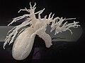
MRCP image showing stones in the distal common bile duct: (a) Gallbladder with stones, (b) Stones in bile duct, (c) Pancreatic duct, (d) Duodenum.
ICD-9-CM 88.97
MeSH D049448
OPS-301 code 3-843

Magnetic resonance cholangiopancreatography (MRCP) is a medical imaging technique. It uses magnetic resonance imaging to visualize the biliary and pancreatic ducts non-invasively. This procedure can be used to determine whether gallstones are lodged in any of the ducts surrounding the gallbladder.

Contents

Uses

MRCP has been slowly replacing endoscopic retrograde cholangiopancreatography (ERCP) as investigation of choice. MRCP is highly accurate in diagnosing the biliary system, pancreatic duct and accessing surrounding solid organs. Several advantages offered by MRCP is its non-invasive nature, less costly, requires less examination time when compared to ERCP (30 minutes), fewer staff required, and does not require any ionising radiation. [1] [2] [3] [4]

MRCP is used to diagnose gallstones. It can also diagnose choledochal cysts very reliably. [5] Besides providing information regarding the biliary system, MRCP also provides information regarding surrounding solid organs and blood vessels, thus useful for planning the resection of pancreatic cancer and look for complications of primary sclerosing cholangitis such as liver cirrhosis and cholangiocarcinoma. [5]

Technique

Subject is needed to fast for at least four hours to ensure the biliary system is maximally distended with fluid while keeping the fluid in the gastrointestinal system at a minimum. [1] However, clear fluid and routine medication is allowed before the scan. [1] Negative oral contrast such as pineapple juice, [1] date syrup, ferumoxsil, Açaí juice and water are useful in decreasing T2 signal intensity, thus minimising signals from stomach and duodenum from interfering with signals from the biliary system. [6]

MRCP makes use of heavily T2-weighted MRI pulse sequences. [3] [7] These sequences show high signal in static or slow moving fluids within the gallbladder, biliary ducts and pancreatic duct, with low signal of surrounding tissue. Secretin is also given to a patient to increase ductal compliance, making imaging easier. [3]

History

It was introduced by Wallner in 1991. [8]

Additional images

See also

Related Research Articles

<span class="mw-page-title-main">Bile</span> Dark greenish-brown fluid aiding in the digestion of fats

Bile, or gall, is a dark-green-to-yellowish-brown fluid produced by the liver of most vertebrates that aids the digestion of lipids in the small intestine. In humans, bile is produced continuously by the liver and stored and concentrated in the gallbladder. After eating, this stored bile is discharged into the duodenum.

<span class="mw-page-title-main">Gallbladder</span> Organ in humans and other vertebrates

In vertebrates, the gallbladder, also known as the cholecyst, is a small hollow organ where bile is stored and concentrated before it is released into the small intestine. In humans, the pear-shaped gallbladder lies beneath the liver, although the structure and position of the gallbladder can vary significantly among animal species. It receives and stores bile, produced by the liver, via the common hepatic duct, and releases it via the common bile duct into the duodenum, where the bile helps in the digestion of fats.

<span class="mw-page-title-main">Cholecystitis</span> Medical condition

Cholecystitis is inflammation of the gallbladder. Symptoms include right upper abdominal pain, pain in the right shoulder, nausea, vomiting, and occasionally fever. Often gallbladder attacks precede acute cholecystitis. The pain lasts longer in cholecystitis than in a typical gallbladder attack. Without appropriate treatment, recurrent episodes of cholecystitis are common. Complications of acute cholecystitis include gallstone pancreatitis, common bile duct stones, or inflammation of the common bile duct.

<span class="mw-page-title-main">Cholecystectomy</span> Surgical removal of the gallbladder

Cholecystectomy is the surgical removal of the gallbladder. Cholecystectomy is a common treatment of symptomatic gallstones and other gallbladder conditions. In 2011, cholecystectomy was the eighth most common operating room procedure performed in hospitals in the United States. Cholecystectomy can be performed either laparoscopically, or via an open surgical technique.

<span class="mw-page-title-main">Klatskin tumor</span> Medical condition

A Klatskin tumor is a cholangiocarcinoma occurring at the confluence of the right and left hepatic bile ducts. The disease was named after Gerald Klatskin, who in 1965 described 15 cases and found some characteristics for this type of cholangiocarcinoma

<span class="mw-page-title-main">Endoscopic retrograde cholangiopancreatography</span> Use of endoscopy and fluoroscopy to treat and diagnose digestive issues.

Endoscopic retrograde cholangiopancreatography (ERCP) is a technique that combines the use of endoscopy and fluoroscopy to diagnose and treat certain problems of the biliary or pancreatic ductal systems. It is primarily performed by highly skilled and specialty trained gastroenterologists. Through the endoscope, the physician can see the inside of the stomach and duodenum, and inject a contrast medium into the ducts in the biliary tree and pancreas so they can be seen on radiographs.

<span class="mw-page-title-main">Primary sclerosing cholangitis</span> Medical condition

Primary sclerosing cholangitis (PSC) is a long-term progressive disease of the liver and gallbladder characterized by inflammation and scarring of the bile ducts, which normally allow bile to drain from the gallbladder. Affected individuals may have no symptoms or may experience signs and symptoms of liver disease, such as yellow discoloration of the skin and eyes, itching, and abdominal pain.

<span class="mw-page-title-main">Cholangiography</span>

Cholangiography is the imaging of the bile duct by x-rays and an injection of contrast medium.

<span class="mw-page-title-main">Cholangiocarcinoma</span> Bile duct adenocarcinoma

Cholangiocarcinoma, also known as bile duct cancer, is a type of cancer that forms in the bile ducts. Symptoms of cholangiocarcinoma may include abdominal pain, yellowish skin, weight loss, generalized itching, and fever. Light colored stool or dark urine may also occur. Other biliary tract cancers include gallbladder cancer and cancer of the ampulla of Vater.

<span class="mw-page-title-main">Acute pancreatitis</span> Medical condition

Acute pancreatitis (AP) is a sudden inflammation of the pancreas. Causes in order of frequency include: 1) a gallstone impacted in the common bile duct beyond the point where the pancreatic duct joins it; 2) heavy alcohol use; 3) systemic disease; 4) trauma; 5) and, in minors, mumps. Acute pancreatitis may be a single event; it may be recurrent; or it may progress to chronic pancreatitis.

<span class="mw-page-title-main">Gastrointestinal disease</span> Medical condition

Gastrointestinal diseases refer to diseases involving the gastrointestinal tract, namely the oesophagus, stomach, small intestine, large intestine and rectum, and the accessory organs of digestion, the liver, gallbladder, and pancreas.

<span class="mw-page-title-main">Common bile duct stone</span> Medical condition

Common bile duct stone, also known as choledocholithiasis, is the presence of gallstones in the common bile duct (CBD). This condition can cause jaundice and liver cell damage. Treatments include choledocholithotomy and endoscopic retrograde cholangiopancreatography (ERCP).

Pancreatic divisum is a congenital anomaly in the anatomy of the ducts of the pancreas in which a single pancreatic duct is not formed, but rather remains as two distinct dorsal and ventral ducts. Most individuals with pancreas divisum remain without symptoms or complications. A minority of people with pancreatic divisum may develop episodes of abdominal pain, nausea or vomiting due to acute or chronic pancreatitis. The presence of pancreas divisum is usually identified with cross sectional diagnostic imaging, such as MRI or computed tomography (CT) imaging. In some cases, endoscopic retrograde cholangiopancreatography (ERCP) is performed, revealing the diagnosis of pancreas divisum. If no symptoms or complications are present, then treatment is not necessary. However, if there is recurrent pancreatitis, then a sphincterotomy of the minor papilla may be indicated.

<span class="mw-page-title-main">Ascending cholangitis</span> Medical condition

Ascending cholangitis, also known as acute cholangitis or simply cholangitis, is inflammation of the bile duct, usually caused by bacteria ascending from its junction with the duodenum. It tends to occur if the bile duct is already partially obstructed by gallstones.

<span class="mw-page-title-main">Biliary colic</span> Medical condition in which gallstones cause acute pain

Biliary colic, also known as symptomatic cholelithiasis, a gallbladder attack or gallstone attack, is when a colic occurs due to a gallstone temporarily blocking the cystic duct. Typically, the pain is in the right upper part of the abdomen, and can be severe. Pain usually lasts from 15 minutes to a few hours. Often, it occurs after eating a heavy meal, or during the night. Repeated attacks are common.

<span class="mw-page-title-main">Sphincter of Oddi dysfunction</span> Medical condition

Sphincter of Oddi dysfunction refers to a group of functional disorders leading to abdominal pain due to dysfunction of the Sphincter of Oddi: functional biliary sphincter of Oddi and functional pancreatic sphincter of Oddi disorder. The sphincter of Oddi is a sphincter muscle, a circular band of muscle at the bottom of the biliary tree which controls the flow of pancreatic juices and bile into the second part of the duodenum. The pathogenesis of this condition is recognized to encompass stenosis or dyskinesia of the sphincter of Oddi ; consequently the terms biliary dyskinesia, papillary stenosis, and postcholecystectomy syndrome have all been used to describe this condition. Both stenosis and dyskinesia can obstruct flow through the sphincter of Oddi and can therefore cause retention of bile in the biliary tree and pancreatic juice in the pancreatic duct.

<span class="mw-page-title-main">Pancreatic cyst</span> Medical condition

A pancreatic cyst is a fluid filled sac within the pancreas.

<span class="mw-page-title-main">Michel Kahaleh</span>

Michel Kahaleh is an American gastroenterologist and an expert in therapeutic endoscopy.

<span class="mw-page-title-main">Choledochoduodenostomy</span>

Choledochoduodenostomy (CDD) is a surgical procedure to create an anastomosis, a surgical connection, between the common bile duct (CBD) and an alternative portion of the duodenum. In healthy individuals, the CBD meets the pancreatic duct at the ampulla of Vater, which drains via the major duodenal papilla to the second part of duodenum. In cases of benign conditions such as narrowing of the distal CBD or recurrent CBD stones, performing a CDD provides the diseased patient with CBD drainage and decompression. A side-to-side anastomosis is usually performed.

<span class="mw-page-title-main">Biliary endoscopic sphincterotomy</span> Use of endoscopy and fluoroscopy to treat and diagnose digestive issues.

Biliary endoscopic sphincterotomy is a procedure where the sphincter of Oddi and the segment of the common bile duct where it enters the duodenum are cannulated and then cut with a sphincterotome, a device that includes a wire which cuts with an electric current (electrocautery).

References

  1. 1 2 3 4 Mandarano G, Sim J (October 2008). "The diagnostic MRCP examination: overcoming technical challenges to ensure clinical success". Biomedical Imaging and Intervention Journal. 4 (4): e28. doi:10.2349/biij.4.4.e28. PMC   3097748 . PMID   21611015.
  2. Prasad, SR; D. Sahani; S. Saini (November 2001). "Clinical applications of magnetic resonance cholangiopancreatography". Journal of Clinical Gastroenterology. 33 (5): 362–6. doi:10.1097/00004836-200111000-00004. PMID   11606850. S2CID   28902645.
  3. 1 2 3 Stevens, Tyler; Freeman, Martin L. (2019-01-01), Chandrasekhara, Vinay; Elmunzer, B. Joseph; Khashab, Mouen A.; Muthusamy, V. Raman (eds.), "57 - Recurrent Acute Pancreatitis", Clinical Gastrointestinal Endoscopy (Third Edition), Philadelphia: Elsevier, pp. 661–673.e3, doi:10.1016/b978-0-323-41509-5.00057-8, ISBN   978-0-323-41509-5 , retrieved 2021-01-28
  4. Hekimoglu K, Ustundag Y, Dusak A, et al. (August 2008). "MRCP vs. ERCP in the evaluation of biliary pathologies: review of current literature". Journal of Digestive Diseases. 9 (3): 162–9. doi:10.1111/j.1751-2980.2008.00339.x. PMID   18956595. S2CID   25726694.
  5. 1 2 Fulcher, Ann S.; Turner, Mary Ann (2008-01-01), Gore, Richard M.; Levine, Marc S. (eds.), "chapter 77 - Magnetic Resonance Cholangiopancreatography", Textbook of Gastrointestinal Radiology (Third Edition), Philadelphia: W.B. Saunders, pp. 1383–1398, doi:10.1016/b978-1-4160-2332-6.50082-8, ISBN   978-1-4160-2332-6 , retrieved 2021-01-28
  6. Al-Atia, Mohassad. "Can oral contrast enhance image quality at MRCP? - A literature review" (PDF). Örebro University, Sweden. Part of Medicine, advanced level, Degree project. Archived from the original (PDF) on 12 June 2020. Retrieved 7 May 2022.
  7. Griffin, Nyree; Charles-Edwards, Geoff; Grant, Lee Alexander (2011-09-28). "Magnetic resonance cholangiopancreatography: the ABC of MRCP". Insights into Imaging. 3 (1): 11–21. doi:10.1007/s13244-011-0129-9. ISSN   1869-4101. PMC   3292642 . PMID   22695995.
  8. Albert L. Baert (13 February 2008). Encyclopedia of Diagnostic Imaging. Springer. pp. 123–. ISBN   978-3-540-35278-5 . Retrieved 3 July 2011.