A transthoracic echocardiogram (TTE) is the most common type of echocardiogram, which is a still or moving image of the internal parts of the heart using ultrasound. In this case, the probe (or ultrasonic transducer) is placed on the chest or abdomen of the subject to get various views of the heart. It is used as a non-invasive assessment of the overall health of the heart, including a patient's heart valves and degree of heart muscle contraction (an indicator of the ejection fraction). The images are displayed on a monitor for real-time viewing and then recorded.
Often abbreviated "TTE", it can be easily confused with transesophageal echocardiography which is abbreviated "TEE". Pronunciation of "TTE" and "TEE" are similar, and full use of "transthoracic" or "transesophageal" can minimize any verbal miscommunication.
Details
A TTE is a clinical tool to evaluate the structure and function of the heart. All four chambers and all four valves can be assessed by TTE, but the quality and visibility of these structures varies from person to person. Other structures visible on TTE include the aorta, the pericardium, pleural effusions, ascites, and inferior vena cava. It can be used to diagnose a heart attack, enlargement/hypertrophy of the heart, infiltration of the heart from an abnormal substance (e.g. amyloidosis), weakness of the heart, and cardiac tumors. With advanced measurements of the movement of the tissue with time (Tissue Doppler), it can measure diastolic function, fluid status,[1] and ventricular dyssynchrony.
TTE in adults is also of limited use for the structures at the back of the heart, such as the left atrial appendage. Transesophageal echocardiography may be more accurate than TTE because it excludes the variables previously mentioned and allows closer visualization of common sites for vegetation and other abnormalities. Transesophageal Echocardiography also affords better visualization of prosthetic heart valves and clots within the four chambers of the heart. This type of Echocardiogram may be a better option for patients with thick chests, abnormal chest walls, chronic obstructive pulmonary disease and the obese. However, transthoracic is often superior to transesophageal for visualization of the apex of the left ventricle (e.g., left ventricular thrombus) and visualization of the ventricular size of mechanical valves.[citation needed]
"Bubble contrast TTE" involves the injection of agitated saline into a vein, followed by an Echocardiographic study. The bubbles are initially detected in the right atrium and right ventricle. If bubbles appear in the left heart, it may indicate a shunt, such as a patent foramen ovale, atrial septal defect, ventricular septal defect or arteriovenous malformations in the lungs.[2]
If a doctor deems it necessary, a stress TTE may be performed. It can be accomplished by either exercising on a bike or treadmill, or by medicine given through an IV along with a contrast agent to make the bodily fluids show up brighter. It allows a comparison between the heart at rest and the heart when it is beating at a faster rate. (Transthoracic Echocardiogram, n.d.)
There are some risks associated when having an echocardiogram performed. It is possible that the images will not show up clearly enough, which can cause a misdiagnosis.
A typical TTE examination is done by either a cardiologist or a cardiac sonographer. It is a non-invasive test that can be done in many settings that include clinic exam room, inpatient rooms, and exam rooms dedicated to echo imaging. Examination involves using an echo probe at various positions or windows to obtain views of the heart (thus capturing images/videos for later playback while formally "reading" the study to come up with the findings). Examination is usually done while lying flat and tilted onto the left side to bring the heart into better view. Ultrasound gel is used to improve the acoustic windows and increase quality of the captured images. Overall, an uncomplicated TTE exam takes less than 30 minutes.
Limited studies (i.e., looking at only specific structures) can be done as a follow-up exam to a full study, or can be done as "point of care" to answer specific questions in the appropriate setting. For example, critically ill patients often have "bedside ultrasounds" performed to assess particular questions the treating team has about their status. This could be looking for cardiac tamponade and acute valve regurgitation. Often, this may include examination of other organ systems such as lungs for effusions or the focused assessment with sonography for trauma.
Interpretation of the exam can be done by anyone trained in reading echocardiograms. However, this is often limited to cardiologists for "formal reading" of these studies. Anesthesiologists can perform intra-operative TEEs during surgical cases and they interpret their own studies. Anyone performing a bedside or "point of care" echocardiogram is expected to interpret their own study as it is performed. Pocket-sized TTE devices are growing in popularity.
Limitations
TTE is inherently limited in what it is capable of doing. Notably, it must be used through the skin, and ultrasound waves must go through skin and soft tissue before reaching the heart. This is in contrast to transesophageal echo (TEE) in which the probe is directly behind the left atrium in the esophagus and has far less tissue to penetrate, which means images from TEE are far superior in quality. Extremes in body sizes (obesity and cachexia) limit the acoustic windows and degrade the image quality of TTEs.
TTE is also limited in its views of structures. Being a surface modality, the structures closest to the skin are better visualized than those deeper to the skin. A common example that demonstrates this is the visibility of the left atrial appendage. This structure is known to form clots in atrial fibrillation and the LAA is rarely seen on TTE but readily seen on TEE. Cardioversion of atrial fibrillation in someone not on anticoagulation would require TEE to best visualize the LAA to rule out a thrombus. (If a thrombus is present, there is a much higher risk of stroke if returned to a sinus rhythm.)
Like all of the kinds of echocardiography, TTE is limited to structure and function. It is not, for example, able to determine perfusion of the myocardium, which would require a metabolic imaging modality such as PET or SPECT stress testing. Perfusion can be inferred based on wall motion, however.
Modalities
2D echo
This is the most common modality used today. A two-dimensional plane is formed by sweeping the ultrasound waves to obtain an image that varies with angle and depth. Evaluation of the structures is done in this view.
M-mode
This modality is obtained at a single angle and then plotted against time to obtain an image. It can be used to watch the movement of structures with time
Doppler
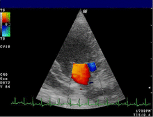
Color doppler
Color doppler over mitral valve
Color doppler is a form of 2D echo in which the doppler shift of the structures is shown as color. Typically, this is shown as red and blue with red indicating movement toward the transducer and blue indicating movement away from the transducer. This can be used to show blood flow through the valves to visually indicate the direction of blood flow. Abnormal blood flow can reflect stenosis and regurgitation of the valve. Color doppler can also show blood flow in abnormal locations such as with septal defects (ASD or VSD).
Color doppler can also be applied to M-mode. Using color doppler in this way gives better visualization of the changes in flow with time due to the higher frame rate with M-mode.
Spectral doppler
Spectral doppler through pulmonary vein
Spectral doppler is presented similarly to M-mode in which the doppler information is plotted as a spectrogram. This can be both "continuous" and "pulse" wave where the former shows the spectrum along a specific line and the latter shows within a small window along that line. Continuous wave is better at showing maximal velocities and pulse wave is better for showing flow through a small volume.
Spectral doppler is often used for quantification of flow. For example, the aortic valve area can be estimated using the continuity equation by measuring the velocity time integral (VTI) of the aortic valve & LV outflow tract; the VTI is calculated by tracing the flow on the spectral doppler curve. Spectral doppler is also useful for calculating the maximum flow and mean flow through a valve (used to grade valve stenosis).
Tissue doppler can be used to determine motion of myocardial tissue. This can be used to measure motion of the septal and lateral mitral annulus to suggest diastolic heart failure.
Axes
The images obtained with echocardiography are in reference to the heart itself. Two key axes of the heart are the long axis and short axis. The long axis is an imaginary line from the apex of the heart through the center of the tricuspid/mitral valve (depending on ventricle of reference). The short axis is perpendicular to the long axis and shows the heart in cross section. Axes are also defined for each valve with long-axis defined as that of blood flow, and short-axis is the plane perpendicular to the flow.
Windows
Evaluation of the heart with echocardiography requires having "acoustic windows" of the heart. Bone reflects the ultrasound waves and so all structures directly behind bone are not visible with ultrasound. This requires that the heart be viewed between bones and, in particular, between ribs. The most common views are the parasternal, apical, subcostal, and suprasternal windows..
Parasternal: adjacent to the sternum. Without qualification, this means the left side of the heart but right parasternal views can be attempted.
Apical: at the apex of the heart.
Subcostal: below the sternum at the top of the abdomen
Suprasternal: above the sternal at the base of the neck.
Views
There are several typical views obtained during a routine TTE. Views outside of the typical views can be considered "off axis" and may be obtained for specific purposes.
Parasternal long axis (PLAX)
Parasternal long axis
This view is obtained to the left of the sternum and views the heart in its long axis. In this view, the mitral valve, aortic valve, right ventricular outflow tract, base of the left ventricle, and the left atrium can be visible. Angulation in this view can bring the right ventricular inflow tract and tricuspid valve into view, and angulation the opposite way can bring the pulmonary valve into view.
In this view, it is possible to appreciate the long-axis cross section of the mitral and aortic valves. The classic "hockey stick" shape of rheumatic mitral stenosis can be appreciated in this view. However, the angle of the probe with these valves can lead to under-appreciation of valve dysfunction.
The parasternal long view of the pulmonary valve is the only view of the posterior leaflet.
Structures visible:
Anterior septal and inferior lateral walls of the left ventricle
Left atrium
Mitral valve in long-axis with chordae
Aortic valve in long-axis
Tricuspid valve in long-axis (angulated) and right ventricular inflow tract
Pulmonary valve in long-axis (angulated) and right ventricular outflow tract
Measurements in this view can be used to quantify the heart:
Left ventricular size and wall thickness
Left atrial linear dimension (as opposed to area)
Left ventricular outflow tract diameter (used to calculate aortic valve area by the continuity equation)
Aortic annulus, sinus of Valsalva, and aortic root sizes
Color doppler of all four valves
Spectral doppler of tricuspid and pulmonary valves
Parasternal short axis (PSAX)
Parasternal short axis at mid-LV showing papillary musclesParasternal short axis showing aortic valve
This view is obtained in the same window as the parasternal long, but with the probe rotated 90°. In this view, the aortic valve is seen in cross-section with the right ventricular inflow & outflow tracts visible with the tricuspid valve as well. Pulmonary valve is not visible in this view. Both the right and left atria are visible.
The standard PSAX view is at the level of the aortic valve, but moving the probe along the long-axis can review the LV outflow tract, LV at the base, and LV at the midsection.
Right ventricle, including inflow and outflow tracts
Left ventricle in short-axis
Closer to the base can reveal the left ventricular outflow tract
At the level of the base can show the movement of the mitral valve leaflets in short-axis
At the level of mid-LV can show papillary muscles
Measurements in this view can be used to quantify the heart:
Aortic valve area by planimetry
Color doppler of all four valves
Spectral doppler of tricuspid and pulmonary valves
Apical four chamber (A4C)
Apical 4 chamber
This view is obtained at the apex of the heart and looking toward the base of the heart (where the valves are). In this view, the mitral valve, tricuspid valve, and all four chambers are visible. This view shows the right ventricle from base to apex and is a useful view to estimate RV systolic function. TAPSE (= tricuspid annular plane systolic excursion) is also measured in this view with M-mode through the lateral tricuspid annulus.
Structures:
Inferior septum and anterior lateral segments of the left ventricle
Right ventricle
Left atrium
Right atrium
Mitral valve
Tricuspid valve
Measurements in this view can be used to quantify the heart:
Mitral valve flow is best seen in this view and has the best angle with probe to estimate flows
Tricuspid valve flow
Tissue doppler at the mitral valve annulus (septum and lateral wall) for diastolic function
Agitated saline bubble study for right to left shunting (PFO, ASD, VSD)
With contrast, apical and mural LV thrombi can be easily seen
Apical three chamber (A3C)
This view is obtained at the same window as the apical four chamber and then rotation of the probe. In this view, the mitral valve and aortic valve are in view and is roughly similar to the parasternal long axis. In this view, the LV outflow tract is best in alignment with the probe and so gives the best estimate of flow through the LVOT, which is commonly used to estimate aortic stenosis.
Structures:
Aortic valve
Mitral valve
Left ventricle
Left atrium
Measurements in this view can be used to quantify the heart:
Left ventricular outflow tract volume-time integral (LVOT VTI) to be used in conjunction with aortic valve VTI for aortic valve area and stenosis
Mitral valve flow
Apical two chamber (A2C)
Apical two chamber
This view is obtained at the same window as the apical four chamber and then rotation of the probe. In this view, the mitral valve is visible with the left atrium and left ventricle.
Structures:
Anterior and inferior segments of the left ventricle
Mitral valve in long-axis
Left atrium
Measurements in this view can be used to quantify the heart:
Mitral valve flow
Spectral doppler of the mitral valve
Subcostal
Subcostal
This view is obtained below the sternum and at the top part of the abdomen. In this view, the junction of the inferior vena cava with the right atrium is best seen. From this window, it is possible in some people to see roughly equivalent views of the apical four chamber and parasternal short views. In some people, this may afford these common views but at a subcostal window that may not be obtained through the parasternal and/or apical windows because of various reasons such as chest wall trauma, open wounds, or poor acoustic windows. However, the subcostal window is the only window to view the inferior vena cava that can help support an estimation of the central venous pressure based on size and collapsibility during respiration.
Other non-cardiac structures are visible in this view and some pathologies — such as ascites — can be observed.
Suprasternal (SSN)
This view is obtained above the sternum in the suprasternal notch. In this view, the aortic arch and portion of the descending aorta can be seen. Color and spectral doppler through the descending aorta can show signs of coarctation of the aorta.
Structures
Examples of TTE views of various structures of the heart.
Aortic valve
PSAX
PLAX zoom
PSAX bicuspid
PLAX regurgitation
A3C regurgitation
PLAX endocarditis
PLAX calcified & stenosis
PSAX calcified & stenosis
Mitral valve
PLAX zoom
A4C mild regurgitation
A4C moderate regurgitation
PLAX stenosis
A4C stenosis
A3C stenosis
Tricuspid valve
PSAX severe regurgitation
A4C severe regurgitation
SUB severe regurgitation
SUB endocarditis
Pulmonary valve
PLAX severe regurgitation
PSAX severe regurgitation
Measurements
Example of quantification of the aortic root
Routine TTE exams can provide a significant wealth of information about the heart's structure and function:
Left ventricular size, thickness, systolic function, and diastolic function
Right ventricular size and systolic function
Aortic valve
Aortic valve sclerosis & stenosis
Aortic valve insufficiency
Mitral valve
Mitral stenosis
Mitral regurgitation
Tricuspid valve
Tricuspid regurgitation (stenosis is possible, but rare)
Pulmonary valve
Pulmonary regurgitation (stenosis is possible, but rare)
Inferior vena cava size as estimate of central venous pressure
Aortic root size for thoracic ascending aortic aneurysm
Pericardial effusion size
All function dysfunction is graded on a scale (normal, trace, mild, moderate, or severe) based on various criteria. Grading of valve function is used for prognosis and helps determine management as valve dysfunction progresses.
Echo societies have published normal ranges for various features of the heart on various views to promote TTE as a quantifiable assessment of the heart.
Equations
Quantitative echo utilizes a number of equations to calculate aspects of the heart structure and function. Simplified Bernoulli equation and continuity equation are two common equations used. Others are used in grading valve function (e.g., EROA, PISA).
Diseases
TTE can be useful for diagnosing many cardiac diseases.
A heart valve is a biological one-way valve that allows blood to flow in one direction through the chambers of the heart. Four valves are usually present in a mammalian heart and together they determine the pathway of blood flow through the heart. A heart valve opens or closes according to differential blood pressure on each side.
Heart sounds are the noises generated by the beating heart and the resultant flow of blood through it. Specifically, the sounds reflect the turbulence created when the heart valves snap shut. In cardiac auscultation, an examiner may use a stethoscope to listen for these unique and distinct sounds that provide important auditory data regarding the condition of the heart.
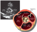
The aortic valve is a valve in the heart of humans and most other animals, located between the left ventricle and the aorta. It is one of the four valves of the heart and one of the two semilunar valves, the other being the pulmonary valve. The aortic valve normally has three cusps or leaflets, although in 1–2% of the population it is found to congenitally have two leaflets. The aortic valve is the last structure in the heart the blood travels through before stopping the flow through the systemic circulation.
The mitral valve, also known as the bicuspid valve or left atrioventricular valve, is one of the four heart valves. It has two cusps or flaps and lies between the left atrium and the left ventricle of the heart. The heart valves are all one-way valves allowing blood flow in just one direction. The mitral valve and the tricuspid valve are known as the atrioventricular valves because they lie between the atria and the ventricles.
Heart murmurs are unique heart sounds produced when blood flows across a heart valve or blood vessel. This occurs when turbulent blood flow creates a sound loud enough to hear with a stethoscope. The sound differs from normal heart sounds by their characteristics. For example, heart murmurs may have a distinct pitch, duration and timing. The major way health care providers examine the heart on physical exam is heart auscultation; another clinical technique is palpation, which can detect by touch when such turbulence causes the vibrations called cardiac thrill. A murmur is a sign found during the cardiac exam. Murmurs are of various types and are important in the detection of cardiac and valvular pathologies.
A ventricle is one of two large chambers located toward the bottom of the heart that collect and expel blood towards the peripheral beds within the body and lungs. The blood pumped by a ventricle is supplied by an atrium, an adjacent chamber in the upper heart that is smaller than a ventricle. Interventricular means between the ventricles, while intraventricular means within one ventricle.
Echocardiography, also known as cardiac ultrasound, is the use of ultrasound to examine the heart. It is a type of medical imaging, using standard ultrasound or Doppler ultrasound. The visual image formed using this technique is called an echocardiogram, a cardiac echo, or simply an echo.
Mitral stenosis is a valvular heart disease characterized by the narrowing of the opening of the mitral valve of the heart. It is almost always caused by rheumatic valvular heart disease. Normally, the mitral valve is about 5 cm2 during diastole. Any decrease in area below 2 cm2 causes mitral stenosis. Early diagnosis of mitral stenosis in pregnancy is very important as the heart cannot tolerate increased cardiac output demand as in the case of exercise and pregnancy. Atrial fibrillation is a common complication of resulting left atrial enlargement, which can lead to systemic thromboembolic complications such as stroke.
A transesophageal echocardiogram is an alternative way to perform an echocardiogram. A specialized probe containing an ultrasound transducer at its tip is passed into the patient's esophagus. This allows image and Doppler evaluation which can be recorded. It is commonly used during cardiac surgery and is an excellent modality for assessing the aorta, although there are some limitations.
A ventricular septal defect (VSD) is a defect in the ventricular septum, the wall dividing the left and right ventricles of the heart. The extent of the opening may vary from pin size to complete absence of the ventricular septum, creating one common ventricle. The ventricular septum consists of an inferior muscular and superior membranous portion and is extensively innervated with conducting cardiomyocytes.
Mitral regurgitation (MR), also known as mitral insufficiency or mitral incompetence, is a form of valvular heart disease in which the mitral valve is insufficient and does not close properly when the heart pumps out blood. It is the abnormal leaking of blood backwards – regurgitation from the left ventricle, through the mitral valve, into the left atrium, when the left ventricle contracts. Mitral regurgitation is the most common form of valvular heart disease.
Valvular heart disease is any cardiovascular disease process involving one or more of the four valves of the heart. These conditions occur largely as a consequence of aging, but may also be the result of congenital (inborn) abnormalities or specific disease or physiologic processes including rheumatic heart disease and pregnancy.
Regurgitation is blood flow in the opposite direction from normal, as the backward flowing of blood into the heart or between heart chambers. It is the circulatory equivalent of backflow in engineered systems. It is sometimes called reflux.
Lutembacher's syndrome is a very rare form of congenital heart disease that affects one of the chambers of the heart as well as a valve. It is commonly known as both congenital atrial septal defect (ASD) and acquired mitral stenosis (MS). Congenital atrial septal defect refers to a hole being in the septum or wall that separates the two atria; this condition is usually seen in fetuses and infants. Mitral stenosis refers to mitral valve leaflets sticking to each other making the opening for blood to pass from the atrium to the ventricles very small. With the valve being so small, blood has difficulty passing from the left atrium into the left ventricle. Septal defects that may occur with Lutembacher's syndrome include: Ostium primum atrial septal defect or ostium secundum which is more prevalent.
The following outline is provided as an overview of and topical guide to cardiology, the branch of medicine dealing with disorders of the human heart. The field includes medical diagnosis and treatment of congenital heart defects, coronary artery disease, heart failure, valvular heart disease and electrophysiology. Physicians who specialize in cardiology are called cardiologists.
The E/A ratio is a marker of the function of the left ventricle of the heart. It represents the ratio of peak velocity blood flow from left ventricular relaxation in early diastole to peak velocity flow in late diastole caused by atrial contraction. It is calculated using Doppler echocardiography, an ultrasound-based cardiac imaging modality. Abnormalities in the E/A ratio suggest that the left ventricle, which pumps blood into the systemic circulation, cannot fill with blood properly in the period between contractions. This phenomenon is referred to as diastolic dysfunction and can eventually lead to the symptoms of heart failure.
Tissue Doppler echocardiography (TDE) is a medical ultrasound technology, specifically a form of echocardiography that measures the velocity of the heart muscle (myocardium) through the phases of one or more heartbeats by the Doppler effect of the reflected ultrasound. The technique is the same as for flow Doppler echocardiography measuring flow velocities. Tissue signals, however, have higher amplitude and lower velocities, and the signals are extracted by using different filter and gain settings. The terms tissue Doppler imaging (TDI) and tissue velocity imaging (TVI) are usually synonymous with TDE because echocardiography is the main use of tissue Doppler.
A quadricuspid aortic valve (QAV) is a rare congenital heart defect characterized by the presence of four cusps, instead of the usual three found normally in the aortic valve. It is a defect that occurs during embryological development of the aortic trunk during gestation. There is an increased risk of developing post-natal aortic regurgitations and other heart-related diseases; therefore patients with the condition should be carefully monitored.
Mitral annular calcification (MAC) is a multifactorial chronic degenerative process in which calcium with lipid is deposited (calcified) in the annular fibrosa ring of the heart's mitral valve. MAC was first discovered and described in 1908 by M. Bonninger in the journal Deutsche Medizinische Wochenschrift. In the majority of cases, affected patients are asymptomatic and the condition is only noted incidentally on echocardiography or computed tomography (CT) scans. However, mitral annular calcification remains clinically significant because while in many cases the calcification is limited to the annulus and proximal leaflet bases, it may also extend further into the valve structure. This may potentially cause mitral regurgitation (MR) or more rarely mitral stenosis (MS), which may produce the classic symptoms of these conditions over time. In addition, calcification of the annulus can inhibit electrical conduction of the AV node, consequently causing various degrees of heart block. While MAC does not usually necessitate treatment independently, the degree of calcification present in the annulus is an important factor in choosing the most appropriate treatment modality for several conditions that do require intervention, particularly those that cause symptomatic obstruction of left ventricular outflow (LVOT).
Intracardiac echocardiography (ICE) is a specialized form of echocardiography that utilizes an ultrasound-tipped catheter to perform imaging of the heart from within the heart. Unlike transthoracic echocardiography (TTE), ICE is not limited by body habitus. An ICE catheter is inserted into the body, typically, through the femoral vein and advanced into the heart.
This page is based on this Wikipedia article Text is available under the CC BY-SA 4.0 license; additional terms may apply. Images, videos and audio are available under their respective licenses.