Abdominal x-ray

Last updated

Abdominal x-ray
Medical X-Ray imaging ALP02 nevit.jpg
ICD-9-CM 87.5,87.9, 88.0-88.1
MedlinePlus 003815

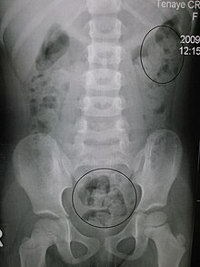
An abdominal x-ray is an x-ray of the abdomen. It is sometimes abbreviated to AXR, or KUB (for kidneys, ureters, and urinary bladder).

Contents

Indications

In children, abdominal x-ray is indicated in the acute setting:

Yet, CT scan is the best alternative for diagnosing intra-abdominal injury. [1]

Computed tomography provides an overall better surgical strategy planning, and possibly fewer unnecessary laparotomies. Abdominal x-ray is therefore not recommended for adults with acute abdominal pain presenting in the emergency department. [2]

Projections

The standard abdominal X-ray protocol is usually a single anteroposterior projection in supine position. [3] Special projections include a PA prone, lateral decubitus, upright AP, and lateral cross-table (with the patient supine). A minimal acute obstructive series (for the purpose of ruling out small bowel obstruction) includes two views: typically, a supine view and an upright view (which are sufficient to detect air-fluid levels), although a lateral decubitus could be substituted for the upright.

Coverage on the x-ray should include from the top of the Liver (or diaphragm) to the pubic symphysis. The abdominal organs included on the xray are the liver, spleen, stomach, intestines, pancreas, kidneys, and bladder.

KUB

Constipation in a young child as seen by KUB X-ray. Circles represent areas of fecal matter (stool is opaque white (not to be confused with white opaque skeletal mass and muscle mass) surrounded by black bowel gas Constipation.JPG
Constipation in a young child as seen by KUB X-ray. Circles represent areas of fecal matter (stool is opaque white (not to be confused with white opaque skeletal mass and muscle mass) surrounded by black bowel gas

KUB stands for Kidneys, Ureters, and Bladder. The KUB projection does not necessarily include the diaphragm. The projection includes the entire urinary system, from the pubic symphysis to the superior aspects of the kidneys. The anteroposterior (AP) abdomen projection, in contrast, includes both halves of the diaphragm. [4] [5] If the patient is large, more than one film loaded in the Bucky in a "landscape" direction may be used for each projection. This is done to ensure that the majority of bowel can be reviewed.

A KUB is a plain frontal supine radiograph of the abdomen. It is often supplemented by an upright PA view of the chest (to rule out air under the diaphragm or thoracic etiologies presenting as abdominal complaints) and a standing view of the abdomen (to differentiate obstruction from ileus by examining gastrointestinal air/water levels).

Despite its name, a KUB is not typically used to investigate pathology of the kidneys, ureters, or bladder, since these structures are difficult to assess (for example, the kidneys may not be visible due to overlying bowel gas.) In order to assess these structures radiographically, a technique called an intravenous pyelogram was historically utilized, and today at many institutions CT urography is the technique of choice. [6]

KUB is typically used to investigate gastrointestinal conditions such as a bowel obstruction and gallstones, and can detect the presence of kidney stones. The KUB is often used to diagnose constipation as stool can be seen readily. The KUB is also used to assess positioning of indwelling devices such as ureteric stents and nasogastric tubes. KUB is also done as a scout film for other procedures such as barium enemas.

Gastrointestinal series

An upper gastrointestinal series is where a contrast medium, usually a radiocontrast agent such as barium sulfate barium salt mixed with water, is ingested or instilled into the gastrointestinal tract, and X-rays are used to create radiographs of the regions of interest. The barium enhances the visibility of the relevant parts of the gastrointestinal tract by coating the inside wall of the tract and appearing white on the film.

A lower gastrointestinal series is where radiographs are taken while barium sulfate, a radiocontrast agent, fills the colon via an enema through the rectum. The term barium enema usually refers to a lower gastrointestinal series, although enteroclysis (an upper gastrointestinal series) is often called a small bowel barium enema.

See also

Related Research Articles

<span class="mw-page-title-main">Bowel obstruction</span> Medical condition

Bowel obstruction, also known as intestinal obstruction, is a mechanical or functional obstruction of the intestines which prevents the normal movement of the products of digestion. Either the small bowel or large bowel may be affected. Signs and symptoms include abdominal pain, vomiting, bloating and not passing gas. Mechanical obstruction is the cause of about 5 to 15% of cases of severe abdominal pain of sudden onset requiring admission to hospital.

<span class="mw-page-title-main">Lower gastrointestinal series</span> Radiographs used to examine abnormalities of the colon

A lower gastrointestinal series is a medical procedure used to examine and diagnose problems with the human colon of the large intestine. Radiographs are taken while barium sulfate, a radiocontrast agent, fills the colon via an enema through the rectum.

<span class="mw-page-title-main">Upper gastrointestinal series</span> Radiographs used to examine abnormalities of the digestive system excluding the colon

An upper gastrointestinal series, also called a barium swallow, barium study, or barium meal, is a series of radiographs used to examine the gastrointestinal tract for abnormalities. A contrast medium, usually a radiocontrast agent such as barium sulfate mixed with water, is ingested or instilled into the gastrointestinal tract, and X-rays are used to create radiographs of the regions of interest. The barium enhances the visibility of the relevant parts of the gastrointestinal tract by coating the inside wall of the tract and appearing white on the film. This in combination with other plain radiographs allows for the imaging of parts of the upper gastrointestinal tract such as the pharynx, larynx, esophagus, stomach, and small intestine such that the inside wall lining, size, shape, contour, and patency are visible to the examiner. With fluoroscopy, it is also possible to visualize the functional movement of examined organs such as swallowing, peristalsis, or sphincter closure. Depending on the organs to be examined, barium radiographs can be classified into "barium swallow", "barium meal", "barium follow-through", and "enteroclysis". To further enhance the quality of images, air or gas is sometimes introduced into the gastrointestinal tract in addition to barium, and this procedure is called double-contrast imaging. In this case the gas is referred to as the negative contrast medium. Traditionally the images produced with barium contrast are made with plain-film radiography, but computed tomography is also used in combination with barium contrast, in which case the procedure is called "CT enterography".

<span class="mw-page-title-main">Abdominal pain</span> Stomach aches

Abdominal pain, also known as a stomach ache, is a symptom associated with both cancer and serious medical issues.

Radiocontrast agents are substances used to enhance the visibility of internal structures in X-ray-based imaging techniques such as computed tomography, projectional radiography, and fluoroscopy. Radiocontrast agents are typically iodine, or more rarely barium sulfate. The contrast agents absorb external X-rays, resulting in decreased exposure on the X-ray detector. This is different from radiopharmaceuticals used in nuclear medicine which emit radiation.

<span class="mw-page-title-main">Gastrointestinal disease</span> Medical condition

Gastrointestinal diseases refer to diseases involving the gastrointestinal tract, namely the esophagus, stomach, small intestine, large intestine and rectum, and the accessory organs of digestion, the liver, gallbladder, and pancreas.

Colic in horses is defined as abdominal pain, but it is a clinical symptom rather than a diagnosis. The term colic can encompass all forms of gastrointestinal conditions which cause pain as well as other causes of abdominal pain not involving the gastrointestinal tract. The most common forms of colic are gastrointestinal in nature and are most often related to colonic disturbance. There are a variety of different causes of colic, some of which can prove fatal without surgical intervention. Colic surgery is usually an expensive procedure as it is major abdominal surgery, often with intensive aftercare. Among domesticated horses, colic is the leading cause of premature death. The incidence of colic in the general horse population has been estimated between 4 and 10 percent over the course of the average lifespan. Clinical signs of colic generally require treatment by a veterinarian. The conditions that cause colic can become life-threatening in a short period of time.

<span class="mw-page-title-main">Chest radiograph</span> Projection X-ray of the chest

A chest radiograph, called a chest X-ray (CXR), or chest film, is a projection radiograph of the chest used to diagnose conditions affecting the chest, its contents, and nearby structures. Chest radiographs are the most common film taken in medicine.

<span class="mw-page-title-main">Intussusception (medical disorder)</span> Medical condition

Intussusception is a medical condition in which a part of the intestine folds into the section immediately ahead of it. It typically involves the small bowel and less commonly the large bowel. Symptoms include abdominal pain which may come and go, vomiting, abdominal bloating, and bloody stool. It often results in a small bowel obstruction. Other complications may include peritonitis or bowel perforation.

<span class="mw-page-title-main">Volvulus</span> Twisting of part of the intestine, causing a bowel obstruction

A volvulus is when a loop of intestine twists around itself and the mesentery that supports it, resulting in a bowel obstruction. Symptoms include abdominal pain, abdominal bloating, vomiting, constipation, and bloody stool. Onset of symptoms may be rapid or more gradual. The mesentery may become so tightly twisted that blood flow to part of the intestine is cut off, resulting in ischemic bowel. In this situation there may be fever or significant pain when the abdomen is touched.

<span class="mw-page-title-main">Diatrizoate</span>

Diatrizoate, also known as amidotrizoate, is a contrast agent used during X-ray imaging. This includes visualizing veins, the urinary system, spleen, and joints, as well as computer tomography. It is given by mouth, injection into a vein, injection into the bladder, through a nasogastric tube, or rectally.

<span class="mw-page-title-main">Pneumoperitoneum</span> Medical condition

Pneumoperitoneum is pneumatosis in the peritoneal cavity, a potential space within the abdominal cavity. The most common cause is a perforated abdominal organ, generally from a perforated peptic ulcer, although any part of the bowel may perforate from a benign ulcer, tumor or abdominal trauma. A perforated appendix seldom causes a pneumoperitoneum.

Pyelogram is a form of imaging of the renal pelvis and ureter.

Gastric volvulus or volvulus of stomach is a twisting of all or part of the stomach by more than 180 degrees with obstruction of the flow of material through the stomach, variable loss of blood supply and possible tissue death. The twisting can occur around the long axis of the stomach: this is called organoaxial or around the axis perpendicular to this, called mesenteroaxial. Obstruction is more likely in organoaxial twisting than with mesenteroaxial while the latter is more associated with ischemia. About one third of the cases are associated with a hiatus hernia. Treatment is surgical.

Abdominal guarding is the tensing of the abdominal wall muscles to guard inflamed organs within the abdomen from the pain of pressure upon them. The tensing is detected when the abdominal wall is pressed. Abdominal guarding is also known as 'défense musculaire'.

<span class="mw-page-title-main">Projectional radiography</span> Formation of 2D images using X-rays

Projectional radiography, also known as conventional radiography, is a form of radiography and medical imaging that produces two-dimensional images by x-ray radiation. The image acquisition is generally performed by radiographers, and the images are often examined by radiologists. Both the procedure and any resultant images are often simply called "X-ray". Plain radiography or roentgenography generally refers to projectional radiography. Plain radiography can also refer to radiography without a radiocontrast agent or radiography that generates single static images, as contrasted to fluoroscopy, which are technically also projectional.

<span class="mw-page-title-main">Ureterostomy</span>

A ureterostomy is the creation of a stoma for a ureter or kidney.

<span class="mw-page-title-main">Abdominopelvic cavity</span>

The abdominopelvic cavity is a body cavity that consists of the abdominal cavity and the pelvic cavity. The upper portion is the abdominal cavity, and it contains the stomach, liver, pancreas, spleen, gallbladder, kidneys, small intestine, and most of the large intestine. The lower portion is the pelvic cavity, and it contains the urinary bladder, the rest of the large intestine, and the internal reproductive organs.

<span class="mw-page-title-main">Double-contrast barium enema</span>

A double-contrast barium enema is a form of contrast radiography in which x-rays of the colon and rectum are taken using two forms of contrast to make the structures easier to see. A liquid containing barium is put into the rectum. Barium is a silver-white metallic compound that outlines the colon and rectum on an x-ray and helps show abnormalities. Air is also put into the rectum and colon to further enhance the x-ray.

Urologic diseases or conditions include urinary tract infections, kidney stones, bladder control problems, and prostate problems, among others. Some urologic conditions do not affect a person for that long and some are lifetime conditions. Kidney diseases are normally investigated and treated by nephrologists, while the specialty of urology deals with problems in the other organs. Gynecologists may deal with problems of incontinence in women.

References

  1. 1 2 3 4 5 "Radiology - Acute indications". Royal Children's Hospital, Melbourne. Retrieved 2017-07-23.
  2. Boermeester, Marie A; Gans, Sarah L.; Stoker, J; Boermeester, Marie A (2012). "Plain abdominal radiography in acute abdominal pain; past, present, and future". International Journal of General Medicine . 5: 525–33. doi:10.2147/IJGM.S17410. ISSN   1178-7074. PMC   3396109 . PMID   22807640.
  3. "Abdomen X-ray system and anatomy - Image data and quality". Radiology Masterclass. Retrieved 27 January 2016.
  4. Frank, Eugene D.; Long, Bruce W.; Smith, Barbara J. (2012). Merrill's Atlas of Radiographic Positioning & Procedures (12 ed.). St. Louis, MO: Mosby Inc. ISBN   978-0-323-07334-9.
  5. Bontrager, Kenneth L.; Lampignano, John P. (2005). Textbook of Radiographic Positioning and Related Anatomy (6 ed.). St. Louis, MO: Mosby, Inc. ISBN   978-0-323-02507-2.
  6. Paul Schmitz, MD, et al. Medscape. Kidneys, ureters, and bladder imaging: plain films of the abdomen. Updated 27 Aug 2015.