Radionuclide angiography

Last updated
Radionuclide angiography
Normal MUGA scan 1.png
Normail MUGA scan
ICD-9-CM 92.05
MeSH D015635 D011875; D015635
OPS-301 code 3-704, 3-708

Radionuclide angiography is an area of nuclear medicine which specialises in imaging to show the functionality of the right and left ventricles of the heart, thus allowing informed diagnostic intervention in heart failure. It involves use of a radiopharmaceutical, injected into a patient, and a gamma camera for acquisition. A MUGA scan (multigated acquisition) involves an acquisition triggered (gated) at different points of the cardiac cycle. MUGA scanning is also called equilibrium radionuclide angiocardiography, radionuclide ventriculography (RNVG), or gated blood pool imaging, as well as SYMA scanning (synchronized multigated acquisition scanning).

Contents

This mode of imaging uniquely provides a cine type of image of the beating heart, and allows the interpreter to determine the efficiency of the individual heart valves and chambers. MUGA/Cine scanning represents a robust adjunct to the now more common echocardiogram. Mathematics regarding acquisition of cardiac output (Q) is well served by both of these methods as well as other inexpensive models supporting ejection fraction as a product of the heart/myocardium in systole. The advantage of a MUGA scan over an echocardiogram or an angiogram is its accuracy. An echocardiogram measures the shortening fraction of the ventricle and is limited by the user's ability. Furthermore, an angiogram is invasive and, often, more expensive. A MUGA scan provides a more accurate representation of cardiac ejection fraction. [1]

History

Normal MUGA scan Normal MUGA scan 2.png
Normal MUGA scan

The MUGA scan was first introduced in the early 1970s and quickly became accepted as the preferred technique for measurement of left ventricular ejection fraction (LVEF) with a high degree of accuracy. Several early studies demonstrated an excellent correlation of MUGA-derived LVEF with values obtained by cardiac catheterization contrast ventriculography. [2]

Purpose

Radionuclide ventriculography is done to evaluate coronary artery disease (CAD), valvular heart disease, congenital heart diseases, cardiomyopathy, and other cardiac disorders. [3] MUGA is typically ordered for the following patients:[ citation needed ]

Radionuclide ventriculography gives a much more precise measurement of left ventricular ejection fraction (LVEF) than a transthoracic echocardiogram (TTE). Transthoracic echocardiogram is highly operator dependant, therefore radionuclide ventriculography is a more reproducible measurement of LVEF. Its primary use today is in monitoring cardiac function in patients receiving certain chemotherapeutic agents (anthracyclines: doxorubicin or daunorubicin) which are cardiotoxic. The chemotherapy dose is often determined by the patient's cardiac function. In this setting, a much more accurate measurement of ejection fraction, than a transthoracic echocardiogram can provide, is necessary. [3]

Procedure

The MUGA scan is performed by labeling the patient's red blood pool with a radioactive tracer, technetium-99m-pertechnetate (Tc-99m), and measuring radioactivity over the anterior chest as the radioactive blood flows through the large vessels and the heart chambers.[ citation needed ]

The introduction of the radioactive marker can either take place in vivo or in vitro . In the in vivo method, stannous (tin) ions are injected into the patient's bloodstream. A subsequent intravenous injection of the radioactive substance, technetium-99m-pertechnetate, labels the red blood cells in vivo . With an administered activity of about 800 MBq, the effective radiation dose is about 6 mSv. [4] [5]

In the in vitro method, some of the patient's blood is drawn and the stannous ions (in the form of stannous chloride) are injected into the drawn blood. The technetium is subsequently added to the mixture as in the in vivo method. In both cases, the stannous chloride reduces the technetium ion and prevents it from leaking out of the red blood cells during the procedure. [6] [7]

The in vivo technique is more convenient for the majority of patients since it is less time-consuming and less costly and more than 80 percent of the injected radionuclide usually binds to red blood cells with this approach. Red blood cell binding of the radioactive tracer is generally more efficient than in vitro labeling, and it is preferred in patients with indwelling intravenous catheters to decrease the adherence of Tc-99m to the catheter wall and increase the efficiency of blood pool labeling. [8]

The patient is placed under a gamma camera, which detects the low-level 140 keV gamma radiation being given off by Technetium-99m (99mTc). As the gamma camera images are acquired, the patient's heart beat is used to 'gate' the acquisition. The final result is a series of images of the heart (usually sixteen), one at each stage of the cardiac cycle.[ citation needed ]

Depending on the objectives of the test, the doctor may decide to perform either a resting or a stress MUGA. During the resting MUGA, the patient lies stationary, whereas during a stress MUGA, the patient is asked to exercise during the scan. The stress MUGA measures the heart performance during exercise and is usually performed to assess the impact of a suspected coronary artery disease. In some cases, a nitroglycerin MUGA may be performed, where nitroglycerin (a vasodilator) is administered prior to the scan.[ citation needed ]

The resulting images show that the volumetrically derived blood pools in the chambers of the heart and timed images may be computationally interpreted to calculate the ejection fraction and injection fraction of the heart. The Massardo method can be used to calculate ventricle volumes. This nuclear medicine scan yields an accurate, inexpensive and easily reproducible means of measuring and monitoring the ejection and injection fractions of the ventricles, which are one of many of the important clinical metrics in assessing global heart performance.[ citation needed ]

Radiation exposure

It exposes patients to less radiation than do comparable chest x-ray studies. However, the radioactive material is retained in the patient for several days after the test, during which sophisticated radiation alarms may be triggered, such as in airports. [3] Radionuclide ventriculography has largely been replaced by echocardiography, which is less expensive, and does not require radiation exposure.[ citation needed ]

Results

Normal results

In normal subjects, the left ventricular ejection fraction (LVEF) should be about 50% [9] (range, 50-80%). There should be no area of abnormal wall motion (hypokinesis, akinesis or dyskinesis). Abnormalities in cardiac function may be manifested as a decrease in LVEF and/or the presence of abnormalities in global and regional wall motion. For normal subjects, peak filling rates should be between 2.4 and 3.6 end diastolic volume (EDV) per second, and the time to peak filling rate should be 135-212 ms. [ citation needed ]

Abnormal results

An uneven distribution of technetium in the heart indicates that the patient has coronary artery disease, a cardiomyopathy, or blood shunting within the heart. Abnormalities in a resting MUGA usually indicate a heart attack, while those that occur during exercise usually indicate ischemia. In a stress MUGA, patients with coronary artery disease may exhibit a decrease in ejection fraction. For a patient that has had a heart attack, or is suspected of having another disease that affects the heart muscle, this scan can help pinpoint the position in the heart that has sustained damage as well as assess the degree of damage. MUGA scans are also used to evaluate heart function prior to and while receiving certain chemotherapies (e.g. doxorubicin (Adriamycin)) or immunotherapy (specifically, herceptin) that have a known effect on heart function.[ citation needed ]

Massardo method

The Massardo method [10] is one of a number of approaches for estimating the volume of the ventricles and thus ultimately the ejection fraction. Recall that a MUGA scan is a nuclear imaging method involving the injection of a radioactive isotope (Tc-99m) that acquires gated 2D images of the heart using a SPECT scanner. The pixel values in such an image represent the number of counts (nuclear decays) detected from within that region in a given time interval. The Massardo method enables a 3D volume to be estimated from such a 2D image of decay counts via:[ citation needed ]

,

where is the pixel dimension and is the ratio of total counts within the ventricle to the number of counts within the brightest (hottest) pixel. The Massardo method relies on two assumptions: (i) the ventricle is spherical and (ii) the radioactivity is homogeneously distributed.[ citation needed ]

The ejection fraction, , can then be calculated:

,

where the EDV (end-diastolic volume) is the volume of blood within the ventricle immediately before a contraction and the ESV (end-systolic volume) is the volume of blood remaining in the ventricle at the end of a contraction. The ejection fraction is hence the fraction of the end-diastolic volume that is ejected with each beat.[ citation needed ]

The Siemens Intevo SPECT scanners employ the Massardo method in their MUGA scans. Other methods for estimating ventricular volume exist, but the Massardo method is sufficiently accurate and simple to perform, avoiding the need for blood samples, attenuation corrections or decay corrections. [11] [12] [13]

Derivation

Define the ratio as the ratio of counts within the chamber of the heart to the counts in the hottest pixel:[ citation needed ]

.

Assuming that the activity is homogeneously distributed, the total count is proportional to the volume. The maximum pixel count is thus proportional to the length of the longest axis perpendicular to the collimator, , times the cross-sectional area of a pixel, . We can thus write:[ citation needed ]

,

where is some constant of proportionality with units counts/cm. The total counts, , can be written where is the volume of the ventricle and is the same constant of proportionality since we are assuming a homogeneous distribution of activity. The Massardo method now makes the simplification that the ventricle is spherical in shape, giving

,

where is the diameter of the sphere and is thus equivalent to above. This allows us to express the ratio as[ citation needed ]

,

finally giving the diameter of the ventricle in terms of , i.e. counts, alone:

.

From this, the volume of the ventricle in terms of counts alone is simply

.

Related Research Articles

The diagnostic tests in cardiology are methods of identifying heart conditions associated with healthy vs. unhealthy, pathologic heart function.

Systole Part of the cardiac cycle when a heart chamber contracts

Systole is the part of the cardiac cycle during which some chambers of the heart contract after refilling with blood. The term originates, via New Latin, from Ancient Greek συστολή (sustolē), from συστέλλειν, and is similar to the use of the English term to squeeze.

Ventricle (heart) Chamber of the heart

A ventricle is one of two large chambers toward the bottom of the heart that collect and expel blood towards the peripheral beds within the body and lungs. The blood pumped by a ventricle is supplied by an atrium, an adjacent chamber in the upper heart that is smaller than a ventricle. Interventricular means between the ventricles, while intraventricular means within one ventricle.

Single-photon emission computed tomography Nuclear medicine tomographic imaging technique

Single-photon emission computed tomography is a nuclear medicine tomographic imaging technique using gamma rays. It is very similar to conventional nuclear medicine planar imaging using a gamma camera, but is able to provide true 3D information. This information is typically presented as cross-sectional slices through the patient, but can be freely reformatted or manipulated as required.

Echocardiography Medical imaging technique of the heart

An echocardiography, echocardiogram, cardiac echo or simply an echo, is an ultrasound of the heart. It is a type of medical imaging of the heart, using standard ultrasound or Doppler ultrasound.

In cardiovascular physiology, stroke volume (SV) is the volume of blood pumped from the left ventricle per beat. Stroke volume is calculated using measurements of ventricle volumes from an echocardiogram and subtracting the volume of the blood in the ventricle at the end of a beat from the volume of blood just prior to the beat. The term stroke volume can apply to each of the two ventricles of the heart, although it usually refers to the left ventricle. The stroke volumes for each ventricle are generally equal, both being approximately 70 mL in a healthy 70-kg man.

An ejection fraction (EF) is the volumetric fraction of fluid ejected from a chamber with each contraction. It can refer to the cardiac atrium, ventricle, gall bladder, or leg veins, although if unspecified it usually refers to the left ventricle of the heart. EF is widely used as a measure of the pumping efficiency of the heart and is used to classify heart failure types. It is also used as an indicator of the severity of heart failure, although it has recognized limitations.

Afterload Pressure in the wall of the left ventricle during ejection

Afterload is the pressure that the heart must work against to eject blood during systole. Afterload is proportional to the average arterial pressure. As aortic and pulmonary pressures increase, the afterload increases on the left and right ventricles respectively. Afterload changes to adapt to the continually changing demands on an animal's cardiovascular system. Afterload is proportional to mean systolic blood pressure and is measured in millimeters of mercury.

Diastole Part of the cardiac cycle

Diastole is the part of the cardiac cycle during which the heart refills with blood after emptying during systole (contraction). Ventricular diastole is the period during which the two ventricles are relaxing from the contortions/wringing of contraction, then dilating and filling; atrial diastole is the period during which the two atria likewise are relaxing under suction, dilating, and filling. The term originates from the Greek word διαστολή (diastolē), meaning "dilation", from διά + στέλλειν.

Aortic regurgitation Medical condition

Aortic regurgitation (AR), also known as aortic insufficiency (AI), is the leaking of the aortic valve of the heart that causes blood to flow in the reverse direction during ventricular diastole, from the aorta into the left ventricle. As a consequence, the cardiac muscle is forced to work harder than normal.

Cardiac stress test Measures the hearts ability to respond to external stress in a controlled clinical environment

A cardiac stress test is a cardiological test that measures the heart's ability to respond to external stress in a controlled clinical environment. The stress response is induced by exercise or by intravenous pharmacological stimulation.

Mitral regurgitation Form of valvular heart disease

Mitral regurgitation(MR), also known as mitral insufficiency or mitral incompetence, is a form of valvular heart disease in which the mitral valve is insufficient and does not close properly when the heart pumps out blood. It is the abnormal leaking of blood backwards – regurgitation from the left ventricle, through the mitral valve, into the left atrium, when the left ventricle contracts. Mitral regurgitation is the most common form of valvular heart disease.
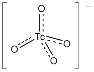
The Dor procedure is a medical technique used as part of heart surgery and originally introduced by the French cardiac surgeon Vincent Dor (b.1932). It is also known as endoventricular circular patch plasty (EVCPP).

Aortic valve area calculation is an indirect method of determining the area of the aortic valve. The calculated aortic valve orifice area is currently one of the measures for evaluating the severity of aortic stenosis. A valve area of less than 1.0 cm2 is considered to be severe aortic stenosis.

Myocardial perfusion imaging Nuclear medicine imaging method

Myocardial perfusion imaging or scanning is a nuclear medicine procedure that illustrates the function of the heart muscle (myocardium).

Cardiac resynchronization therapy

Cardiac resynchronisation therapy is the insertion of electrodes in the left and right ventricles of the heart, as well as on occasion the right atrium, to treat heart failure by coordinating the function of the left and right ventricles via a pacemaker, a small device inserted into the interior chest wall.

Cardiac ventriculography

Cardiac ventriculography is a medical imaging test used to determine a person's heart function in the right, or left ventricle. Cardiac ventriculography involves injecting contrast media into the heart's ventricle(s) to measure the volume of blood pumped. Cardiac ventriculography can be performed with a radionuclide in radionuclide ventriculography or with an iodine-based contrast in cardiac chamber catheterization.

A plot of a system's pressure versus volume has long been used to measure the work done by the system and its efficiency. This analysis can be applied to heat engines and pumps, including the heart. A considerable amount of information on cardiac performance can be determined from the pressure vs. volume plot. A number of methods have been determined for measuring PV-loop values experimentally.

Heart failure with preserved ejection fraction Medical condition

Heart failure with preserved ejection fraction (HFpEF) is a form of heart failure in which the ejection fraction – the percentage of the volume of blood ejected from the left ventricle with each heartbeat divided by the volume of blood when the left ventricle is maximally filled – is normal, defined as greater than 50%; this may be measured by echocardiography or cardiac catheterization. Approximately half of people with heart failure have preserved ejection fraction, while the other half have a reduction in ejection fraction, called heart failure with reduced ejection fraction (HFrEF).

In clinical cardiology the term "diastolic function" is most commonly referred as how the heart fills. Parallel to "diastolic function", the term "systolic function" is usually referenced in terms of the left ventricular ejection fraction (LVEF), which is the ratio of stroke volume and end-diastolic volume. Due to the epidemic of heart failure, particularly the cases determined as diastolic heart failure, it is increasingly urgent and crucial to understand the meaning of “diastolic function”. Unlike "systolic function", which can be simply evaluated by LVEF, there are no established dimensionless parameters for "diastolic function" assessment. Hence to further study "diastolic function" the complicated and speculative physiology must be taken into consideration.

References

  1. Gillam, Linda D.; Otto, Catherine M. (2011). Advanced Approaches in Echocardiography. Elsevier Health Sciences. p. 224. ISBN   978-1437726978.
  2. Folland, ED; Hamilton GW; Larson SM; Kennedy JW; Williams DL; Ritchie JL (1977). "The radionuclide ejection fraction: a comparison of three radionuclide techniques with contrast angiography". J Nucl Med. 18 (12): 1159–66. PMID   606737.
  3. 1 2 3 Merck manuals > Radionuclide Imaging Last full review/revision May 2009 by Michael J. Shea, MD. Content last modified May 2009
  4. "Procedure Guideline for Planar Radionuclide Cardiac Ventriculogram for the Assessment of Left Ventricular Systolic Function" (PDF). BNMS. 2016. Retrieved 25 September 2017.
  5. "Society of Nuclear Medicine Procedure Guideline for Gated Equilibrium Radionuclide Ventriculography" (PDF). SNMMI. 15 June 2002. Retrieved 25 September 2017.
  6. Saha, Gopal B. (2010). "Characteristics of Specific Radiopharmaceuticals". Fundamentals of nuclear pharmacy (6th ed.). New York: Springer. pp. 115–152. doi:10.1007/978-1-4419-5860-0_7. ISBN   978-1-4419-5859-4.
  7. Callahan, R J (2006). "Radiolabeled Red Blood Cells: Method and Mechanisms" (PDF). Continuing Education for Nuclear Pharmacists and Nuclear Medicine Professionals. University of New Mexico Health Sciences Center. Retrieved 25 September 2017.
  8. Waterstram-Rich, Kristen M.; Gilmore, David (2016). Nuclear Medicine and PET/CT: Technology and Techniques. Elsevier Health Sciences. p. 512. ISBN   9780323400350.
  9. "MUGA Scan". Cancer.Net.
  10. Massardo, Teresa; Gal, Rami A.; Grenier, Raymond P.; Schmidt, Donald H.; Port, Steven C. (1990). "Left Ventricular Volume Calculation Using a Count-Based Ratio Method Applied to Multigated Radionuclide Angiography". Journal of Nuclear Medicine. 31 (4): 450–456. PMID   2324820.
  11. Levy, Wayne C.; Cerqueira, Manuel D.; Matsuoka, Dale T.; Harp, George D.; Sheehan, Florence H.; Stratton, John R. (1992). "Four Radionuclide Methods for Left Ventricular Volume Determination: Comparison of a Manual and an Automated Technique". Journal of Nuclear Medicine. 33 (5): 763–770. PMID   1569488.
  12. Gal, Rami A.; Grenier, Raymond P.; Port, Steven C.; Dymond, Duncan S.; Schmidt, Donald H. (1992). "Left Ventricular Volume Calculation Using a Count-Based Ration Method Applied too First-Pass Radionuclide Angiography". Journal of Nuclear Medicine. 33 (12): 2124–2132. PMID   1460504.
  13. Sobic-Saranovic, D; et al. (2005). "Methods for the quantification of left ventricular volumes assessed by radionuclide ventriculography (first part)". Glas SRP Akad Nauka Med. 4: 11–30.