
Varicose veins, also known as varicoses, are a medical condition in which superficial veins become enlarged and twisted. These veins typically develop in the legs, just under the skin. Varicose veins usually cause few symptoms. However, some individuals may experience fatigue or pain in the area. Complications can include bleeding or superficial thrombophlebitis. Varices in the scrotum are known as a varicocele, while those around the anus are known as hemorrhoids. Due to the various physical, social, and psychological effects of varicose veins, they can negatively affect one's quality of life.
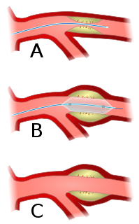
Angioplasty, is also known as balloon angioplasty and percutaneous transluminal angioplasty (PTA), is a minimally invasive endovascular procedure used to widen narrowed or obstructed arteries or veins, typically to treat arterial atherosclerosis. A deflated balloon attached to a catheter is passed over a guide-wire into the narrowed vessel and then inflated to a fixed size. The balloon forces expansion of the blood vessel and the surrounding muscular wall, allowing an improved blood flow. A stent may be inserted at the time of ballooning to ensure the vessel remains open, and the balloon is then deflated and withdrawn. Angioplasty has come to include all manner of vascular interventions that are typically performed percutaneously.

Interventional radiology (IR) is a medical subspecialty that performs various minimally-invasive procedures using medical imaging guidance, such as x-ray fluoroscopy, computed tomography, magnetic resonance imaging, or ultrasound. IR performs both diagnostic and therapeutic procedures through very small incisions or body orifices. Diagnostic IR procedures are those intended to help make a diagnosis or guide further medical treatment, and include image-guided biopsy of a tumor or injection of an imaging contrast agent into a hollow structure, such as a blood vessel or a duct. By contrast, therapeutic IR procedures provide direct treatment—they include catheter-based medicine delivery, medical device placement, and angioplasty of narrowed structures.
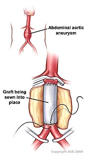
Vascular surgery is a surgical subspecialty in which diseases of the vascular system, or arteries, veins and lymphatic circulation, are managed by medical therapy, minimally-invasive catheter procedures and surgical reconstruction. The specialty evolved from general and cardiac surgery and includes treatment of the body's other major and essential veins and arteries. Open surgery techniques, as well as endovascular techniques are used to treat vascular diseases. The vascular surgeon is trained in the diagnosis and management of diseases affecting all parts of the vascular system excluding the coronaries and intracranial vasculature. Vascular surgeons often assist other physicians to address traumatic vascular injury, hemorrhage control, and safe exposure of vascular structures.

Telangiectasias, also known as spider veins, are small dilated blood vessels that can occur near the surface of the skin or mucous membranes, measuring between 0.5 and 1 millimeter in diameter. These dilated blood vessels can develop anywhere on the body but are commonly seen on the face around the nose, cheeks and chin. Dilated blood vessels can also develop on the legs, although when they occur on the legs, they often have underlying venous reflux or "hidden varicose veins". When found on the legs, they are found specifically on the upper thigh, below the knee joint and around the ankles.
Endovenous laser treatment (ELT) is a minimally invasive ultrasound-guided technique used for treating varicose veins using laser energy commonly performed by a phlebologist, interventional radiologist or vascular surgeon.

Sclerotherapy is a procedure used to treat blood vessel malformations and also malformations of the lymphatic system. A medicine is injected into the vessels, which makes them shrink. It is used for children and young adults with vascular or lymphatic malformations. In adults, sclerotherapy is often used to treat spider veins, smaller varicose veins, hemorrhoids, and hydroceles.

Venous ulcer is defined by the American Venous Forum as "a full-thickness defect of skin, most frequently in the ankle region, that fails to heal spontaneously and is sustained by chronic venous disease, based on venous duplex ultrasound testing." Venous ulcers are wounds that are thought to occur due to improper functioning of venous valves, usually of the legs. They are an important cause of chronic wounds, affecting 1% of the population. Venous ulcers develop mostly along the medial distal leg, and can be painful with negative effects on quality of life.
Ambulatory phlebectomy is a minisurgical treatment for superficial varicose veins and so-called side branches.
The term venous translucence has been used in phlebology since 1996 by surgeon Pedro Fernandes Neto during ambulatory clinical exams in Brazil. His results were published in the annals of the national and international congresses of angiology. Venous translucence is the process of reflective image visualization of veins by light, which reaches up to the superficial venous system. It is a non-invasive method. Since it is a simple, low-cost technique it can be repeated as needed, which is useful in disease-process monitoring. It is a new diagnostic procedure, still undergoing investigation; more analysis is necessary to hone its technical aspects. Venous translucence is based on optical physics. It is caused by the refraction, absorption and reflection of light. The color which is not absorbed is reflected, and is the one that is seen. Therefore, venous translumination is based on the incidence of luminosity on the vein, where part of the light is absorbed and another reflected.

Chronic venous insufficiency (CVI) is a medical condition in which blood pools in the veins, straining the walls of the vein. The most common cause of CVI is superficial venous reflux which is a treatable condition. As functional venous valves are required to provide for efficient blood return from the lower extremities, this condition typically affects the legs. If the impaired vein function causes significant symptoms, such as swelling and ulcer formation, it is referred to as chronic venous disease. It is sometimes called chronic peripheral venous insufficiency and should not be confused with post-thrombotic syndrome in which the deep veins have been damaged by previous deep vein thrombosis.

Klippel–Trénaunay syndrome, formerly Klippel–Trénaunay–Weber syndrome and sometimes angioosteohypertrophy syndrome and hemangiectatic hypertrophy, is a rare congenital medical condition in which blood vessels and/or lymph vessels fail to form properly. The three main features are nevus flammeus, venous and lymphatic malformations, and soft-tissue hypertrophy of the affected limb. It is similar to, though distinctly separate from, the less common Parkes Weber syndrome.
A vein disorder is a class of disease involving veins of the circulatory system.

Paolo Zamboni is an Italian doctor included among the Top Italian Scientists. He is full Professor and Director of the School of Vascular Surgery at the University of Ferrara in Italy.

Pelvic congestion syndrome, also known as pelvic vein incompetence, is a long term condition believed to be due to enlarged veins in the lower abdomen. The condition may cause chronic pain, such as a constant dull ache, which can be worsened by standing or sex. Pain in the legs or lower back may also occur.

Perforator veins are so called because they perforate the deep fascia of muscles, to connect the superficial veins to the deep veins where they drain.

Ultrasonography of suspected or previously confirmed chronic venous insufficiency of leg veins is a risk-free, non-invasive procedure. It gives information about the anatomy, physiology and pathology of mainly superficial veins. As with heart ultrasound (echocardiography) studies, venous ultrasonography requires an understanding of hemodynamics in order to give useful examination reports. In chronic venous insufficiency, sonographic examination is of most benefit; in confirming varicose disease, making an assessment of the hemodynamics, and charting the progression of the disease and its response to treatment. It has become the reference standard for examining the condition and hemodynamics of the lower limb veins. Particular veins of the deep venous system (DVS), and the superficial venous system (SVS) are looked at. The great saphenous vein (GSV), and the small saphenous vein (SSV) are superficial veins which drain into respectively, the common femoral vein and the popliteal vein. These veins are deep veins. Perforator veins drain superficial veins into the deep veins. Three anatomic compartments are described, (N1) containing the deep veins, (N2) containing the perforator veins, and (N3) containing the superficial veins, known as the saphenous compartment. This compartmentalisation makes it easier for the examiner to systematize and map. The GSV can be located in the saphenous compartment where together with the Giacomini vein and the accessory saphenous vein (ASV) an image resembling an eye, known as the 'eye sign' can be seen. The ASV which is often responsible for varicose veins, can be located at the 'alignment sign', where it is seen to align with the femoral vessels.

Claude Franceschi is an angiologist French MD.
CHIVA method is a type of surgery used to treat varicose veins that occur as a result of long term venous insufficiency. The term is a French acronym for Conservatrice Hémodynamique de l'Insuffisance Veineuse en Ambulatoire.

The American Venous Forum (AVF) is the major national academic society focused on venous and lymphatic disease in the United States. Its mission includes education, research, and advocacy.The AVF is the sponsor organization for the Journal of Vascular Surgery Venous and Lymphatic and for the American Venous Forum meeting.