Related Research Articles
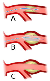
Angioplasty, also known as balloon angioplasty and percutaneous transluminal angioplasty (PTA), is a minimally invasive endovascular procedure used to widen narrowed or obstructed arteries or veins, typically to treat arterial atherosclerosis. A deflated balloon attached to a catheter is passed over a guide-wire into the narrowed vessel and then inflated to a fixed size. The balloon forces expansion of the blood vessel and the surrounding muscular wall, allowing an improved blood flow. A stent may be inserted at the time of ballooning to ensure the vessel remains open, and the balloon is then deflated and withdrawn. Angioplasty has come to include all manner of vascular interventions that are typically performed percutaneously.
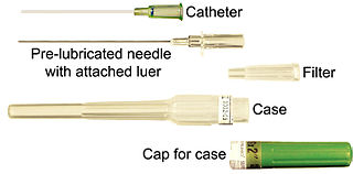
In medicine, a catheter is a thin tube made from medical grade materials serving a broad range of functions. Catheters are medical devices that can be inserted in the body to treat diseases or perform a surgical procedure. By modifying the material or adjusting the way catheters are manufactured, it is possible to tailor catheters for cardiovascular, urological, gastrointestinal, neurovascular, and ophthalmic applications.

Angiography or arteriography is a medical imaging technique used to visualize the inside, or lumen, of blood vessels and organs of the body, with particular interest in the arteries, veins, and the heart chambers. This is traditionally done by injecting a radio-opaque contrast agent into the blood vessel and imaging using X-ray based techniques such as fluoroscopy.

Interventional radiology (IR) is a group of techniques where medical imaging guidance, such as x-ray fluoroscopy, computed tomography, magnetic resonance imaging, or ultrasound, are used to precisely guide medical therapies to the internal structures of the body through very small incisions or body orifices. The range of techniques is broadly classified into two main types of procedures; diagnostic and therapeutic.
In surgery, a percutaneous procedure is any medical procedure or method where access to inner organs or other tissue is done via needle-puncture of the skin, rather than by using an "open" approach where inner organs or tissue are exposed.
Vascular surgery is a surgical subspecialty in which diseases of the vascular system, or arteries, veins and lymphatic circulation, are managed by medical therapy, minimally-invasive catheter procedures, and surgical reconstruction. The specialty evolved from general and cardiac surgery as well as minimally invasive techniques pioneered by interventional radiology. The vascular surgeon is trained in the diagnosis and management of diseases affecting all parts of the vascular system excluding the coronaries and intracranial vasculature.

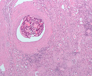
A pseudoaneurysm, also known as a false aneurysm, is a collection of blood that forms between the two outer layers of an artery, the tunica media and the tunica adventitia. It is usually caused by a penetrating injury to the vessel, which then bleeds, but forms a space between the above two layers, rather than exiting the vessel. It may be pulsatile and can resemble a true aneurysm. A true aneurysm involves all three layers of the blood vessel. A dissecting aneurysm is when blood from the vessel lumen tracks between the two inner layers, the intima and the tunica media. This can cause blockage of the flow. A perivascular hematoma is a collection of blood that is external to the three vessel layers. Due to being close to the vessel, it can also be pulsatile, and can be mistaken for a pseudoaneurysm or aneurysm.
Cerebral angiography is a form of angiography which provides images of blood vessels in and around the brain, thereby allowing detection of abnormalities such as arteriovenous malformations and aneurysms. It was pioneered in 1927 by the Portuguese neurologist Egas Moniz at the University of Lisbon, who also helped develop thorotrast for use in the procedure.

Cardiac catheterization is the insertion of a catheter into a chamber or vessel of the heart. This is done both for diagnostic and interventional purposes. A common example of cardiac catheterization is coronary catheterization that involves catheterization of the coronary arteries for coronary artery disease and myocardial infarctions. Catheterization is most often performed in special laboratories with fluoroscopy and highly maneuverable tables. These "cath labs" are often equipped with cabinets of catheters, stents, balloons, etc. of various sizes to increase efficiency. Monitors show the fluoroscopy imaging, EKG, pressure waves, and more.
Embolization refers to the passage and lodging of an embolus within the bloodstream. It may be of natural origin (pathological), in which sense it is also called embolism, for example a pulmonary embolism; or it may be artificially induced (therapeutic), as a hemostatic treatment for bleeding or as a treatment for some types of cancer by deliberately blocking blood vessels to starve the tumor cells.
Vascular access refers to a rapid, direct method of introducing or removing devices or chemicals from the bloodstream. In hemodialysis, vascular access is used to remove the patient's blood so that it can be filtered through the dialyzer. Three primary methods are used to gain access to the blood: an intravenous catheter, an arteriovenous fistula (AV) or a synthetic graft. In the latter two, needles are used to puncture the graft or fistula each time dialysis is performed.

Carotid artery stenting (CAS) is an endovascular procedure where a stent is deployed within the lumen of the carotid artery to treat narrowing of the carotid artery and decrease the risk of stroke. CAS is used to treat narrowing of the carotid artery in high-risk patients, when carotid endarterectomy is considered too risky. Carotid artery stenosis can present with no symptoms or with symptoms such as transient ischemic attacks (TIAs) or strokes.
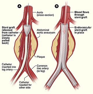
Endovascular aneurysm repair (EVAR), is a type of endovascular surgery used to treat pathology of the aorta, most commonly an abdominal aortic aneurysm (AAA). When used to treat thoracic aortic disease, the procedure is then specifically termed TEVAR for "thoracic endovascular aortic/aneurysm repair." The procedure involves the placement of an expandable stent graft within the aorta to treat aortic disease without operating directly on the aorta. In 2003, EVAR surpassed open aortic surgery as the most common technique for repair of AAA, and in 2010, EVAR accounted for 78% of all intact AAA repair in the United States.
Terumo Corporation was founded in 1921 as Sekisen Ken-onki Corporation by a group of medical scientists led by Dr. Kitasato Shibasaburō to produce medical thermometers in Japan.

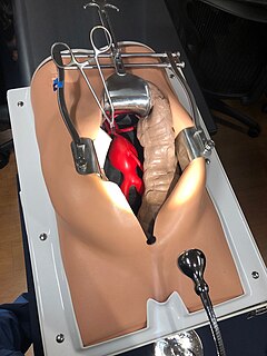
Atrial septostomy is a surgical procedure in which a small hole is created between the upper two chambers of the heart, the atria. This procedure is primarily used to palliate dextro-Transposition of the great arteries or d-TGA, a life-threatening cyanotic congenital heart defect seen in infants. It is performed prior to an arterial switch operation. Atrial septostomy has also seen limited use as a surgical treatment for pulmonary hypertension. One common technique was developed in 1966 by American cardiologist William Rashkind at the Children's Hospital of Philadelphia. The first atrial septectomy was developed by Vivien Thomas in a canine model and performed in humans by Alfred Blalock.
Dr. Thomas J. "Tom" Fogarty is an American surgeon and medical device inventor. He is best known for the invention of the embolectomy catheter, which revolutionised the treatment of blood clots (embolus).

Atherectomy is a minimally invasive endovascular surgery technique for removing atherosclerosis from blood vessels within the body. It is an alternative to angioplasty for the treatment of peripheral artery disease, but the studies that exist are not adequate to determine if it is superior to angioplasty. It has also been used to treat coronary artery disease, albeit ineffectively.
Vascular closure devices (VCDs) are medical devices used to achieve hemostasis of the small hole in the artery after a cardiovascular procedure of endovascular surgery requiring a catheterization.
The Mynx Vascular Closure Device is an extravascular vascular closure device (VCD) whose deployment system is designed to minimize the discomfort commonly associated with closing the small hole in the artery following catheterization procedure. The device is manufactured by AccessClosure, Inc., a medical device company located in Mountain View, California.
Endovascular Surgical Neuroradiology (ESN), also known as Interventional Neuroradiology (INR), and Endovascular Neurosurgery, is a medical subspecialty of Neurology, Neurosurgery, and radiology specializing in minimally invasive image-based technologies and procedures used in diagnosis and treatment of diseases of the head, neck, and spine.
References
- ↑ Mitchell, WB; Bonn, J (July 2005). "Percutaneous retrieval of a Greenfield filter after migration to the left pulmonary artery". Journal of Vascular and Interventional Radiology. 16 (7): 1013–7. doi:10.1097/01.RVI.0000160344.50022.75. PMID 16002510.