Contrast-enhanced ultrasound

Last updated
Contrast-enhanced renal ultrasonograph, showing a renal cell carcinoma successfully treated with thermal ablation, as no contrast enhancement is seen Contrast-enhanced ultrasonography of renal cell carcinoma successfully treated with thermal ablation.jpg
Contrast-enhanced renal ultrasonograph, showing a renal cell carcinoma successfully treated with thermal ablation, as no contrast enhancement is seen
Unspecific cortical lesion on CT is confirmed cystic and benign with contrast-enhanced renal ultrasonography using image fusion. Contrast-enhanced ultrasonography of benign lesion.jpg
Unspecific cortical lesion on CT is confirmed cystic and benign with contrast-enhanced renal ultrasonography using image fusion.

Contrast-enhanced ultrasound (CEUS) is the application of ultrasound contrast medium to traditional medical sonography. Ultrasound contrast agents rely on the different ways in which sound waves are reflected from interfaces between substances. This may be the surface of a small air bubble or a more complex structure. Commercially available contrast media are gas-filled microbubbles that are administered intravenously to the systemic circulation. Microbubbles have a high degree of echogenicity (the ability of an object to reflect ultrasound waves). There is a great difference in echogenicity between the gas in the microbubbles and the soft tissue surroundings of the body. Thus, ultrasonic imaging using microbubble contrast agents enhances the ultrasound backscatter, (reflection) of the ultrasound waves, to produce a sonogram with increased contrast due to the high echogenicity difference. Contrast-enhanced ultrasound can be used to image blood perfusion in organs, measure blood flow rate in the heart and other organs, and for other applications.

Contents

Targeting ligands that bind to receptors characteristic of intravascular diseases can be conjugated to microbubbles, enabling the microbubble complex to accumulate selectively in areas of interest, such as diseased or abnormal tissues. This form of molecular imaging, known as targeted contrast-enhanced ultrasound, will only generate a strong ultrasound signal if targeted microbubbles bind in the area of interest. Targeted contrast-enhanced ultrasound may have many applications in both medical diagnostics and medical therapeutics. However, the targeted technique has not yet been approved by the FDA for clinical use in the United States.

Contrast-enhanced ultrasound is regarded as safe in adults, comparable to the safety of MRI contrast agents, and better than radiocontrast agents used in contrast CT scans. The more limited safety data in children suggests that such use is as safe as in the adult population. [2]

Bubble echocardiogram

An echocardiogram is a study of the heart using ultrasound. A bubble echocardiogram is an extension of this that uses simple air bubbles as a contrast medium during this study and often has to be requested specifically.

Although colour Doppler can be used to detect abnormal flows between the chambers of the heart (e.g., persistent (patent) foramen ovale), it has a limited sensitivity. When specifically looking for a defect such as this, small air bubbles can be used as a contrast medium and injected intravenously, where they travel to the right side of the heart. The test would be positive for an abnormal communication if the bubbles are seen passing into the left side of the heart. (Normally, they would exit the heart through the pulmonary artery and be stopped by the lungs.) This form of bubble contrast medium is generated on an ad hoc basis by the testing clinician by agitating normal saline (e.g., by rapidly and repeatedly transferring the saline between two connected syringes) immediately prior to injection.[ citation needed ]

Microbubble contrast agents

General features

There are a variety of microbubble contrast agents. Microbubbles differ in their shell makeup, gas core makeup, and whether or not they are targeted.[ citation needed ]

Regardless of the shell or gas core composition, microbubble size is fairly uniform. They lie within a range of 1–4 micrometres in diameter. That makes them smaller than red blood cells, which allows them to flow easily through the circulation as well as the microcirculation.

Specific agents

Targeted microbubbles

Targeted microbubbles are under preclinical development. They retain the same general features as untargeted microbubbles, but they are outfitted with ligands that bind specific receptors expressed by cell types of interest, such as inflamed cells or cancer cells. Current microbubbles in development are composed of a lipid monolayer shell with a perfluorocarbon gas core. The lipid shell is also covered with a polyethylene glycol (PEG) layer. PEG prevents microbubble aggregation and makes the microbubble more non-reactive. It temporarily "hides" the microbubble from the immune system uptake, increasing the amount of circulation time, and hence, imaging time. [11] In addition to the PEG layer, the shell is modified with molecules that allow for the attachment of ligands that bind certain receptors. These ligands are attached to the microbubbles using carbodiimide, maleimide, or biotin-streptavidin coupling. [11] Biotin-streptavidin is the most popular coupling strategy because biotin's affinity for streptavidin is very strong and it is easy to label the ligands with biotin. Currently, these ligands are monoclonal antibodies produced from animal cell cultures that bind specifically to receptors and molecules expressed by the target cell type. Since the antibodies are not humanized, they will elicit an immune response when used in human therapy. Humanizing antibodies is an expensive and time-intensive process, so it would be ideal to find an alternative source of ligands, such as synthetically manufactured targeting peptides that perform the same function, but without the immune issues.[ citation needed ]

Types

There are two forms of contrast-enhanced ultrasound, untargeted (used in the clinic today) and targeted (under preclinical development). The two methods slightly differ from each other.

Untargeted CEUS

Untargeted microbubbles, such as the aforementioned SonoVue, Optison, or Levovist, are injected intravenously into the systemic circulation in a small bolus. The microbubbles will remain in the systemic circulation for a certain period of time. During that time, ultrasound waves are directed on the area of interest. When microbubbles in the blood flow past the imaging window, the microbubbles' compressible gas cores oscillate in response to the high frequency sonic energy field, as described in the ultrasound article. The microbubbles reflect a unique echo that stands in stark contrast to the surrounding tissue due to the orders of magnitude mismatch between microbubble and tissue echogenicity. The ultrasound system converts the strong echogenicity into a contrast-enhanced image of the area of interest. In this way, the bloodstream's echo is enhanced, thus allowing the clinician to distinguish blood from surrounding tissues.[ citation needed ]

Targeted CEUS

Targeted contrast-enhanced ultrasound works in a similar fashion, with a few alterations. Microbubbles targeted with ligands that bind certain molecular markers that are expressed by the area of imaging interest are still injected systemically in a small bolus. Microbubbles theoretically travel through the circulatory system, eventually finding their respective targets and binding specifically. Ultrasound waves can then be directed on the area of interest. If a sufficient number of microbubbles have bound in the area, their compressible gas cores oscillate in response to the high frequency sonic energy field, as described in the ultrasound article. The targeted microbubbles also reflect a unique echo that stands in stark contrast to the surrounding tissue due to the orders of magnitude mismatch between microbubble and tissue echogenicity. The ultrasound system converts the strong echogenicity into a contrast-enhanced image of the area of interest, revealing the location of the bound microbubbles. [12] Detection of bound microbubbles may then show that the area of interest is expressing that particular molecular marker, which can be indicative of a certain disease state, or identify particular cells in the area of interest.[ citation needed ]

Applications

Untargeted contrast-enhanced ultrasound is currently applied in echocardiography and radiology. Targeted contrast-enhanced ultrasound is being developed for a variety of medical applications.

Untargeted CEUS

Untargeted microbubbles like Optison and Levovist are currently used in echocardiography. In addition, SonoVue [13] ultrasound contrast agent is used in radiology for lesion characterization.

Targeted CEUS

Advantages

On top of the strengths mentioned in the medical sonography entry, contrast-enhanced ultrasound adds these additional advantages:

Disadvantages

In addition to the weaknesses mentioned in the medical sonography entry, contrast-enhanced ultrasound has the following disadvantages:

See also


Related Research Articles

<span class="mw-page-title-main">Medical ultrasound</span> Diagnostic and therapeutic technique

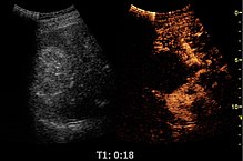
Medical ultrasound includes diagnostic techniques using ultrasound, as well as therapeutic applications of ultrasound. In diagnosis, it is used to create an image of internal body structures such as tendons, muscles, joints, blood vessels, and internal organs, to measure some characteristics or to generate an informative audible sound. The usage of ultrasound to produce visual images for medicine is called medical ultrasonography or simply sonography, or echography. The practice of examining pregnant women using ultrasound is called obstetric ultrasonography, and was an early development of clinical ultrasonography. The machine used is called an ultrasound machine, a sonograph or an echograph. The visual image formed using this technique is called an ultrasonogram, a sonogram or an echogram.

<span class="mw-page-title-main">Cell adhesion</span> Process of cell attachment

Cell adhesion is the process by which cells interact and attach to neighbouring cells through specialised molecules of the cell surface. This process can occur either through direct contact between cell surfaces such as cell junctions or indirect interaction, where cells attach to surrounding extracellular matrix, a gel-like structure containing molecules released by cells into spaces between them. Cells adhesion occurs from the interactions between cell-adhesion molecules (CAMs), transmembrane proteins located on the cell surface. Cell adhesion links cells in different ways and can be involved in signal transduction for cells to detect and respond to changes in the surroundings. Other cellular processes regulated by cell adhesion include cell migration and tissue development in multicellular organisms. Alterations in cell adhesion can disrupt important cellular processes and lead to a variety of diseases, including cancer and arthritis. Cell adhesion is also essential for infectious organisms, such as bacteria or viruses, to cause diseases.

Cell adhesion molecules (CAMs) are a subset of cell surface proteins that are involved in the binding of cells with other cells or with the extracellular matrix (ECM), in a process called cell adhesion. In essence, CAMs help cells stick to each other and to their surroundings. CAMs are crucial components in maintaining tissue structure and function. In fully developed animals, these molecules play an integral role in generating force and movement and consequently ensuring that organs are able to execute their functions normally. In addition to serving as "molecular glue", CAMs play important roles in the cellular mechanisms of growth, contact inhibition, and apoptosis. Aberrant expression of CAMs may result in a wide range of pathologies, ranging from frostbite to cancer.

<span class="mw-page-title-main">P-selectin glycoprotein ligand-1</span> Protein-coding gene in the species Homo sapiens

Selectin P ligand, also known as SELPLG or CD162, is a human gene.

<span class="mw-page-title-main">Selectin</span> Family of cell adhesion molecules

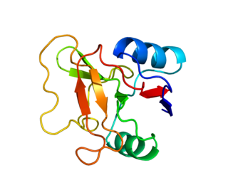
The selectins are a family of cell adhesion molecules. All selectins are single-chain transmembrane glycoproteins that share similar properties to C-type lectins due to a related amino terminus and calcium-dependent binding. Selectins bind to sugar moieties and so are considered to be a type of lectin, cell adhesion proteins that bind sugar polymers.

A contrast agent is a substance used to increase the contrast of structures or fluids within the body in medical imaging. Contrast agents absorb or alter external electromagnetism or ultrasound, which is different from radiopharmaceuticals, which emit radiation themselves. In x-ray imaging, contrast agents enhance the radiodensity in a target tissue or structure. In magnetic resonance imaging, contrast agents shorten the relaxation times of nuclei within body tissues in order to alter the contrast in the image.

<span class="mw-page-title-main">L-selectin</span> Mammalian protein found in Homo sapiens

L-selectin, also known as CD62L, is a cell adhesion molecule found on the cell surface of leukocytes, and the blastocyst. It is coded for in the human by the SELL gene. L-selectin belongs to the selectin family of proteins, which recognize sialylated carbohydrate groups containing a Sialyl LewisX (sLeX) determinant. L-selectin plays an important role in both the innate and adaptive immune responses by facilitating leukocyte-endothelial cell adhesion events. These tethering interactions are essential for the trafficking of monocytes and neutrophils into inflamed tissue as well as the homing of lymphocytes to secondary lymphoid organs. L-selectin is also expressed by lymphoid primed hematopoietic stem cells and may participate in the migration of these stem cells to the primary lymphoid organs. In addition to its function in the immune response, L-selectin is expressed on embryonic cells and facilitates the attachment of the blastocyst to the endometrial endothelium during human embryo implantation.

<span class="mw-page-title-main">E-selectin</span> Mammalian protein found in Homo sapiens

E-selectin, also known as CD62 antigen-like family member E (CD62E), endothelial-leukocyte adhesion molecule 1 (ELAM-1), or leukocyte-endothelial cell adhesion molecule 2 (LECAM2), is a selectin cell adhesion molecule expressed only on endothelial cells activated by cytokines. Like other selectins, it plays an important part in inflammation. In humans, E-selectin is encoded by the SELE gene.

Targeted drug delivery, sometimes called smart drug delivery, is a method of delivering medication to a patient in a manner that increases the concentration of the medication in some parts of the body relative to others. This means of delivery is largely founded on nanomedicine, which plans to employ nanoparticle-mediated drug delivery in order to combat the downfalls of conventional drug delivery. These nanoparticles would be loaded with drugs and targeted to specific parts of the body where there is solely diseased tissue, thereby avoiding interaction with healthy tissue. The goal of a targeted drug delivery system is to prolong, localize, target and have a protected drug interaction with the diseased tissue. The conventional drug delivery system is the absorption of the drug across a biological membrane, whereas the targeted release system releases the drug in a dosage form. The advantages to the targeted release system is the reduction in the frequency of the dosages taken by the patient, having a more uniform effect of the drug, reduction of drug side-effects, and reduced fluctuation in circulating drug levels. The disadvantage of the system is high cost, which makes productivity more difficult, and the reduced ability to adjust the dosages.

Lymphocyte homing receptors are cell adhesion molecules expressed on lymphocyte cell membranes that recognize addressins on target tissues. Lymphocyte homing refers to adhesion of the circulating lymphocytes in blood to specialized endothelial cells within lymphoid organs. These diverse tissue-specific adhesion molecules on lymphocytes and on endothelial cells contribute to the development of specialized immune responses.

<span class="mw-page-title-main">Leukocyte extravasation</span>

Leukocyte extravasation is the movement of leukocytes out of the circulatory system and towards the site of tissue damage or infection. This process forms part of the innate immune response, involving the recruitment of non-specific leukocytes. Monocytes also use this process in the absence of infection or tissue damage during their development into macrophages.

<span class="mw-page-title-main">Echogenicity</span> Ability to bounce an echo

Echogenicity or echogeneity is the ability to bounce an echo, e.g. return the signal in medical ultrasound examinations. In other words, echogenicity is higher when the surface bouncing the sound echo reflects increased sound waves. Tissues that have higher echogenicity are called "hyperechogenic" and are usually represented with lighter colors on images in medical ultrasonography. In contrast, tissues with lower echogenicity are called "hypoechogenic" and are usually represented with darker colors. Areas that lack echogenicity are called "anechogenic" and are usually displayed as completely dark.

<span class="mw-page-title-main">Sonoporation</span> Technique in molecular biology

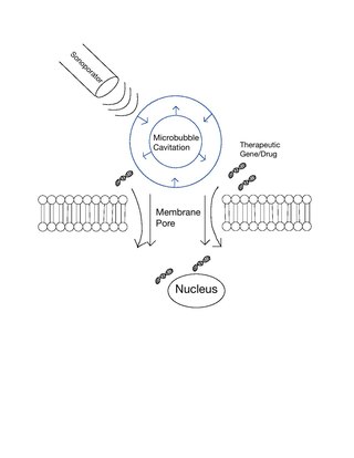
Sonoporation, or cellular sonication, is the use of sound in the ultrasonic range for increasing the permeability of the cell plasma membrane. This technique is usually used in molecular biology and non-viral gene therapy in order to allow uptake of large molecules such as DNA into the cell, in a cell disruption process called transfection or transformation. Sonoporation employs the acoustic cavitation of microbubbles to enhance delivery of these large molecules. The exact mechanism of sonoporation-mediated membrane translocation remains unclear, with a few different hypotheses currently being explored.

Microbubbles are bubbles smaller than one hundredth of a millimetre in diameter, but larger than one micrometre. They have widespread application in industry, medicine, life science, and food technology. The composition of the bubble shell and filling material determine important design features such as buoyancy, crush strength, thermal conductivity, and acoustic properties.

Preclinical imaging is the visualization of living animals for research purposes, such as drug development. Imaging modalities have long been crucial to the researcher in observing changes, either at the organ, tissue, cell, or molecular level, in animals responding to physiological or environmental changes. Imaging modalities that are non-invasive and in vivo have become especially important to study animal models longitudinally. Broadly speaking, these imaging systems can be categorized into primarily morphological/anatomical and primarily molecular imaging techniques. Techniques such as high-frequency micro-ultrasound, magnetic resonance imaging (MRI) and computed tomography (CT) are usually used for anatomical imaging, while optical imaging, positron emission tomography (PET), and single photon emission computed tomography (SPECT) are usually used for molecular visualizations.

Alexander L. (Sasha) Klibanov is associate professor in the Division of Cardiovascular Medicine and Department of Biomedical Engineering at the University of Virginia. He specializes in the study of ultrasound and medical imaging techniques.

A specific branch of contrast-enhanced ultrasound, acoustic angiography is a minimally invasive and non-ionizing medical imaging technique used to visualize vasculature. Acoustic angiography was first developed by the Dayton Laboratory at North Carolina State University and provides a safe, portable, and inexpensive alternative to the most common methods of angiography such as Magnetic Resonance Angiography and Computed Tomography Angiography. Although ultrasound does not traditionally exhibit the high resolution of MRI or CT, high-frequency ultrasound (HFU) achieves relatively high resolution by sacrificing some penetration depth. HFU typically uses waves between 20 and 100 MHz and achieves resolution of 16-80μm at depths of 3-12mm. Although HFU has exhibited adequate resolution to monitor things like tumor growth in the skin layers, on its own it lacks the depth and contrast necessary for imaging blood vessels. Acoustic angiography overcomes the weaknesses of HFU by combining contrast-enhanced ultrasound with the use of a dual-element ultrasound transducer to achieve high resolution visualization of blood vessels at relatively deep penetration levels.

<span class="mw-page-title-main">Focused ultrasound for intracranial drug delivery</span> Medical technique

Focused ultrasound for intracrainial drug delivery is a non-invasive technique that uses high-frequency sound waves to disrupt tight junctions in the blood–brain barrier (BBB), allowing for increased passage of therapeutics into the brain. The BBB normally blocks nearly 98% of drugs from accessing the central nervous system, so FUS has the potential to address a major challenge in intracranial drug delivery by providing targeted and reversible BBB disruption. Using FUS to enhance drug delivery to the brain could significantly improve patient outcomes for a variety of diseases including Alzheimer's disease, Parkinson's disease, and brain cancer.

<span class="mw-page-title-main">Ligand-targeted liposome</span> Ligand-targeted liposomes for use in medical applications

A ligand-targeted liposome (LTL) is a nanocarrier with specific ligands attached to its surface to enhance localization for targeted drug delivery. The targeting ability of LTLs enhances cellular localization and uptake of these liposomes for therapeutic or diagnostic purposes. LTLs have the potential to enhance drug delivery by decreasing peripheral systemic toxicity, increasing in vivo drug stability, enhancing cellular uptake, and increasing efficiency for chemotherapeutics and other applications. Liposomes are beneficial in therapeutic manufacturing because of low batch-to-batch variability, easy synthesis, favorable scalability, and strong biocompatibility. Ligand-targeting technology enhances liposomes by adding targeting properties for directed drug delivery.

Theranostics, also known as theragnostics, is a technique in personalised medicine and nuclear medicine where a one radioactive drug is used to identify (diagnose) and a second radioactive drug is used to treat cancerous tumors.

References

  1. 1 2 Content initially copied from: Hansen, Kristoffer; Nielsen, Michael; Ewertsen, Caroline (2015). "Ultrasonography of the Kidney: A Pictorial Review". Diagnostics. 6 (1): 2. doi: 10.3390/diagnostics6010002 . ISSN   2075-4418. PMC   4808817 . PMID   26838799. (CC-BY 4.0)
  2. Sidhu, Paul; Cantisani, Vito; Deganello, Annamaria; Dietrich, Christoph; Duran, Carmina; Franke, Doris; Harkanyi, Zoltan; Kosiak, Wojciech; Miele, Vittorio; Ntoulia, Aikaterini; Piskunowicz, Maciej; Sellars, Maria; Gilja, Odd (2016). "Role of Contrast-Enhanced Ultrasound (CEUS) in Paediatric Practice: An EFSUMB Position Statement". Ultraschall in der Medizin – European Journal of Ultrasound. 38 (1): 33–43. doi: 10.1055/s-0042-110394 . ISSN   0172-4614. PMID   27414980.
  3. 1 2 McCulloch M.; Gresser C.; Moos S.; Odabashian J.; Jasper S.; Bednarz J.; Burgess P.; Carney D.; Moore V.; Sisk E.; Waggoner A.; Witt S.; Adams D. (2000). "Ultrasound contrast physics: A series on contrast echocardiography, article 3". J Am Soc Echocardiogr. 13 (10): 959–67. doi:10.1067/mje.2000.107004. PMID   11029724.
  4. 1 2 3 4 5 Lindner J.R. (2004). "Microbubbles in medical imaging: current applications and future directions". Nat Rev Drug Discov. 3 (6): 527–32. doi:10.1038/nrd1417. PMID   15173842. S2CID   29807146.
  5. Anderton N, Carlson CS, Matsumoto R, Shimizu RI, Poortinga AT, Kudo N, Postema M (2022). "On the rigidity of four hundred Pickering-stabilised microbubbles". Japanese Journal of Applied Physics. 61 (SG): SG8001. Bibcode:2022JaJAP..61G8001A. doi: 10.35848/1347-4065/ac4adc . S2CID   245915590.
  6. Panfilova A, Chen P, van Sloun R, Wijkstra H, Postema M, Mischi M (2021). "Experimental acoustic characterisation of an endoskeletal antibubble contrast agent: first results". Medical Physics. 48 (11): 6765–6780. Bibcode:2021MedPh..48.6765P. doi:10.1002/mp.15242. PMC   9293338 . PMID   34580883.
  7. "Definity- perflutren injection, suspension". DailyMed. 19 August 2020. Retrieved 22 October 2020.
  8. "Luminity EPAR". European Medicines Agency (EMA). 17 September 2018. Retrieved 22 October 2020.
  9. "SonoVue, INN-sulphur hexafluoride - Annex I - Summary of Product Characteristics" (PDF). European Medicines Agency . Retrieved 2019-02-24.
  10. "Perflexane: (AF0150, AFO 150, Imagent, Imavist™)". Drugs in R&D, Volume 3, Number 5, 2002, pp. 306–309(4). Adis International . Retrieved 2010-03-08.
  11. 1 2 3 4 Klibanov A.L. (2005). "Ligand-carrying gas-filled microbubbles: ultrasound contrast agents for targeted molecular imaging". Bioconjug. Chem. 16 (1): 9–17. doi:10.1021/bc049898y. PMID   15656569.
  12. 1 2 3 4 Klibanov A.L. (1999). "Targeted delivery of gas-filled microspheres, contrast agents for ultrasound imaging". Adv Drug Deliv Rev. 37 (1–3): 139–157. doi:10.1016/s0169-409x(98)00104-5. PMID   10837732.
  13. Schneider, M (November 1999). "SonoVue, a new ultrasound contrast agent". Eur. Radiol. 9 (3 Supplement): S347–S348. doi:10.1007/pl00014071. PMID   10602926. S2CID   19613214.
  14. Rognin, NG; Frinking, P.; Costa, M.; Arditi, M. (November 2008). "In-vivo perfusion quantification by contrast ultrasound: Validation of the use of linearized video data vs. Raw RF data". 2008 IEEE Ultrasonics Symposium. pp. 1690–1693. doi:10.1109/ULTSYM.2008.0413. ISBN   978-1-4244-2428-3. S2CID   45679140.
  15. Claudon, M; Dietrich, CF.; Choi, BI.; Cosgrove, DO.; Kudo, M.; Nolsøe, CP.; Piscaglia, F.; Wilson, SR.; Barr, RG.; Chammas, MC.; Chaubal, NG.; Chen, MH.; Clevert, DA.; Correas, JM.; Ding, H.; Forsberg, F.; Fowlkes, JB.; Gibson, RN.; Goldberg, BB.; Lassau, N.; Leen, EL.; Mattrey, RF.; Moriyasu, F.; Solbíatí, L.; Weskott, HP.; Xu, HX (February 2013). "Guidelines and Good Clinical Practice Recommendations for Contrast Enhanced Ultrasound (CEUS) in the Liver-Update 2012: A WFUMB-EFSUMB Initiative in Cooperation With Representatives of AFSUMB, AIUM, ASUM, FLAUS and ICUS". Ultrasound Med Biol. 39 (2): 187–210. doi:10.1016/j.ultrasmedbio.2012.09.002. hdl:11585/144895. PMID   23137926. S2CID   2224370.
  16. Rognin, NG; Arditi, M.; Mercier, L.; Frinking, P.J.A.; Schneider, M.; Perrenoud, G.; Anaye, A.; Meuwly, J.; Tranquart, F. (November 2010). "Parametric imaging for characterizing focal liver lesions in contrast-enhanced ultrasound". IEEE Transactions on Ultrasonics, Ferroelectrics and Frequency Control. 57 (11): 2503–2511. doi:10.1109/TUFFC.2010.1716. PMID   21041137. S2CID   19339331.
  17. Anaye, A; Perrenoud, G.; Rognin, N.; Arditi, M.; Mercier, L.; Frinking, P.; Ruffieux, C.; Peetrons, P.; Meuli, R.; Meuwly, JY. (October 2011). "Differentiation of focal liver lesions: usefulness of parametric imaging with contrast-enhanced US". Radiology. 261 (1): 300–310. doi:10.1148/radiol.11101866. PMID   21746815.
  18. 1 2 Takalkar A.M.; Klibanov A.L.; Rychak J.J.; Lindner J.R.; Ley K. (2004). "Binding and detachment dynamics of microbubbles targeted to P-selectin under controlled shear flow". J. Control. Release. 96 (3): 473–482. doi:10.1016/j.jconrel.2004.03.002. PMID   15120903.
  19. Eniola A.O.; Willcox P.J.; Hammer D.A. (2003). "Interplay between rolling and firm adhesion elucidated with a cell-free system engineered with two distinct receptor-ligand pairs". Biophys. J. 85 (4): 2720–31. Bibcode:2003BpJ....85.2720E. doi:10.1016/s0006-3495(03)74695-5. PMC   1303496 . PMID   14507735.
  20. Eniola A.O.; Hammer D.A. (2005). "In vitro characterization of leukocyte mimetic for targeting therapeutics to the endothelium using two receptors". Biomaterials. 26 (34): 7136–44. doi:10.1016/j.biomaterials.2005.05.005. PMID   15953632.
  21. Weller G.E.; Villanueva F.S.; Tom E.M.; Wagner W.R. (2005). "Targeted ultrasound contrast agents: In vitro assessment of endothelial dysfunction and multi-targeting to ICAM-1 and sialyl Lewis(x)". Biotechnol. Bioeng. 92 (6): 780–8. doi:10.1002/bit.20625. PMID   16121392.
  22. Rychak J.J., A.L. Klibanov, W. Yang, B. Li, S. Acton, A. Leppanen, R.D. Cummings, and K. Ley. "Enhanced Microbubble Adhesion to P-selectin with a Physiologically-tuned Targeting Ligand," 10th Ultrasound Contrast Research Symposium in Radiology, San Diego, CA, March 2005.
  23. Wang, X; Hagemeyer, CE; Hohmann, JD; Leitner, E; Armstrong, PC; Jia, F; Olschewski, M; Needles, A; Peter, K; Ingo, A (June 2012). "Novel single-chain antibody-targeted microbubbles for molecular ultrasound imaging of thrombosis: Validation of a unique non-invasive method for rapid and sensitive detection of thrombi and monitoring of success or failure of thrombolysis in mice". Circulation. 125 (25): 3117–3126. doi: 10.1161/CIRCULATIONAHA.111.030312 . PMID   22647975.
  24. Yap, ML; McFadyen, JD; Wang, X; Hohmann, JD; Ziegler, M; Yao, Y; Pham, A; Harris, M; Donnelly, PS; Hogarth, PM; Pietersz, GA; Lim, B; Peter, K (June 2017). "Targeting Activated Platelets: A Unique and Potentially Universal Approach for Cancer Imaging". Theranostics. 7 (10): 2565–2574. doi: 10.7150/thno.19900 . PMC   5558553 . PMID   28819447.
  25. Wang, X; Searle, AK; Hohmann, JD; Liu, AO; Abraham, MK; Palasubramaniam, J; Lim, B; Yao, Y; Wallert, M; Yu, E; Chen, YC; Peter, K (April 2018). "Dual-Targeted Theranostic Delivery of miRs Arrests Abdominal Aortic Aneurysm Development". Molecular Therapy. 25 (4): 1056–1065. doi: 10.1016/j.ymthe.2018.02.010 . PMC   6080135 . PMID   2952574.
  26. Wang, X; Gkanatsas, Y; Palasubramaniam, J; Hohmann, JD; Chen, YC; Lim, B; Hagemeyer, CE; Peter, K (March 2016). "Thrombus-Targeted Theranostic Microbubbles: A New Technology towards Concurrent Rapid Ultrasound Diagnosis and Bleeding-free Fibrinolytic Treatment of Thrombosis". Theranostics. 6 (5): 726–738. doi: 10.7150/thno.14514 . PMC   4805666 . PMID   27022419.
  27. Dimcevski G, Kotopoulis S, Bjånes T, Hoem D, Schjött J, Gjertsen BT, Biermann M, Molven A, Sorbye H, Mc Cormack E, Postema M, Gilja OH (2016). "A human clinical trial using ultrasound and microbubbles to enhance gemcitabine treatment of inoperable pancreatic cancer". Journal of Controlled Release. 243: 172–181. doi: 10.1016/j.jconrel.2016.10.007 . hdl: 1956/16668 . PMID   27744037.
  28. Søreide K, Stättner S (2021). Søreide K, Stättner S (eds.). Textbook of Pancreatic Cancer: Principles and Practice of Surgical Oncology. Heidelberg: Springer. doi:10.1007/978-3-030-53786-9. ISBN   978-3-030-53786-9. S2CID   231793989.
  29. 1 2 Lindner, J.R., A.L. Klibanov, and K. Ley. Targeting inflammation, In: Biomedical aspects of drug targeting. (Muzykantov, V.R., Torchilin, V.P., eds.) Kluwer, Boston, 2002; pp. 149–172.
  30. Wei, K; Jayaweera, AR; Firoozan, S; Linka, A; Skyba, DM; Kaul, S (February 1998). "Quantification of myocardial blood flow with ultrasound-induced destruction of microbubbles administered as a constant venous infusion". Circulation. 97 (5): 473–483. doi: 10.1161/01.cir.97.5.473 . PMID   9490243.
  31. Arditi, M; Frinking, PJA.; Zhou, X.; Rognin, NG. (June 2006). "A new formalism for the quantification of tissue perfusion by the destruction-replenishment method in contrast ultrasound imaging". IEEE Transactions on Ultrasonics, Ferroelectrics and Frequency Control. 53 (6): 1118–1129. doi:10.1109/TUFFC.2006.1642510. PMID   16846144. S2CID   6328131.
  32. Rychak, JJ; Klibanov, AL; Hossack, JA (March 2005). "Acoustic radiation force enhances targeted delivery of ultrasound contrast microbubbles: in vitro verification". IEEE Transactions on Ultrasonics, Ferroelectrics and Frequency Control. 52 (3): 421–433. doi:10.1109/TUFFC.2005.1417264. PMID   15857050. S2CID   25032596.
  33. Dayton, P; Klibanov, A; Brandenburger, G; Ferrara, K (October 1999). "Acoustic radiation force in vivo: a mechanism to assist targeting of microbubbles". Ultrasound Med Biol. 25 (8): 1195–1201. doi:10.1016/s0301-5629(99)00062-9. PMID   10576262.
  34. Frinking, PJ; Tardy, I; Théraulaz, M; Arditi, M; Powers, J; Pochon, S; Tranquart, F (August 2012). "Effects of acoustic radiation force on the binding efficiency of BR55, a VEGFR2-specific ultrasound contrast agent". Ultrasound Med Biol. 38 (8): 1460–1469. doi:10.1016/j.ultrasmedbio.2012.03.018. PMID   22579540.
  35. Gessner, RC; Streeter, JE; Kothadia, R; Feingold, S; Dayton, PA (April 2012). "An in vivo validation of the application of acoustic radiation force to enhance the diagnostic utility of molecular imaging using 3-d ultrasound". Ultrasound Med Biol. 38 (4): 651–660. doi:10.1016/j.ultrasmedbio.2011.12.005. PMC   3355521 . PMID   22341052.
  36. Rognin, NG; Unnikrishnan, S.; Klibanov, AL. (September 2013). "Molecular Ultrasound Imaging Enhancement by Volumic Acoustic Radiation Force (VARF): Pre-clinical in vivo Validation in a Murine Tumor Model". Abstracts of the 2013 World Molecular Imaging Congress. Archived from the original on 2013-10-11.