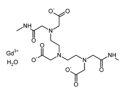
Gadodiamide

Last updated

Gadodiamide
Gadodiamide.png
Clinical data
Trade names Omniscan
Other names2-[bis[2-(carboxylatomethyl-(methylcarbamoylmethyl)amino)ethyl]amino]acetate; gadolinium(+3) cation
AHFS/Drugs.com Micromedex Detailed Consumer Information
License data
Routes of
administration
Intravenous
ATC code
Legal status
Legal status
Pharmacokinetic data
Protein binding negligible
Metabolism not metabolized
Elimination half-life 77.8 minutes
Excretion Kidney
Identifiers
  • gadolinium(III) 5,8-bis(carboxylatomethyl)-2-[2-(methylamino)-2-oxoethyl]-10-oxo-2,5,8,11-tetraazadodecane-1-carboxylate hydrate
CAS Number
PubChem CID
DrugBank
ChemSpider
UNII
KEGG
ChEBI
ChEMBL
CompTox Dashboard (EPA)
Chemical and physical data
Formula C16H28GdN5O9
Molar mass 591.68 g·mol−1
3D model (JSmol)
  • [Gd+3].[O-]C(=O)CN(CC(=O)NC)CCN(CCN(CC([O-])=O)CC(=O)NC)CC([O-])=O
  • InChI=1S/C16H29N5O8.Gd/c1-17-12(22)7-20(10-15(26)27)5-3-19(9-14(24)25)4-6-21(11-16(28)29)8-13(23)18-2;/h3-11H2,1-2H3,(H,17,22)(H,18,23)(H,24,25)(H,26,27)(H,28,29);/q;+3/p-3 Yes check.svgY
  • Key:HZHFFEYYPYZMNU-UHFFFAOYSA-K Yes check.svgY
 X mark.svgNYes check.svgY  (what is this?)    (verify)

Gadodiamide, sold under the brand name Omniscan, is a gadolinium-based MRI contrast agent (GBCA), used in magnetic resonance imaging (MRI) procedures to assist in the visualization of blood vessels.

Contents

Medical uses

A bottle of Omniscan contrast agent. Omniscan nima.JPG
A bottle of Omniscan contrast agent.

Gadodiamide is a contrast medium used for cranial and spinal magnetic resonance imaging (MRI) and for general MRI of the body after intravenous administration. It provides contrast enhancement and facilitates visualisation of abnormal structures or lesions in various parts of the body including the central nervous system (CNS). It crosses intact the blood brain barrier. [5]

Adverse effects

Gadodiamide is one of the main GBCA associated with nephrogenic systemic fibrosis (NSF), a toxic reaction occurring in some people with kidney problems. [6] No cases have been seen in people with normal kidney function. [7]

A 2015 study found gadolinium deposited in the brain tissue of people who had received gadodiamide. [8] Other studies using post-mortem mass spectrometry found most of the deposit remained at least 2 years after an injection and deposit also in individuals with no kidney issues.

In vitro studies found it to be neurotoxic. [9]

An Italian task force recommended that breastfeeding mothers precautionally avoid any contrast agent, such as gadodiamide, that has been associated with nephrogenic systemic fibrosis. [10]

Society and culture

Gadodiamide was suspended along with gadopentetic acid (Magnevist) by the European Medicines Agency in 2017. [11]

Related Research Articles

<span class="mw-page-title-main">Gadolinium</span> Chemical element, symbol Gd and atomic number 64

Gadolinium is a chemical element; it has symbol Gd and atomic number 64. Gadolinium is a silvery-white metal when oxidation is removed. It is a malleable and ductile rare-earth element. Gadolinium reacts with atmospheric oxygen or moisture slowly to form a black coating. Gadolinium below its Curie point of 20 °C (68 °F) is ferromagnetic, with an attraction to a magnetic field higher than that of nickel. Above this temperature it is the most paramagnetic element. It is found in nature only in an oxidized form. When separated, it usually has impurities of the other rare-earths because of their similar chemical properties.

<span class="mw-page-title-main">Magnetic resonance imaging</span> Medical imaging technique

Magnetic resonance imaging (MRI) is a medical imaging technique used in radiology to form pictures of the anatomy and the physiological processes inside the body. MRI scanners use strong magnetic fields, magnetic field gradients, and radio waves to generate images of the organs in the body. MRI does not involve X-rays or the use of ionizing radiation, which distinguishes it from computed tomography (CT) and positron emission tomography (PET) scans. MRI is a medical application of nuclear magnetic resonance (NMR) which can also be used for imaging in other NMR applications, such as NMR spectroscopy.

<span class="mw-page-title-main">Arthrogram</span>

An arthrogram is a series of images of a joint after injection of a contrast medium, usually done by fluoroscopy or MRI. The injection is normally done under a local anesthetic such as Novocain or lidocaine. The radiologist or radiographer performs the study using fluoroscopy or x-ray to guide the placement of the needle into the joint and then injects around 10 ml of contrast based on age. There is some burning pain from the anesthetic and a painful bubbling feeling in the joint after the contrast is injected. This only lasts 20 – 30 hours until the Contrast is absorbed. During this time, while it is allowed, it is painful to use the limb for around 10 hours. After that the radiologist can more clearly see what is going on under your skin and can get results out within 24 to 48 hours.

<span class="mw-page-title-main">Magnetic resonance angiography</span> Group of techniques based on magnetic resonance imaging (MRI) to image blood vessels.

Magnetic resonance angiography (MRA) is a group of techniques based on magnetic resonance imaging (MRI) to image blood vessels. Magnetic resonance angiography is used to generate images of arteries in order to evaluate them for stenosis, occlusions, aneurysms or other abnormalities. MRA is often used to evaluate the arteries of the neck and brain, the thoracic and abdominal aorta, the renal arteries, and the legs.

A contrast agent is a substance used to increase the contrast of structures or fluids within the body in medical imaging. Contrast agents absorb or alter external electromagnetism or ultrasound, which is different from radiopharmaceuticals, which emit radiation themselves. In x-ray imaging, contrast agents enhance the radiodensity in a target tissue or structure. In magnetic resonance imaging, contrast agents shorten the relaxation times of nuclei within body tissues in order to alter the contrast in the image.

<span class="mw-page-title-main">Gadopentetic acid</span> Complex of gadolinium by DTPA

Gadopentetic acid, sold under the brand name Magnevist, is a gadolinium-based MRI contrast agent.

<span class="mw-page-title-main">Breast MRI</span> Form of breast imaging

One alternative to mammography, breast MRI or contrast-enhanced magnetic resonance imaging (MRI), has shown substantial progress in the detection of breast cancer.

Magnetic resonance elastography (MRE) is a form of elastography that specifically leverages MRI to quantify and subsequently map the mechanical properties of soft tissue. First developed and described at Mayo Clinic by Muthupillai et al. in 1995, MRE has emerged as a powerful, non-invasive diagnostic tool, namely as an alternative to biopsy and serum tests for staging liver fibrosis.

Nephrogenic systemic fibrosis is a rare syndrome that involves fibrosis of the skin, joints, eyes, and internal organs. NSF is caused by exposure to gadolinium in gadolinium-based MRI contrast agents (GBCAs) in patients with impaired kidney function. Epidemiological studies suggest that the incidence of NSF is unrelated to gender or ethnicity and it is not thought to have a genetic basis. After GBCAs were identified as a cause of the disorder in 2006, and screening and prevention measures put in place, it is now considered rare.

MRI contrast agents are contrast agents used to improve the visibility of internal body structures in magnetic resonance imaging (MRI). The most commonly used compounds for contrast enhancement are gadolinium-based contrast agents (GBCAs). Such MRI contrast agents shorten the relaxation times of nuclei within body tissues following oral or intravenous administration.

<span class="mw-page-title-main">Cardiac magnetic resonance imaging</span>

Cardiac magnetic resonance imaging, also known as cardiovascular MRI, is a magnetic resonance imaging (MRI) technology used for non-invasive assessment of the function and structure of the cardiovascular system. Conditions in which it is performed include congenital heart disease, cardiomyopathies and valvular heart disease, diseases of the aorta such as dissection, aneurysm and coarctation, coronary heart disease. It can also be used to look at pulmonary veins. Patient information may be found here.

<span class="mw-page-title-main">Gadobutrol</span> Chemical compound

Gadobutrol (INN) (Gd-DO3A-butrol) is a gadolinium-based MRI contrast agent (GBCA).

<span class="mw-page-title-main">Gadoteric acid</span> Chemical compound

Gadoteric acid, sold under the brand name Dotarem among others, is a macrocycle-structured gadolinium-based MRI contrast agent (GBCA). It consists of the organic acid DOTA as a chelating agent, and gadolinium (Gd3+), and is used in form of the meglumine salt (gadoterate meglumine). The paramagnetic property of gadoteric acid reduces the T1 relaxation time (and to some extent the T2 and T2* relaxation times) in MRI, which is the source of its clinical utility. Because it has magnetic properties, gadoteric acid develops a magnetic moment when put under a magnetic field, which increases the signal intensity (brightness) of tissues during MRI imaging.

<span class="mw-page-title-main">Physics of magnetic resonance imaging</span> Overview article

Magnetic resonance imaging (MRI) is a medical imaging technique mostly used in radiology and nuclear medicine in order to investigate the anatomy and physiology of the body, and to detect pathologies including tumors, inflammation, neurological conditions such as stroke, disorders of muscles and joints, and abnormalities in the heart and blood vessels among others. Contrast agents may be injected intravenously or into a joint to enhance the image and facilitate diagnosis. Unlike CT and X-ray, MRI uses no ionizing radiation and is, therefore, a safe procedure suitable for diagnosis in children and repeated runs. Patients with specific non-ferromagnetic metal implants, cochlear implants, and cardiac pacemakers nowadays may also have an MRI in spite of effects of the strong magnetic fields. This does not apply on older devices, and details for medical professionals are provided by the device's manufacturer.

<span class="mw-page-title-main">Cardiac magnetic resonance imaging perfusion</span>

Cardiac magnetic resonance imaging perfusion, also known as stress CMR perfusion, is a clinical magnetic resonance imaging test performed on patients with known or suspected coronary artery disease to determine if there are perfusion defects in the myocardium of the left ventricle that are caused by narrowing of one or more of the coronary arteries.

<span class="mw-page-title-main">Intravoxel incoherent motion</span> Concept and a method initially introduced and developed by Le Bihan et al

Intravoxel incoherent motion (IVIM) imaging is a concept and a method initially introduced and developed by Le Bihan et al. to quantitatively assess all the microscopic translational motions that could contribute to the signal acquired with diffusion MRI. In this model, biological tissue contains two distinct environments: molecular diffusion of water in the tissue, and microcirculation of blood in the capillary network (perfusion). The concept introduced by D. Le Bihan is that water flowing in capillaries mimics a random walk (Fig.1), as long as the assumption that all directions are represented in the capillaries is satisfied.

<span class="mw-page-title-main">Val Murray Runge</span> American professor of radiology

Val Murray Runge is an American and Swiss professor of radiology and the editor-in-chief of Investigative Radiology. Runge was one of the early researchers to investigate the use of gadolinium-based contrast agents for magnetic resonance imaging (MRI), giving the first presentation in this field, followed two years later by the first presentation of efficacy. His research also pioneered many early innovations in MRI, including the use of tilted planes and respiratory gating. His publication on multiple sclerosis in 1984 represented the third and largest clinical series investigating the role of MRI in this disease, and the first to show characteristic abnormalities on MRI in patients whose CT was negative.

<span class="mw-page-title-main">Perfusion MRI</span>

Perfusion MRI or perfusion-weighted imaging (PWI) is perfusion scanning by the use of a particular MRI sequence. The acquired data are then post-processed to obtain perfusion maps with different parameters, such as BV, BF, MTT and TTP.

Magnetic resonance enterography is a magnetic resonance imaging technique used to evaluate bowel wall features of both upper and lower gastro-intestinal tract, although it is usually used for small bowel evaluation. It is a less invasive technique with the advantages of no ionizing radiation exposure, multiplanarity and high contrast resolution for soft tissue.

<span class="mw-page-title-main">Gadopiclenol</span> MRI contrast agent

Gadopiclenol, sold under the brand name Elucirem among others, is a contrast agent used with magnetic resonance imaging (MRI) to detect and visualize lesions with abnormal vascularity in the central nervous system and in the body. Gadopiclenol is a paramagnetic macrocyclic non-ionic complex of gadolinium.

References

  1. "Product monograph brand safety updates". Health Canada. February 2024. Retrieved 24 March 2024.
  2. "FDA-sourced list of all drugs with black box warnings (Use Download Full Results and View Query links.)". nctr-crs.fda.gov. FDA . Retrieved 22 October 2023.
  3. "Omniscan- gadodiamide injection". DailyMed. Retrieved 29 August 2021.
  4. "Active substance: gadodiamide" (PDF). List of nationally authorised medicinal products. European Medicine Agency. 14 January 2021.
  5. Rasschaert M, Weller RO, Schroeder JA, Brochhausen C, Idée JM (2020). "Retention of Gadolinium in Brain Parenchyma: Pathways for Speciation, Access, and Distribution. A Critical Review". Journal of Magnetic Resonance Imaging. 52 (5): 1293–1305. doi:10.1002/jmri.27124. PMC   7687192 . PMID   32246802.
  6. Ibrahim MA, Hazhirkarzar B, Dublin AB (January 2018). "Magnetic Resonance Imaging (MRI), Gadolinium". StatPearls [Internet]. Treasure Island (FL): StatPearls Publishing. PMID   29494094.
  7. Canavese C, Mereu MC, Aime S, Lazzarich E, Fenoglio R, Quaglia M, Stratta P (2008). "Gadolinium-associated nephrogenic systemic fibrosis: the need for nephrologists' awareness". Journal of Nephrology. 21 (3): 324–36. PMID   18587720.
  8. Anderson P (26 March 2015). "Gadolinium Found in Brain Tissue". Medscape. Retrieved 14 April 2015.
  9. Bower DV, Richter JK, von Tengg-Kobligk H, Heverhagen JT, Runge VM (August 2019). "Gadolinium-Based MRI Contrast Agents Induce Mitochondrial Toxicity and Cell Death in Human Neurons, and Toxicity Increases With Reduced Kinetic Stability of the Agent". Investigative Radiology. 54 (8): 453–463. doi:10.1097/RLI.0000000000000567. PMID   31265439. S2CID   164486744.
  10. Cova MA, Stacul F, Quaranta R, Guastalla P, Salvatori G, Banderali G, et al. (August 2014). "Radiological contrast media in the breastfeeding woman: a position paper of the Italian Society of Radiology (SIRM), the Italian Society of Paediatrics (SIP), the Italian Society of Neonatology (SIN) and the Task Force on Breastfeeding, Ministry of Health, Italy". European Radiology. 24 (8): 2012–22. doi:10.1007/s00330-014-3198-6. PMID   24838733. S2CID   24502257.
  11. "Gadolinium-containing contrast agents: removal of Omniscan and iv Magnevist, restrictions to the use of other linear agents". GOV.UK. 14 December 2017. Retrieved 29 August 2021.