Metrizoic acid

Last updated
Metrizoic acid
Metrizoic acid.png
Metrizoic-acid-3D-spacefill.png
Clinical data
ATC code
Identifiers
  • 3-(acetylamino)-5-[acetyl(methyl)amino]-2,4,6-triiodobenzoic acid
CAS Number
PubChem CID
DrugBank
ChemSpider
UNII
KEGG
ChEBI
ChEMBL
CompTox Dashboard (EPA)
ECHA InfoCard 100.016.147 OOjs UI icon edit-ltr-progressive.svg
Chemical and physical data
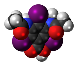
Formula C12H11I3N2O4
Molar mass 627.943 g·mol−1
3D model (JSmol)
  • CC(=O)NC1=C(C(=C(C(=C1I)C(=O)O)I)N(C)C(=O)C)I
  • InChI=1S/C12H11I3N2O4/c1-4(18)16-10-7(13)6(12(20)21)8(14)11(9(10)15)17(3)5(2)19/h1-3H3,(H,16,18)(H,20,21)
  • Key:GGGDNPWHMNJRFN-UHFFFAOYSA-N

Metrizoic acid is a pharmaceutical drug that was used as an iodinated contrast medium for X-ray imaging. Its uses included angiography [1] (imaging of blood vessels and heart chambers) and urography [2] (imaging of the urinary tract), but it has been discontinued, at least in the US. [3]

Contents

It was used in form of its salts, metrizoates. Due to its high osmolality, metrizoic acid had a risk of inducing allergic reactions higher than that of lower osmolar contrast media. [4]

Chemistry

The iodine content of metrizoate ranged from 370 mg/ml to 440 mg/ml, with osmolarity has high as 2100 mOsm/kg. The viscosity is 3.4 cP at 37 degree Celsius (human body temperature). [5]

Adverse effects

Side effects of metrizoate are: urticaria, headache, nausea, vomiting, dizziness, and hypotension. Other side effects include minor electrocardiographic changes such as tachycardia, bradycardia, and inversion of T waves. [6]

Related Research Articles

<span class="mw-page-title-main">Aspirin</span> Medication

Aspirin, also known as acetylsalicylic acid (ASA), is a nonsteroidal anti-inflammatory drug (NSAID) used to reduce pain, fever, and/or inflammation, and as an antithrombotic. Specific inflammatory conditions which aspirin is used to treat include Kawasaki disease, pericarditis, and rheumatic fever.

<span class="mw-page-title-main">Hypercholesterolemia</span> High levels of cholesterol in the blood

Hypercholesterolemia, also called high cholesterol, is the presence of high levels of cholesterol in the blood. It is a form of hyperlipidemia, hyperlipoproteinemia, and dyslipidemia.

<span class="mw-page-title-main">Iodinated contrast</span> Substance to enhance X-ray imaging

Iodinated contrast is a form of water-soluble, intravenous radiocontrast agent containing iodine, which enhances the visibility of vascular structures and organs during radiographic procedures. Some pathologies, such as cancer, have particularly improved visibility with iodinated contrast.

<span class="mw-page-title-main">Iotrolan</span> Chemical compound

Iotrolan is an iodine-containing radiocontrast agent, a substance used to improve the visibility of body structures on images obtained by X-ray techniques.

<span class="mw-page-title-main">Digital subtraction angiography</span> Method for delineating blood vessels using contrast medium

Digital subtraction angiography (DSA) is a fluoroscopy technique used in interventional radiology to clearly visualize blood vessels in a bony or dense soft tissue environment. Images are produced using contrast medium by subtracting a "pre-contrast image" or mask from subsequent images, once the contrast medium has been introduced into a structure. Hence the term "digital subtraction angiography. Subtraction angiography was first described in 1935 and in English sources in 1962 as a manual technique. Digital technology made DSA practical starting in the 1970s.

<span class="mw-page-title-main">Iohexol</span> Chemical compound

Iohexol, sold under the trade name Omnipaque among others, is a contrast agent used for X-ray imaging. This includes when visualizing arteries, veins, ventricles of the brain, the urinary system, and joints, as well as during computed tomography. It is given by mouth, injection into a vein, or into a body cavity.

Iodixanol, sold under the brand name Visipaque, is an iodine-containing non-ionic radiocontrast agent.

<span class="mw-page-title-main">Venography</span>

Venography is a procedure in which an X-ray of the veins, a venogram, is taken after a special dye is injected into the bone marrow or veins. The dye has to be injected constantly via a catheter, making it an invasive procedure. Normally the catheter is inserted by the groin and moved to the appropriate site by navigating through the vascular system.

<span class="mw-page-title-main">Computed tomography angiography</span> Medical investigation technique

Computed tomography angiography is a computed tomography technique used for angiography—the visualization of arteries and veins—throughout the human body. Using contrast injected into the blood vessels, images are created to look for blockages, aneurysms, dissections, and stenosis. CTA can be used to visualize the vessels of the heart, the aorta and other large blood vessels, the lungs, the kidneys, the head and neck, and the arms and legs. CTA can also be used to localise arterial or venous bleed of the gastrointestinal system.

<span class="mw-page-title-main">CT pulmonary angiogram</span>

A CT pulmonary angiogram (CTPA) is a medical diagnostic test that employs computed tomography (CT) angiography to obtain an image of the pulmonary arteries. Its main use is to diagnose pulmonary embolism (PE). It is a preferred choice of imaging in the diagnosis of PE due to its minimally invasive nature for the patient, whose only requirement for the scan is an intravenous line.

<span class="mw-page-title-main">Cardiac magnetic resonance imaging</span>

Cardiac magnetic resonance imaging, also known as cardiovascular MRI, is a magnetic resonance imaging (MRI) technology used for non-invasive assessment of the function and structure of the cardiovascular system. Conditions in which it is performed include congenital heart disease, cardiomyopathies and valvular heart disease, diseases of the aorta such as dissection, aneurysm and coarctation, coronary heart disease. It can also be used to look at pulmonary veins. Patient information may be found here.

<span class="mw-page-title-main">Iopamidol</span> Contrast agent

Iopamidol (INN), sold under the brand name Isovue among others, is a nonionic, low-osmolar iodinated contrast agent, developed by Bracco Diagnostics.

<span class="mw-page-title-main">Iodamide</span> Chemical compound

Iodamide is a pharmaceutical drug used as an iodinated contrast medium for X-ray imaging. Its uses include imaging of the uterus and fallopian tubes.

<span class="mw-page-title-main">Iopromide</span> Chemical compound

Iopromide is an iodinated contrast medium for X-ray imaging. It is marketed under the name Ultravist which is produced by Bayer Healthcare. It is a low osmolar, non-ionic contrast agent for intravascular use; i.e., it is injected into blood vessels.

<span class="mw-page-title-main">Iopentol</span> Chemical compound

Iopentol is a pharmaceutical drug that was used as a radiocontrast agent for X-ray imaging in Europe.

<span class="mw-page-title-main">Iomeprol</span> Chemical compound

Iomeprol is a pharmaceutical drug used as a radiocontrast agent in X-ray imaging. It is sold under the trade names Imeron and Iomeron.

<span class="mw-page-title-main">Iobitridol</span> Pharmaceutical drug

Iobitridol is a pharmaceutical drug used as an iodine-based radiocontrast agent in X-ray imaging. It is injected into blood vessels, joints, or body cavities such as the uterus, and filtered out by the kidneys. Its most common adverse effect is nausea. Severe allergic reactions are rare.

<span class="mw-page-title-main">Ioglicic acid</span> Chemical compound

Ioglicic acid is a pharmaceutical drug that was used as an iodinated contrast medium for X-ray imaging, in form of its salt meglumine ioglicate. Uses included imaging of the brain, the aorta and femoral arteries, and the urinary system.

<span class="mw-page-title-main">Ioxaglic acid</span> Chemical compound

Ioxaglic acid is pharmaceutical drug used as an iodinated contrast medium for X-ray imaging. It has low osmolality, typically resulting in fewer side effects than high-osmolality media. It is manufactured by Guerbet, but marketing in the US has been discontinued. As of 2021, it may still be available in some European countries.

<span class="mw-page-title-main">Cardiac magnetic resonance imaging perfusion</span>

Cardiac magnetic resonance imaging perfusion, also known as stress CMR perfusion, is a clinical magnetic resonance imaging test performed on patients with known or suspected coronary artery disease to determine if there are perfusion defects in the myocardium of the left ventricle that are caused by narrowing of one or more of the coronary arteries.

References

  1. Vik-Mo H, Danielsen R, Skinningsrud K, Haider T, Bjørkhaug A (1997). "Cardiovascular and electrocardiographic effects of iopentol in left ventricular angiography. Comparison of the low-osmolar, non-ionic iopentol (Imagopaque 350) and the hyper-osmolar, ionic metrizoate meglumine-Na-Ca (Isopaque Coronar 370) in patients with coronary heart disease". European Radiology. 7 (Suppl 4): S156–S161. doi:10.1007/pl00006885. PMID   9204361. S2CID   27742637.
  2. Zachrisson BE, Jagenburg R (1983). "Comparison of iohexol with metrizoate in urography. A single blind parallel investigation". Acta Radiologica. Supplementum. 366: 30–37. PMID   6147958.
  3. "Metrizoic acid". DrugBank. Retrieved 3 March 2017.
  4. "Metrizoic acid - C12H11I3N2O4". PubChem. U.S. National Library of Medicine. Retrieved 3 March 2017.
  5. Patel R. "Applications in contrast imaging: contrast media basics - important considerations for the pharmacist" (PDF). Braco Diagnostics, Inc. Archived from the original (PDF) on 15 December 2022. Retrieved 15 December 2022.
  6. Steinberg I, Evans JA (September 1967). "Isopaque 440 (metrizoate); a new cardiovascular contrast medium. Experience with 100 consecutive cases". The American Journal of Roentgenology, Radium Therapy, and Nuclear Medicine. 101 (1): 229–233. doi:10.2214/ajr.101.1.229. PMID   4166780.