Related Research Articles

Emergency medical services (EMS), also known as ambulance services or paramedic services, are emergency services that provide urgent pre-hospital treatment and stabilisation for serious illness and injuries and transport to definitive care. They may also be known as a first aid squad, FAST squad, emergency squad, ambulance squad, ambulance corps, life squad or by other initialisms such as EMAS or EMARS.
An emergency medical technician is a medical professional that provides emergency medical services. EMTs are most commonly found serving on ambulances and in fire departments in the US and Canada, as full-time and some part-time departments require their firefighters to at least be EMT certified.

Medical ultrasound includes diagnostic techniques using ultrasound, as well as therapeutic applications of ultrasound. In diagnosis, it is used to create an image of internal body structures such as tendons, muscles, joints, blood vessels, and internal organs, to measure some characteristics or to generate an informative audible sound. The usage of ultrasound to produce visual images for medicine is called medical ultrasonography or simply sonography, or echography. The practice of examining pregnant women using ultrasound is called obstetric ultrasonography, and was an early development of clinical ultrasonography. The machine used is called an ultrasound machine, a sonograph or an echograph. The visual image formed using this technique is called an ultrasonogram, a sonogram or an echogram.

Airway management includes a set of maneuvers and medical procedures performed to prevent and relieve airway obstruction. This ensures an open pathway for gas exchange between a patient's lungs and the atmosphere. This is accomplished by either clearing a previously obstructed airway; or by preventing airway obstruction in cases such as anaphylaxis, the obtunded patient, or medical sedation. Airway obstruction can be caused by the tongue, foreign objects, the tissues of the airway itself, and bodily fluids such as blood and gastric contents (aspiration).

Major trauma is any injury that has the potential to cause prolonged disability or death. There are many causes of major trauma, blunt and penetrating, including falls, motor vehicle collisions, stabbing wounds, and gunshot wounds. Depending on the severity of injury, quickness of management, and transportation to an appropriate medical facility may be necessary to prevent loss of life or limb. The initial assessment is critical, and involves a physical evaluation and also may include the use of imaging tools to determine the types of injuries accurately and to formulate a course of treatment.

Traumatic cardiac arrest (TCA) is a condition in which the heart has ceased to beat due to blunt or penetrating trauma, such as a stab wound to the thoracic area. It is a medical emergency which will always result in death without prompt advanced medical care. Even with prompt medical intervention, survival without neurological complications is rare. In recent years, protocols have been proposed to improve survival rate in patients with traumatic cardiac arrest, though the variable causes of this condition as well as many coexisting injuries can make these protocols difficult to standardize. Traumatic cardiac arrest is a complex form of cardiac arrest often derailing from advanced cardiac life support in the sense that the emergency team must first establish the cause of the traumatic arrest and reverse these effects, for example hypovolemia and haemorrhagic shock due to a penetrating injury.
Advanced trauma life support (ATLS) is a training program for medical providers in the management of acute trauma cases, developed by the American College of Surgeons. Similar programs exist for immediate care providers such as paramedics. The program has been adopted worldwide in over 60 countries, sometimes under the name of Early Management of Severe Trauma, especially outside North America. Its goal is to teach a simplified and standardized approach to trauma patients. Originally designed for emergency situations where only one doctor and one nurse are present, ATLS is now widely accepted as the standard of care for initial assessment and treatment in trauma centers. The premise of the ATLS program is to treat the greatest threat to life first. It also advocates that the lack of a definitive diagnosis and a detailed history should not slow the application of indicated treatment for life-threatening injury, with the most time-critical interventions performed early.

Paracentesis is a form of body fluid sampling procedure, generally referring to peritoneocentesis in which the peritoneal cavity is punctured by a needle to sample peritoneal fluid.

A trauma team is a multidisciplinary group of healthcare workers under the direction of a team leader that works together to assess and treat the severely injured. This team typically meets before the patient reaches the trauma center. Upon arrival, the team does an initial assessment and necessary resuscitation, adhering to a defined protocol.

Blunt trauma, also known as blunt force trauma or non-penetrating trauma, describes a physical trauma due to a forceful impact without penetration of the body's surface. Blunt trauma stands in contrast with penetrating trauma, which occurs when an object pierces the skin, enters body tissue, and creates an open wound. Blunt trauma occurs due to direct physical trauma or impactful force to a body part. Such incidents often occur with road traffic collisions, assaults, and sports-related injuries, and are notably common among the elderly who experience falls.

A cricothyrotomy is an incision made through the skin and cricothyroid membrane to establish a patient airway during certain life-threatening situations, such as airway obstruction by a foreign body, angioedema, or massive facial trauma. Cricothyrotomy is nearly always performed as a last resort in cases where other means of tracheal intubation are impossible or impractical. Compared with tracheotomy, cricothyrotomy is quicker and easier to perform, does not require manipulation of the cervical spine, and is associated with fewer complications. However, while cricothyrotomy may be life-saving in extreme circumstances, this technique is only intended to be a temporizing measure until a definitive airway can be established.
In the United States, the paramedic is an allied health professional whose primary focus is to provide advanced emergency medical care for patients who access Emergency Medical Services (EMS). This individual possesses the complex knowledge and skills necessary to provide patient care and transportation. Paramedics function as part of a comprehensive EMS response under physician medical direction. Paramedics often serve in a prehospital role, responding to Public safety answering point (9-1-1) calls in an ambulance. The paramedic serves as the initial entry point into the health care system. A standard requirement for state licensure involves successful completion of a nationally accredited Paramedic program at the certificate or associate degree level.
Advanced Emergency Medical Technician - Critical Care (AEMT-CC) is a former Emergency Medical Services (EMS) certification that was unique to New York. The curriculum for AEMT-CC's in New York was similar to that of the national standard EMT-I/99 but with a broader scope of practice. EMT-CCs are fully classified as Advanced Life Support (ALS) providers within New York and are trained in advanced airway management, including intubation, IV fluid administration, cardiac monitoring, cardiac pacing, and both synchronized and unsynchronized cardioversion, and medication usage/administration in adult and pediatric patients.
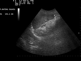
Focused assessment with sonography in trauma is a rapid bedside ultrasound examination performed by surgeons, emergency physicians, and paramedics as a screening test for blood around the heart or abdominal organs (hemoperitoneum) after trauma. There is also the extended FAST (eFAST) which includes some additional ultrasound views to assess for pneumothorax.
Emergency ultrasound employing point-of-care ultrasound (POCUS) is the application of ultrasound at the point of care to make immediate patient-care decisions. It is performed by the health care professional caring for the injured or ill persons. This point-of-care use of ultrasound is often to evaluate an emergency medical condition, in settings such as an emergency department, critical care unit, ambulance, or combat zone.
An advanced emergency medical technician (AEMT) is a provider of emergency medical services in the United States. A transition to this level of training from the emergency medical technician-intermediate, which have somewhat less training, began in 2013 and has been implemented by most states. AEMTs are not intended to deliver definitive medical care in most cases, but rather to augment prehospital critical care and provide rapid on-scene treatment. AEMTs are usually employed in ambulance services, working in conjunction with EMTs and paramedics; however they are also commonly found in fire departments and law enforcement agencies as non-transporting first responders. Ambulances operating at the AEMT level of care are commonplace in rural areas, and occasionally found in larger cities as part of a tiered-response system, but are overall much less common than EMT- and paramedic-level ambulances. The AEMT provides a low-cost, high-benefit option to provide advanced-level care when the paramedic level of care is not feasible. The AEMT is authorized to provide limited advanced life support, which is beyond the scope of an EMT.
The following outline is provided as an overview of and topical guide to emergency medicine:

A resuscitative thoracotomy (sometimes referred to as an emergency department thoracotomy (EDT), trauma thoracotomy or, colloquially, as "cracking the chest") is a thoracotomy performed to aid in the resuscitation of a major trauma patient who has sustained severe thoracic or abdominal trauma. The procedure allows immediate direct access to the thoracic cavity, permitting rescuers to control hemorrhage, relieve cardiac tamponade, repair or control major injuries to the heart, lungs or thoracic vasculature, and perform direct cardiac massage or defibrillation. The procedure is rarely performed and is a procedure of last resort.

Spinal precautions, also known as spinal immobilization and spinal motion restriction, are efforts to prevent movement of the bones of the spine in those with a risk of a spine injury. This is done as an effort to prevent injury to the spinal cord in unstable spinal fractures. About 0.5-3% of people with blunt trauma will have a spine injury, with 42-50% of injuries due to motor vehicle accidents, 27-43% from falls or work injuries, and the rest due to sports injuries (9%) or assault (11%). The majority of spinal cord injuries are to the cervical spine (52%), followed by the thoracic and lumbar spine. Cervical spinal cord injuries can result in tetraplegia or paraplegia, depending on severity. Of spine injuries, only 0.01% are unstable and require intervention.

Pre-hospital emergency medicine, also referred to as pre-hospital care, immediate care, or emergency medical services medicine, is a medical subspecialty which focuses on caring for seriously ill or injured patients before they reach hospital, and during emergency transfer to hospital or between hospitals. It may be practised by physicians from various backgrounds such as anaesthesiology, emergency medicine, intensive care medicine and acute medicine, after they have completed initial training in their base specialty.
References
- ↑ Bonadonna, Peter. "Paramedic Ultrasound" . Retrieved 12 May 2011.
- ↑ von Foerster, Nicholas; Radomski, Marek A.; Martin-Gill, Christian (2 January 2024). "Prehospital Ultrasound: A Narrative Review". Prehospital Emergency Care. 28 (1): 1–13. doi:10.1080/10903127.2022.2132332.
- 1 2 3 Mallinson, T (2024). Prehospital & Emergency Ultrasound: Logbook & Guide. London, England: Caladrius Press. ISBN 978-1917521062.
- ↑ Emergency Ultrasound Made Easy. Justin Bowra, Russell E. McLaughlin. Elsevier Churchill Livingstone, 2006 ISBN 0-443-10150-7, ISBN 978-0-443-10150-2 [ page needed ]
- ↑ Emergency Ultrasound: Principles and Practice. Romolo Joseph Gaspari, J. Christian Fox, Paul R. Sierzenski. Mosby, 2005. ISBN 0-323-03750-X, 9780323037501[ page needed ]
- ↑ Atlas of Emergency Medicine. Kevin J. Knoop, Lawrence B. Stack, Alan B. Storrow. McGraw-Hill Professional, 2002. ISBN 0-07-135294-5, ISBN 978-0-07-135294-9 [ page needed ]
- ↑ Kundra, Pankaj; Mishra, SandeepKumar; Ramesh, Anathakrishnan (2011). "Ultrasound of the airway". Indian Journal of Anaesthesia. 55 (5): 456–462. doi: 10.4103/0019-5049.89868 . PMC 3237144 . PMID 22174461.
- ↑ "EMS Pre-Hospital Ultrasound".
- ↑ Thomas, Bruce; Falcone, Robert E.; Vasquez, Donald; Santanello, Steven; Townsend, Michael; Hockenberry, Scott; Innes, Jeffrey; Wanamaker, Steven (March 1997). "Ultrasound Evaluation of Blunt Abdominal Trauma: Program Implementation, Initial Experience, and Learning Curve". The Journal of Trauma: Injury, Infection, and Critical Care. 42 (3): 384–390. doi:10.1097/00005373-199703000-00004. PMID 9095104.
- ↑ Introduction To Emergency Ultrasound: A Review Of Justifications, Indications And Significant Findings. Steven A. Godwin M.D. March, 1999. Jacksonville Medicine Journal. http://www.dcmsonline.org/jax-medicine/1999journals/march99/ultrasound.htm
- ↑ Mateer, James R.; Ogata, Masaaki; Kefer, Michael P.; Wittmann, Dietmar; Aprahamian, Charles (June 1995). "Prospective Analysis of a Rapid Trauma Ultrasound Examination Performed by Emergency Physicians". The Journal of Trauma: Injury, Infection, and Critical Care. 38 (6): 879–885. doi:10.1097/00005373-199506000-00009. PMID 7602628.
- ↑ Boitnott, J. Optic Nerve Sheath Ultrasound. EMSPOCUS. http://emspocus.com/2015/12/07/optic-nerve-sheath-ultrasound/