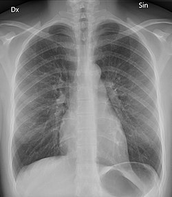
A chest radiograph, chest X-ray (CXR), or chest film is a projection radiograph of the chest used to diagnose conditions affecting the chest, its contents, and nearby structures. Chest radiographs are the most common film taken in medicine.
Like all methods of radiography, chest radiography employs ionizing radiation in the form of X-rays to generate images of the chest. The mean radiation dose to an adult from a chest radiograph is around 0.02 mSv (2 mrem) for a front view (PA, or posteroanterior) and 0.08 mSv (8 mrem) for a side view (LL, or latero-lateral).[1] Together, this corresponds to a background radiation equivalent time of about 10 days.[2]
Medical uses
Dedicated chest radiography room
Conditions commonly identified by chest radiography
Chest radiographs are used to diagnose many conditions involving the chest wall, including its bones, and also structures contained within the thoracic cavity including the lungs, heart, and great vessels. Pneumonia and congestive heart failure are very commonly diagnosed by chest radiograph. Chest radiographs are also used to screen for job-related lung disease in industries such as mining where workers are exposed to dust.[3]
For some conditions of the chest, radiography is good for screening but poor for diagnosis. When a condition is suspected based on chest radiography, additional imaging of the chest can be obtained to definitively diagnose the condition or to provide evidence in favor of the diagnosis suggested by initial chest radiography. Unless a fractured rib is suspected of being displaced, and therefore likely to cause damage to the lungs and other tissue structures, x-ray of the chest is not necessary as it will not alter patient management.
The main regions where a chest X-ray may identify problems may be summarized as ABCDEF by their first letters:[4]
Airways, including hilar adenopathy or enlargement
Breast shadows
Bones, e.g. rib fractures and lytic bone lesions
Cardiac silhouette, detecting cardiac enlargement
Costophrenic angles, including pleural effusions
Diaphragm, e.g. evidence of free air, indicative of perforation of an abdominal viscus
Edges, e.g. apices for fibrosis, pneumothorax, pleural thickening or plaques
Extrathoracic tissues
Fields (lung parenchyma), being evidence of alveolar flooding
Positioning for a PA chest x-rayNormal lateral chest radiograph.
Different views (also known as projections) of the chest can be obtained by changing the relative orientation of the body and the direction of the x-ray beam. The most common views are posteroanterior, anteroposterior, and lateral. In a posteroanterior (PA) view, the x-ray source is positioned so that the x-ray beam enters through the posterior (back) aspect of the chest and exits out of the anterior (front) aspect, where the beam is detected. To obtain this view, the patient stands facing a flat surface behind which is an x-ray detector. A radiation source is positioned behind the patient at a standard distance (most often 6 feet, 1,8m), and the x-ray beam is fired toward the patient.
In anteroposterior (AP) views, the positions of the x-ray source and detector are reversed: the x-ray beam enters through the anterior aspect and exits through the posterior aspect of the chest. AP chest x-rays are harder to read than PA x-rays [citation needed] and are therefore generally reserved for situations where it is difficult for the patient to get an ordinary chest x-ray, such as when the patient is bedridden. In this situation, mobile X-ray equipment is used to obtain a lying down chest x-ray (known as a "supine film"). As a result, most supine films are also AP.
Lateral views of the chest are obtained in a similar fashion as the posteroanterior views, except in the lateral view, the patient stands with both arms raised and the left side of the chest pressed against a flat surface.
Typical views
Required projections can vary by country and hospital, although an erect posteroanterior (PA) projection is typically the first preference. If this is not possible, then an anteroposterior view will be taken. Further imaging depends on local protocols which is dependent on the hospital protocols, the availability of other imaging modalities and the preference of the image interpreter. In the UK, the standard chest radiography protocol is to take an erect posteroanterior view only and a lateral one only on request by a radiologist.[5] In the US, chest radiography includes a PA and Lateral with the patient standing or sitting up. Special projections include an AP in cases where the image needs to be obtained stat (immediately) and with a portable device, particularly when a patient cannot be safely positioned upright. Lateral decubitus may be used for visualization of air-fluid levels if an upright image cannot be obtained. Anteroposterior (AP) Axial Lordotic projects the clavicles above the lung fields, allowing better visualization of the apices (which is extremely useful when looking for evidence of primary tuberculosis).
Additional views
Decubitus – taken while the patient is lying down, typically on their side. Useful for differentiating pleural effusions from consolidation (e.g. pneumonia) and loculated effusions from free fluid in the pleural space. In effusions, the fluid layers out (by comparison to an up-right view, when it often accumulates in the costophrenic angles).
Oblique view – useful for the visualization of the ribs and sternum. Although it is necessary to do the appropriate adaptations to the x-ray dosage to be used.
Landmarks
A chest radiograph with the angle parts of the ribs and some other landmarks labeled.Mediastinal structures on a chest radiograph.
In the average person, the diaphragm should be intersected by the 5th to 7th anterior ribs at the mid-clavicular line, and 9 to 10 posterior ribs should be viewable on a normal PA inspiratory film. An increase in the number of viewable ribs implies hyperinflation, as can occur, for example, with obstructive lung disease or foreign body aspiration. A decrease implies hypoventilation, as can occur with restrictive lung disease, pleural effusions or atelectasis. Underexpansion can also cause interstitial markings due to parenchymal crowding, which can mimic the appearance of interstitial lung disease. Enlargement of the right descending pulmonary artery can indirectly reflect changes of pulmonary hypertension, with a size greater than 16mm abnormal in men and 15mm in women.[6]
Appropriate penetration of the film can be assessed by faint visualization of the thoracic spines and lung markings behind the heart. The right diaphragm is usually higher than the left, with the liver being situated beneath it in the abdomen. The minor fissure can sometimes be seen on the right as a thin horizontal line at the level of the fifth or sixth rib. Splaying of the carina can also suggest a tumor or process in the middle mediastinum or enlargement of the left atrium, with a normal angle of approximately 60 degrees. The right paratracheal stripe is also important to assess, as it can reflect a process in the posterior mediastinum, in particular the spine or paraspinal soft tissues; normally it should measure 3mm or less. The left paratracheal stripe is more variable and only seen in 25% of normal patients on posteroanterior views.[7]
Localization of lesions or inflammatory and infectious processes can be difficult to discern on chest radiograph, but can be inferred by silhouetting and the hilum overlay sign with adjacent structures. If either hemidiaphragm is blurred, for example, this suggests the lesion to be from the corresponding lower lobe. If the right heart border is blurred, than the pathology is likely in the right middle lobe, though a cavum deformity can also blur the right heard border due to indentation of the adjacent sternum. If the left heart border is blurred, this implies a process at the lingula.[8]
Abnormalities
Nodule
A lung nodule is a discrete opacity in the lung which may be caused by:
Fluid in space between the lung and the chest wall is termed a pleural effusion. There needs to be at least 75 mL of pleural fluid in order to blunt the costophrenic angle on the lateral chest radiograph and 200 mL of pleural fluid in order to blunt the costophrenic angle on the posteroanterior chest radiograph. On a lateral decubitus, amounts as small as 50ml of fluid are possible. Pleural effusions typically have a meniscus visible on an erect chest radiograph, but loculated effusions (as occur with an empyema) may have a lenticular shape (the fluid making an obtuse angle with the chest wall).
Pleural thickening may cause blunting of the costophrenic angle, but is distinguished from pleural fluid by the fact that it occurs as a linear shadow ascending vertically and clinging to the ribs.
Diffuse shadowing
The differential for diffuse shadowing is very broad and can defeat even the most experienced radiologist. It is seldom possible to reach a diagnosis on the basis of the chest radiograph alone: high-resolution CT of the chest is usually required and sometimes a lung biopsy. The following features are usually noted:
Pleural effusions may occur with cancer, sarcoid, connective tissue diseases and lymphangioleiomyomatosis. The presence of a pleural effusion argues against pneumocystis pneumonia.
Reticular (linear) pattern
(sometimes called "reticulonodular" because of the appearance of nodules at the intersection of the lines, even though there are no true nodules present)
The silhouette sign is especially helpful in localizing lung lesions. (e.g., loss of right heart border in right middle lobe pneumonia),[9]
The air bronchogram sign, where branching radiolucent columns of air corresponding to bronchi is seen, usually indicates air-space (alveolar) disease, as from blood, pus, mucus, cells, protein surrounding the air bronchograms. This is seen in Respiratory distress syndrome[9]
Disease mimics
Disease mimics are visual artifacts, normal anatomic structures or harmless variants that may simulate diseases and abnormalities.
While chest radiographs are a relatively cheap and safe method of investigating diseases of the chest, there are a number of serious chest conditions that may be associated with a normal chest radiograph and other means of assessment may be necessary to make the diagnosis. For example, a patient with an acute myocardial infarction may have a completely normal chest radiograph.
Gallery
Chest X-ray PA inverted and enhanced.
Projectionally rendered CT scan, showing the transition of thoracic structures between the anteroposterior and lateral view
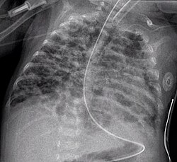
Chest film showing increased opacity in both lungs, indicative of pneumonia
A chest film after insertion of an implantable cardioverter-defibrillator, showing the shock generator in the upper left chest and the electrical lead inside the right heart. Note both radio-opaque coils along the device lead.
Portable chest (ie, antero-posterior view) showing endotracheal tube and Levin tube, both properly in place.
References
↑Fred A. Mettler, Walter Huda, Terry T. Yoshizumi, Mahadevappa Mahesh: "Effective Doses in Radiology and Diagnostic Nuclear Medicine: A Catalog" – Radiology 2008;248:254–263
↑Gibbs, JM; Chandrasekhar, CA; Ferguson, EC; Oldham, SA (2007). "Lines and stripes: where did they go?--From conventional radiography to CT". Radiographics. 27 (1): 33–48. doi:10.1148/rg.271065073. PMID17234997.
↑Khan, Nausheen; Thebe, Dimakatso C.; Suleman, Farhanah; Van de Werke, Irma (2015). "Pitfalls and mimics: The many facets of normal paediatric thymus". South African Journal of Radiology. 19 (1). doi:10.4102/sajr.v19i1.803. ISSN2078-6778. (CC BY 4.0)
This page is based on this Wikipedia article Text is available under the CC BY-SA 4.0 license; additional terms may apply. Images, videos and audio are available under their respective licenses.