A 'Jedi' helmet, on display at the Science Museum:Medicine:The Wellcome Galleries
In the early 1980s to the early 1990s, 'Jedi' helmets, inspired by the 'Return of the Jedi' Star Wars film, were sometimes worn by children in order to obtain good image quality. The copper coils of the helmet were used as a radio aerial to detect the signals while the 'Jedi' association encouraged children to wear the helmets and not be frightened by the procedure. These helmets were no longer needed as MR scanners improved.
In the early 1990s, Peter Basser and Le Bihan, working at NIH, and Aaron Filler, Franklyn Howe, and colleagues developed diffusion tensor imaging (DTI).[6][7][8][9] The first DTI Brain Tractogram was published by the United States as Figure 17 of US Patent 5,560,360 (filed March 9, 1993, granted Oct. 1, 1996 - Filler, Howe, Richards & Tsuruda). Joseph Hajnal, Young and Graeme Bydder described the use of FLAIR pulse sequence to demonstrate high signal regions in normal white matter in 1992.[10] In the same year, John Detre, Alan P. Koretsky and coworkers developed arterial spin labeling.[11] In 1997, Jürgen R. Reichenbach, E. Mark Haacke and coworkers at Washington University in St. Louis developed Susceptibility weighted imaging.[12]
The first study of the human brain at 3.0 T was published in 1994,[13] and in 1998 at 8 T.[14] Studies of the human brain have been performed at 9.4 T (2006)[15] and up to 10.5 T (2019).[16]
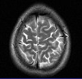
This axial T2-weighted (CSF white) MR scan shows a normal brain at the level of the lateral ventricles.
The record for the highest spatial resolution of a whole intact brain (postmortem) is 100 microns, from Massachusetts General Hospital. The data was published in Scientific Data on 30 October 2019.[17][18]
In the case of a concussion, an MRI should be avoided unless there are progressive neurological symptoms, focal neurological findings or concern of skull fracture on exam.[22] In the analysis of a concussion, measurements of Fractional Anisotropy, Mean Diffusivity, Cerebral Blood Flow, and Global Connectivity can be taken to observe the pathophysiological mechanisms being made while in recovery.[23]
↑Le Bihan D, Breton E (1987). "Method to Measure the Molecular Diffusion and/or Perfusion Parameters of Live Tissue". US Patent # 4,809,701.
↑Villringer A, Rosen BR, Belliveau JW, Ackerman JL, Lauffer RB, Buxton RB, Chao YS, Wedeen VJ, Brady TJ (February 1988). "Dynamic imaging with lanthanide chelates in normal brain: contrast due to magnetic susceptibility effects". Magnetic Resonance in Medicine. 6 (2): 164–74. doi:10.1002/mrm.1910060205. PMID3367774. S2CID41228095.
↑Hajnal JV, De Coene B, Lewis PD, Baudouin CJ, Cowan FM, Pennock JM, Young IR, Bydder GM (July 1992). "High signal regions in normal white matter shown by heavily T2-weighted CSF nulled IR sequences". Journal of Computer Assisted Tomography. 16 (4): 506–13. doi:10.1097/00004728-199207000-00002. PMID1629405. S2CID42727826.
↑Reichenbach JR, Venkatesan R, Schillinger DJ, Kido DK, Haacke EM (July 1997). "Small vessels in the human brain: MR venography with deoxyhemoglobin as an intrinsic contrast agent". Radiology. 204 (1): 272–7. doi:10.1148/radiology.204.1.9205259. PMID9205259.
↑Mansfield P, Coxon R, Glover P (May 1994). "Echo-planar imaging of the brain at 3.0 T: first normal volunteer results". Journal of Computer Assisted Tomography. 18 (3): 339–43. doi:10.1097/00004728-199405000-00001. PMID8188896. S2CID20221062.
This page is based on this Wikipedia article Text is available under the CC BY-SA 4.0 license; additional terms may apply. Images, videos and audio are available under their respective licenses.