Related Research Articles

Magnetic resonance imaging (MRI) is a medical imaging technique used in radiology to form pictures of the anatomy and the physiological processes inside the body. MRI scanners use strong magnetic fields, magnetic field gradients, and radio waves to generate images of the organs in the body. MRI does not involve X-rays or the use of ionizing radiation, which distinguishes it from computed tomography (CT) and positron emission tomography (PET) scans. MRI is a medical application of nuclear magnetic resonance (NMR) which can also be used for imaging in other NMR applications, such as NMR spectroscopy.
In neuroscience, tractography is a 3D modeling technique used to visually represent nerve tracts using data collected by diffusion MRI. It uses special techniques of magnetic resonance imaging (MRI) and computer-based diffusion MRI. The results are presented in two- and three-dimensional images called tractograms.

Perfusion is the passage of fluid through the circulatory system or lymphatic system to an organ or a tissue, usually referring to the delivery of blood to a capillary bed in tissue. Perfusion may also refer to fixation via perfusion, used in histological studies. Perfusion is measured as the rate at which blood is delivered to tissue, or volume of blood per unit time per unit tissue mass. The SI unit is m3/(s·kg), although for human organs perfusion is typically reported in ml/min/g. The word is derived from the French verb perfuser, meaning to "pour over or through". All animal tissues require an adequate blood supply for health and life. Poor perfusion (malperfusion), that is, ischemia, causes health problems, as seen in cardiovascular disease, including coronary artery disease, cerebrovascular disease, peripheral artery disease, and many other conditions.

Diffusion-weighted magnetic resonance imaging is the use of specific MRI sequences as well as software that generates images from the resulting data that uses the diffusion of water molecules to generate contrast in MR images. It allows the mapping of the diffusion process of molecules, mainly water, in biological tissues, in vivo and non-invasively. Molecular diffusion in tissues is not random, but reflects interactions with many obstacles, such as macromolecules, fibers, and membranes. Water molecule diffusion patterns can therefore reveal microscopic details about tissue architecture, either normal or in a diseased state. A special kind of DWI, diffusion tensor imaging (DTI), has been used extensively to map white matter tractography in the brain.
Magnetic resonance elastography (MRE) is a form of elastography that specifically leverages MRI to quantify and subsequently map the mechanical properties of soft tissue. First developed and described at Mayo Clinic by Muthupillai et al. in 1995, MRE has emerged as a powerful, non-invasive diagnostic tool, namely as an alternative to biopsy and serum tests for staging liver fibrosis.

In physics, the spin–spin relaxation is the mechanism by which Mxy, the transverse component of the magnetization vector, exponentially decays towards its equilibrium value in nuclear magnetic resonance (NMR) and magnetic resonance imaging (MRI). It is characterized by the spin–spin relaxation time, known as T2, a time constant characterizing the signal decay. It is named in contrast to T1, the spin–lattice relaxation time. It is the time it takes for the magnetic resonance signal to irreversibly decay to 37% (1/e) of its initial value after its generation by tipping the longitudinal magnetization towards the magnetic transverse plane. Hence the relation
Kenneth Kin Man Kwong is a Hong Kong-born American nuclear physicist. He is a pioneer in human brain imaging. He received his bachelor's degree in Political Science in 1972 from the University of California, Berkeley. He went on to receive his Ph.D. in physics from the University of California, Riverside studying photon-photon collision interactions.

A connectome is a comprehensive map of neural connections in the brain, and may be thought of as its "wiring diagram". An organism's nervous system is made up of neurons which communicate through synapses. A connectome is constructed by tracing the neuron in a nervous system and mapping where neurons are connected through synapses.

Magnetic resonance neurography (MRN) is the direct imaging of nerves in the body by optimizing selectivity for unique MRI water properties of nerves. It is a modification of magnetic resonance imaging. This technique yields a detailed image of a nerve from the resonance signal that arises from in the nerve itself rather than from surrounding tissues or from fat in the nerve lining. Because of the intraneural source of the image signal, the image provides a medically useful set of information about the internal state of the nerve such as the presence of irritation, nerve swelling (edema), compression, pinch or injury. Standard magnetic resonance images can show the outline of some nerves in portions of their courses but do not show the intrinsic signal from nerve water. Magnetic resonance neurography is used to evaluate major nerve compressions such as those affecting the sciatic nerve (e.g. piriformis syndrome), the brachial plexus nerves (e.g. thoracic outlet syndrome), the pudendal nerve, or virtually any named nerve in the body. A related technique for imaging neural tracts in the brain and spinal cord is called magnetic resonance tractography or diffusion tensor imaging.
Robert Turner is a British neuroscientist, physicist, and social anthropologist. He has been a director and professor at the Max Planck Institute for Human Cognitive and Brain Sciences in Leipzig, Germany, and is an internationally recognized expert in brain physics and magnetic resonance imaging (MRI). Coils inside every MRI scanner owe their shape to his ideas.

Magnetic resonance imaging of the brain uses magnetic resonance imaging (MRI) to produce high-quality two- or three-dimensional images of the brain, brainstem, and cerebellum without ionizing radiation (X-rays) or radioactive tracers.
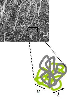
Intravoxel incoherent motion (IVIM) imaging is a concept and a method initially introduced and developed by Le Bihan et al. to quantitatively assess all the microscopic translational motions that could contribute to the signal acquired with diffusion MRI. In this model, biological tissue contains two distinct environments: molecular diffusion of water in the tissue, and microcirculation of blood in the capillary network (perfusion). The concept introduced by D. Le Bihan is that water flowing in capillaries mimics a random walk (Fig.1), as long as the assumption that all directions are represented in the capillaries is satisfied.
Medical image computing (MIC) is an interdisciplinary field at the intersection of computer science, information engineering, electrical engineering, physics, mathematics and medicine. This field develops computational and mathematical methods for solving problems pertaining to medical images and their use for biomedical research and clinical care.

Val Murray Runge is an American and Swiss professor of radiology and the editor-in-chief of Investigative Radiology. Runge was one of the early researchers to investigate the use of gadolinium-based contrast agents for magnetic resonance imaging (MRI), giving the first presentation in this field, followed two years later by the first presentation of efficacy. His research also pioneered many early innovations in MRI, including the use of tilted planes and respiratory gating. His publication on multiple sclerosis in 1984 represented the third and largest clinical series investigating the role of MRI in this disease, and the first to show characteristic abnormalities on MRI in patients whose CT was negative.

Perfusion MRI or perfusion-weighted imaging (PWI) is perfusion scanning by the use of a particular MRI sequence. The acquired data are then post-processed to obtain perfusion maps with different parameters, such as BV, BF, MTT and TTP.

Wolfgang Grodd is a German neuroradiologist and professor emeritus of the University hospital at the University of Tübingen. He is known for his scientific works on the development and application of structural and functional magnetic resonance imaging in metabolic diseases, sensorimotor representation, language production, and cognitive processing, cerebellum, thalamus, and basal ganglia. Currently, Wolfgang Grodd is a research scientist at the Department of the High-Field MR at the Max Planck Institute for Biological Cybernetics.

The history of magnetic resonance imaging (MRI) includes the work of many researchers who contributed to the discovery of nuclear magnetic resonance (NMR) and described the underlying physics of magnetic resonance imaging, starting early in the twentieth century. One researcher was American physicist Isidor Isaac Rabi who won the Nobel Prize in Physics in 1944 for his discovery of nuclear magnetic resonance, which is used in magnetic resonance imaging. MR imaging was invented by Paul C. Lauterbur who developed a mechanism to encode spatial information into an NMR signal using magnetic field gradients in September 1971; he published the theory behind it in March 1973.

An MRI pulse sequence in magnetic resonance imaging (MRI) is a particular setting of pulse sequences and pulsed field gradients, resulting in a particular image appearance.
Arterial spin labeling (ASL), also known as arterial spin tagging, is a magnetic resonance imaging technique used to quantify cerebral blood perfusion by labelling blood water as it flows throughout the brain. ASL specifically refers to magnetic labeling of arterial blood below or in the imaging slab, without the need of gadolinium contrast. A number of ASL schemes are possible, the simplest being flow alternating inversion recovery (FAIR) which requires two acquisitions of identical parameters with the exception of the out-of-slice saturation; the difference in the two images is theoretically only from inflowing spins, and may be considered a 'perfusion map'. The ASL technique was developed by John S. Leigh Jr, John A. Detre, Donald S. Williams, and Alan P. Koretsky in 1992.
Hyperpolarized gas MRI, also known as hyperpolarized helium-3 MRI or HPHe-3 MRI, is a medical imaging technique that uses hyperpolarized gases to improve the sensitivity and spatial resolution of magnetic resonance imaging (MRI). This technique has many potential applications in medicine, including the imaging of the lungs and other areas of the body with low tissue density.
References
- ↑ "Biographie, Denis Le Bihan, Institut de France" (PDF). Archived from the original (PDF) on 3 November 2013. Retrieved 12 December 2019.
- ↑ "ISMRM Awards". Archived from the original on 3 November 2013. Retrieved 12 December 2019.
- ↑ "Louis D. Prize" (in French). Institut de France.
- ↑ "Honda Award 2012".
- ↑ "Denis Le Bihan: 'Water, the molecule of the mind?'". 26 November 2010. Retrieved 10 June 2013.
- ↑ Guy Aubert, Petite histoire des grands instruments : de l’astronomie à la recherche..., Publications de l'AUEG, 2007
- ↑ Le Bihan D et Breton E, « Imagerie de diffusion in vivo par résonance magnétique nucléaire » C.R. Acad. Sc. Paris, T.301, Série II:1109–1112, 1985
- ↑ Le Bihan, D; Breton, E; Lallemand, D; Aubin, M L; Vignaud, J; Laval-Jeantet, M (1988). "Separation of diffusion and perfusion in intravoxel incoherent motion MR imaging". Radiology. 168 (2). Radiological Society of North America (RSNA): 497–505. doi:10.1148/radiology.168.2.3393671. ISSN 0033-8419. PMID 3393671.
- ↑ Le Bihan, Denis (2003). "Looking into the functional architecture of the brain with diffusion MRI". Nature Reviews Neuroscience. 4 (6). Springer Science and Business Media LLC: 469–480. doi:10.1038/nrn1119. ISSN 1471-003X. PMID 12778119. S2CID 14884501.
- ↑ Le Bihan, Denis (2008). "Intravoxel Incoherent Motion Perfusion MR Imaging: A Wake-Up Call". Radiology. 249 (3). Radiological Society of North America (RSNA): 748–752. doi:10.1148/radiol.2493081301. ISSN 0033-8419. PMID 19011179.
- ↑ Denis Le Bihan, Le cerveau de cristal : Ce que nous révèle la neuro-imagerie, Odile Jacob, Paris, 2012
- ↑ Douek, Philippe; Turner, Robert; Pekar, James; Patronas, Nichoias; Bihan, Denis Le (1991). "MR Color Mapping of Myelin Fiber Orientation". Journal of Computer Assisted Tomography. 15 (6). Ovid Technologies (Wolters Kluwer Health): 923–929. doi:10.1097/00004728-199111000-00003. ISSN 0363-8715. PMID 1939769.
- ↑ Basser, P.J.; Mattiello, J.; LeBihan, D. (1994). "MR diffusion tensor spectroscopy and imaging". Biophysical Journal. 66 (1). Elsevier BV: 259–267. Bibcode:1994BpJ....66..259B. doi:10.1016/s0006-3495(94)80775-1. ISSN 0006-3495. PMC 1275686 . PMID 8130344.
- ↑ Basser, P.J.; Mattiello, J.; Lebihan, D. (1994). "Estimation of the Effective Self-Diffusion Tensor from the NMR Spin Echo". Journal of Magnetic Resonance, Series B. 103 (3). Elsevier BV: 247–254. Bibcode:1994JMRB..103..247B. doi:10.1006/jmrb.1994.1037. ISSN 1064-1866. PMID 8019776.
- ↑ Le Bihan, Denis; Mangin, Jean-Francois; Poupon, Cyril; Clark, Chris A.; Pappata, Sabina; Molko, Nicolas; Chabriat, Hughes (2001). "Diffusion tensor imaging: Concepts and applications". Journal of Magnetic Resonance Imaging. 13 (4). Wiley: 534–546. doi: 10.1002/jmri.1076 . ISSN 1053-1807. PMID 11276097. S2CID 7269302.
- ↑ Le Bihan D, Johansen-Berg H, "Diffusion MRI at 25: Exploring brain tissue structure and function", NeuroImage 2011.
- ↑ Iima, Mami; Le Bihan, Denis; Okumura, Ryosuke; Okada, Tomohisa; Fujimoto, Koji; Kanao, Shotaro; Tanaka, Shiro; Fujimoto, Masakazu; Sakashita, Hiromi; Togashi, Kaori (2011). "Apparent Diffusion Coefficient as an MR Imaging Biomarker of Low-Risk Ductal Carcinoma in Situ: A Pilot Study". Radiology. 260 (2). Radiological Society of North America (RSNA): 364–372. doi:10.1148/radiol.11101892. hdl: 2433/188640 . ISSN 0033-8419. PMID 21633054.
- ↑ "Intérêt de l'IRM de diffusion dans le diagnostic et la quantification de la fibrose hépatique" (PDF). pe.sfrnet.org. Archived from the original (PDF) on 12 May 2014. Retrieved 10 June 2013.
- ↑ "Meteore Service (D. Le Bihan)". www.meteoreservice.com. Retrieved 12 May 2023.
- ↑ Eduard Rhein Foundation – Technology Award 2021
| International | |
|---|---|
| National | |
| Academics | |
| Other | |