Cardiac resynchronization therapy

Last updated
Cardiac resynchronization therapy
Cardiac resynchronisation therapy.png
ICD-9-CM 00.51, 00.54
MeSH D058409
eMedicine 1839506-devices

Cardiac resynchronisation therapy (CRT or CRT-P) is the insertion of electrodes in the left and right ventricles of the heart, as well as on occasion the right atrium, to treat heart failure by coordinating the function of the left and right ventricles via a pacemaker, a small device inserted into the anterior chest wall. [1]

Contents

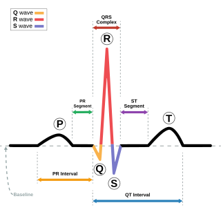
CRT is indicated in patients with a low ejection fraction (typically <35%) indicating heart failure, where electrical activity has been compromised, with prolonged QRS duration to >120 ms. [2]

The insertion of electrodes into the ventricles is done under local anesthetic, with access to the ventricles most commonly via the subclavian vein, although access may be conferred from the axillary or cephalic veins. Right ventricular access is direct, while left ventricular access is conferred via the coronary sinus (CS).

CRT defibrillators (CRT-D) also incorporate the additional function of an implantable cardioverter-defibrillator (ICD), to quickly terminate an abnormally fast, life-threatening heart rhythm. CRT and CRT-D have become increasingly important therapeutic options for patients with moderate and severe heart failure. [3] CRT with pacemaker only is often termed "CRT-P" to help distinguish it from CRT with defibrillator (CRT-D).

Indications

The key indication for CRT is left bundle branch block (LBBB) of the heart, a cardiac abnormality leading to delayed left ventricular contraction. LBBB causes a QRS prolongation of ≥120 ms on the electrocardiogram, contributing to poor left ventricular coordination and reduced systolic function, thereby reduced ejection fraction (<35%). This reduction in ejection fraction is considered heart failure. [2]

Heart failure patients are generally considered if in New York Heart Association (NYHA) class II or III heart failure. Current National Institute for Health and Care Excellence (NICE) guidelines state that CRT-D device placement is inappropriate for class IV heart failure, but placement of CRT-P devices may be appropriate in certain circumstances. [4] [5] [6]

NICE Guidelines for Treatment Options with ICD or CRT for Heart Failure Patients with Left Ventricular Dysfunction with an LVEF of 35% or less [4]
NYHA Class
QRS intervalIIIIIIIV
<120 millisecondsICD only if high risk of sudden cardiac deathNot indicated
120–149 milliseconds without LBBBICD onlyICD onlyICD onlyCRT-P
120–149 milliseconds with LBBBICD onlyCRT-DCRT-P or CRT-DCRT-P
≥150 milliseconds with or without LBBBCRT-DCRT-DCRT-P or CRT-DCRT-P

Method

Chest radiographs of cardiac resynchronization therapy with defibrillator (CRT-D) in an individual with dilated cardiomyopathy after mitral valve replacement (MVR). The leads are:
- Atrial lead at the right appendage
- Right ventricular lead at the apex
- Left ventricular lead through the coronary sinus. CRT in dilated cardiomyopathy and mitral valve replacement.png
Chest radiographs of cardiac resynchronization therapy with defibrillator (CRT-D) in an individual with dilated cardiomyopathy after mitral valve replacement (MVR). The leads are:
- Atrial lead at the right appendage
- Right ventricular lead at the apex
- Left ventricular lead through the coronary sinus.

CRT requires the placement of an electrical device for biventricular pacing, along with placement of (at least) two pacing leads, to facilitate stable left ventricular and right ventricular pacing. For all elements, the first stage of the process is local anaesthetic followed by incision to allow for approach from the appropriate vein. From here, the leads and device can be inserted. [1]

Right ventricular lead placement

A venipuncture is made, and a guide wire inserted into the vein, where it is guided, with use of real time X-ray imaging, through to the right ventricle. The guide wire is then used to assist in the placement of the electrode lead, which travels through the venous system into the right ventricle where the electrode is embedded. [1]

Left ventricular lead placement

This is generally performed subsequent to RV lead placement, with the RV lead providing a backup in case of accidental damage to the electric fibers of the heart, causing an asystolic event. As with the RV lead, a guide wire is first inserted, allowing for the insertion of a multi-delivery catheter. The catheter is subsequently maneuvered to the opening of the coronary sinus in the right atrium. From here a contrast media is injected, allowing the surgical team to obtain a coronary sinus phlebogram to direct the placement of the lead into the most suitable coronary vein. [1]

Once the phlebogram has been obtained, the multi-delivery catheter is used to guide in the lead, from the chosen vein of entry, into the right atrium, through the coronary sinus and into the relevant cardiac vein. [1]

Left ventricular lead placement is the most complicated and potentially hazardous element of the operation, due to the significant variability of coronary venous structure. Alterations in heart structure, fatty deposits, valves and natural variations all cause additional complications in the process of cannulation. [1]

Device placement

The device is inserted in a subcutaneous pocket created by the surgeon, the choice of left or right side of the chest wall is determined mainly by the patient's preference or location of preexisting device. The device, similar to that of a traditional pacemaker, is generally no larger than a pocket watch and has inserts for the electrode leads. [1]

Benefits

Several studies have also shown that CRT can decrease mortality, reverse left ventricular remodeling, and improve quality of life, walking distance, and peak oxygen uptake (VO2 max). [8] A 2013 study showed that CRT improved the left ventricular ejection fraction (LVEF) by an average of 10.6% 12 months after placement. [9]

Complications

Key complications include: [2]

Technology

Several research papers [11] [12] have proposed software platforms for planning and guiding the implantation of CRT devices. This research proposes using pre-operative images to characterize tissue and left ventricle activation to identify potential target regions for deploying the CRT leads.

Related Research Articles

<span class="mw-page-title-main">Electrocardiography</span> Examination of the hearts electrical activity

Electrocardiography is the process of producing an electrocardiogram, a recording of the heart's electrical activity through repeated cardiac cycles. It is an electrogram of the heart which is a graph of voltage versus time of the electrical activity of the heart using electrodes placed on the skin. These electrodes detect the small electrical changes that are a consequence of cardiac muscle depolarization followed by repolarization during each cardiac cycle (heartbeat). Changes in the normal ECG pattern occur in numerous cardiac abnormalities, including cardiac rhythm disturbances, inadequate coronary artery blood flow, and electrolyte disturbances.

<span class="mw-page-title-main">Artificial cardiac pacemaker</span> Medical device

An artificial cardiac pacemaker is a medical device, nowadays always implanted, that generates electrical pulses delivered by electrodes to one or more of the chambers of the heart, the upper atria or lower ventricles. Each pulse causes the targeted chamber(s) to contract and pump blood, thus regulating the function of the electrical conduction system of the heart.

<span class="mw-page-title-main">Cardioversion</span> Abnormally fast heart rate or arrhythmia is converted to a normal rhythm using electricity

Cardioversion is a medical procedure by which an abnormally fast heart rate (tachycardia) or other cardiac arrhythmia is converted to a normal rhythm using electricity or drugs. Synchronized electrical cardioversion uses a therapeutic dose of electric current to the heart at a specific moment in the cardiac cycle, restoring the activity of the electrical conduction system of the heart. Pharmacologic cardioversion, also called chemical cardioversion, uses antiarrhythmia medication instead of an electrical shock.

<span class="mw-page-title-main">Defibrillation</span> Treatment for life-threatening cardiac arrhythmias

Defibrillation is a treatment for life-threatening cardiac arrhythmias, specifically ventricular fibrillation (V-Fib) and non-perfusing ventricular tachycardia (V-Tach). A defibrillator delivers a dose of electric current to the heart. Although not fully understood, this process depolarizes a large amount of the heart muscle, ending the arrhythmia. Subsequently, the body's natural pacemaker in the sinoatrial node of the heart is able to re-establish normal sinus rhythm. A heart which is in asystole (flatline) cannot be restarted by a defibrillator, but would be treated only by cardiopulmonary resuscitation (CPR) and medication. Like this asystole sometimes converts into a shockable rhythm, which can be treated by cardioversion or defibrillation.

<span class="mw-page-title-main">Heart failure</span> Failure of the heart to provide sufficient blood flow

Heart failure (HF), also known as congestive heart failure (CHF), is a syndrome, a group of signs and symptoms, caused by an impairment of the heart's blood pumping function. Symptoms typically include shortness of breath, excessive fatigue, and leg swelling. The shortness of breath may occur with exertion or while lying down, and may wake people up during the night. Chest pain, including angina, is not usually caused by heart failure, but may occur if the heart failure was caused by a heart attack. The severity of the heart failure is mainly decided based on ejection fraction and also measured by the severity of symptoms. Other conditions that may have symptoms similar to heart failure include obesity, kidney failure, liver disease, anemia, and thyroid disease.

<span class="mw-page-title-main">Echocardiography</span> Medical imaging technique of the heart

Echocardiography, also known as cardiac ultrasound, is the use of ultrasound to examine the heart. It is a type of medical imaging, using standard ultrasound or Doppler ultrasound. The visual image formed using this technique is called an echocardiogram, a cardiac echo, or simply an echo.

<span class="mw-page-title-main">Implantable cardioverter-defibrillator</span> Medical device

An implantable cardioverter-defibrillator (ICD) or automated implantable cardioverter defibrillator (AICD) is a device implantable inside the body, able to perform defibrillation, and depending on the type, cardioversion and pacing of the heart. The ICD is the first-line treatment and prophylactic therapy for patients at risk for sudden cardiac death due to ventricular fibrillation and ventricular tachycardia.

<span class="mw-page-title-main">Dilated cardiomyopathy</span> Medical condition

Dilated cardiomyopathy (DCM) is a condition in which the heart becomes enlarged and cannot pump blood effectively. Symptoms vary from none to feeling tired, leg swelling, and shortness of breath. It may also result in chest pain or fainting. Complications can include heart failure, heart valve disease, or an irregular heartbeat.

<span class="mw-page-title-main">Cardiac catheterization</span> Insertion of a catheter into a chamber or vessel of the heart

Cardiac catheterization is the insertion of a catheter into a chamber or vessel of the heart. This is done both for diagnostic and interventional purposes.

In cardiology, ventricular remodeling refers to changes in the size, shape, structure, and function of the heart. This can happen as a result of exercise or after injury to the heart muscle. The injury is typically due to acute myocardial infarction, but may be from a number of causes that result in increased pressure or volume, causing pressure overload or volume overload on the heart. Chronic hypertension, congenital heart disease with intracardiac shunting, and valvular heart disease may also lead to remodeling. After the insult occurs, a series of histopathological and structural changes occur in the left ventricular myocardium that lead to progressive decline in left ventricular performance. Ultimately, ventricular remodeling may result in diminished contractile (systolic) function and reduced stroke volume.

<span class="mw-page-title-main">Coronary sinus</span> Set of veins which drain blood from the myocardium (heart muscle)

The coronary sinus is the largest vein of the heart. It drains over half of the deoxygenated blood from the heart muscle into the right atrium. It begins on the backside of the heart, in between the left atrium, and left ventricle; it begins at the junction of the great cardiac vein, and oblique vein of the left atrium. It receives multiple tributaries. It passes across the backside of the heart along a groove between left atrium and left ventricle, then drains into the right atrium at the orifice of the coronary sinus.

<span class="mw-page-title-main">T wave alternans</span>

T wave alternans (TWA) is a periodic beat-to-beat variation in the amplitude or shape of the T wave in an electrocardiogram TWA was first described in 1908. At that time, only large variations could be detected. Those large TWAs were associated with increased susceptibility to lethal ventricular tachycardias.

The Dor procedure is a medical technique used as part of heart surgery and originally introduced by the French cardiac surgeon Vincent Dor (b.1932). It is also known as endoventricular circular patch plasty (EVCPP).

Morton Maimon Mower was an American cardiologist specializing in electrophysiology and the co-inventor of the automatic implantable cardioverter defibrillator. He served in several professional capacities at Sinai Hospital and Cardiac Pacemakers Inc. In 1996, he became the chairman and chief executive officer of Mower Research Associates. He was inducted into the National Inventors Hall of Fame in 2002 for the development of the automatic implantable cardioverter defibrillator with Michel Mirowski in the 1970s. He continued his research in the biomechanical engineering laboratories at Johns Hopkins University.

Management of heart failure requires a multimodal approach. It involves a combination of lifestyle modifications, medications, and possibly the use of devices or surgery.

A wearable cardioverter defibrillator (WCD) is a non-invasive, external device for patients at risk of sudden cardiac arrest (SCA). It allows physicians time to assess their patient's arrhythmic risk and make appropriate plans. It is a leased device. A summary of the device, its technology and indications was published in 2017 and reviewed by the EHRA Scientific Documents Committee.

Cardiac contractility modulation is a therapy which is intended for the treatment of patients with moderate to severe heart failure with symptoms despite optimal medical therapy who can benefit from an improvement in cardiac output. The short- and long-term use of this therapy enhances the strength of ventricular contraction and therefore the heart's pumping capacity by modulating (adjusting) the myocardial contractility. This is provided by a pacemaker-like device that applies non-excitatory electrical signals adjusted to and synchronized with the electrical action in the cardiac cycle.

<span class="mw-page-title-main">Heart failure with preserved ejection fraction</span> Medical condition

Heart failure with preserved ejection fraction (HFpEF) is a form of heart failure in which the ejection fraction – the percentage of the volume of blood ejected from the left ventricle with each heartbeat divided by the volume of blood when the left ventricle is maximally filled – is normal, defined as greater than 50%; this may be measured by echocardiography or cardiac catheterization. Approximately half of people with heart failure have preserved ejection fraction, while the other half have a reduction in ejection fraction, called heart failure with reduced ejection fraction (HFrEF).

In cardiology, ventricular dyssynchrony is a difference in the timing, or lack of synchrony, of contractions in different ventricles in the heart. Large differences in timing of contractions can reduce cardiac efficiency and is correlated with heart failure.
<span class="mw-page-title-main">Yaariv Khaykin</span> Canadian cardiologist

Yaariv Khaykin is a Canadian cardiologist and a clinical researcher in the area of electrophysiology. He is the director of the Newmarket Electrophysiology Research Group at the Southlake Regional Health Centre. He has published research into complex ablation and pioneered cardiac ablation methods.

References

  1. 1 2 3 4 5 6 7 "Cardiac Resynchronization Therapy Technique: Approach Considerations, Placement of Pacing Leads, Programming of Device". emedicine.medscape.com. Retrieved 2018-07-17.
  2. 1 2 3 Goldman, Lee (2012). Goldman-Cecil Medicine. USA: Elsevier Saunders. p. 379. ISBN   978-9996096563.
  3. Choi, Anthony J.; Thomas, Sunu S.; Singh, Jagmeet P. (2016). "Cardiac Resynchronization Therapy and Implantable Cardioverter Defibrillator Therapy in Advanced Heart Failure". Heart Failure Clinics. 12 (3): 423–436. doi:10.1016/j.hfc.2016.03.010. ISSN   1551-7136. PMID   27371518.
  4. 1 2 "Implantable cardioverter defibrillators and cardiac resynchronisation therapy for arrhythmias and heart failure". National Institute for Health and Care Excellence. 25 June 2014. Retrieved 22 August 2022.
  5. Normand, C; Linde, C; Singh, J; Dickstein, K (2018). "Indications for Cardiac Resynchronization Therapy: A Comparison of the Major International Guidelines". JACC: Heart Failure. 6 (4): 308–316. doi: 10.1016/j.jchf.2018.01.022 . ISSN   2213-1779. PMID   29598935.
  6. Henin, M; Ragy, H; Mannion, J; David, S; Refila, B; Boles, U (2020). "Indications of Cardiac Resynchronization in Non-Left Bundle Branch Block: Clinical Review of Available Evidence". Cardiology Research. 11 (1): 1–8. doi:10.14740/cr989. ISSN   1923-2829. PMC   7011924 . PMID   32095190.
  7. Mima, Takahiro; Baba, Shiro; Yokoo, Noritaka; Kaichi, Shinji; Doi, Takahiro; Doi, Hiraku; Heike, Toshio (2010). "Effective cardiac resynchronization therapy for an adolescent patient with dilated cardiomyopathy seven years after mitral valve replacement and septal anterior ventricular exclusion". Journal of Cardiothoracic Surgery. 5 (1): 47. doi:10.1186/1749-8090-5-47. ISSN   1749-8090. PMC   2898667 . PMID   20525228.
  8. Leyva, Francisco; Nisam, Seah; Auricchio, Angelo (2014). "20 Years of Cardiac Resynchronization Therapy". Journal of the American College of Cardiology. Elsevier BV. 64 (10): 1047–1058. doi: 10.1016/j.jacc.2014.06.1178 . ISSN   0735-1097. PMID   25190241.
  9. Brambatti, M.; Guerra, F.; Matassini, M. V.; Cipolletta, L.; Barbarossa, A.; Urbinati, A.; Marchesini, M.; Capucci, A. (5 February 2013). "Cardiac resynchronization therapy improves ejection fraction and cardiac remodelling regardless of patients' age". Europace. Oxford University Press (OUP). 15 (5): 704–710. doi: 10.1093/europace/eus376 . ISSN   1099-5129. PMID   23385052.
  10. Auricchio, Angelo; Gasparini, Maurizio; Linde, Cecilia; Dobreanu, Dan; Cano, Óscar; Sterlinski, Maciej; Bogale, Nigussie; Stellbrink, Christoph; Refaat, Marwan M.; Blomström-Lundqvist, Carina; Lober, Christiane (September 2019). "Sex-Related Procedural Aspects and Complications in CRT Survey II". JACC: Clinical Electrophysiology. 5 (9): 1048–1058. doi: 10.1016/j.jacep.2019.06.003 . PMID   31537334. S2CID   201159537.
  11. Mountney, P.; Behar, J. M.; Toth, D.; Panayiotou, M.; Reiml, S.; Jolly, M. P.; Karim, R.; Zhang, L.; Brost, A. (November 2017). "A Planning and Guidance Platform for Cardiac Resynchronization Therapy". IEEE Transactions on Medical Imaging. 36 (11): 2366–2375. doi:10.1109/TMI.2017.2720158. ISSN   0278-0062. PMID   28678701. S2CID   3871779.
  12. Zhou, Weihua; Hou, Xiaofeng; Piccinelli, Marina; Tang, Xiangyang; Tang, Lijun; Cao, Kejiang; Garcia, Ernest V.; Zou, Jiangang; Chen, Ji (December 2014). "3D fusion of LV venous anatomy on fluoroscopy venograms with epicardial surface on SPECT myocardial perfusion images for guiding CRT LV lead placement". JACC: Cardiovascular Imaging. 7 (12): 1239–1248. doi:10.1016/j.jcmg.2014.09.002. ISSN   1876-7591. PMID   25440593.