Bowel obstruction

Last updated

Bowel obstruction
Other namesIntestinal obstruction, intestinal occlusion
Upright X-ray demonstrating small bowel obstruction.jpg
Upright abdominal X-ray demonstrating a small bowel obstruction. Note multiple air fluid levels.
Specialty General surgery
Symptoms Abdominal pain, vomiting, bloating, not passing gas [1]
Complications Sepsis, bowel ischemia, bowel perforation [1]
Causes Adhesions, hernias, volvulus, endometriosis, inflammatory bowel disease, appendicitis, tumors, diverticulitis, ischemic bowel, tuberculosis, intussusception [2] [1]
Diagnostic method Medical imaging [1]
Treatment Conservative care, surgery [2]
Frequency3.2 million (2015) [3]
Deaths264,000 (2015) [4]

Bowel obstruction, also known as intestinal obstruction, is a mechanical or functional obstruction of the intestines which prevents the normal movement of the products of digestion. [2] [5] Either the small bowel or large bowel may be affected. [1] Signs and symptoms include abdominal pain, vomiting, bloating and not passing gas. [1] Mechanical obstruction is the cause of about 5 to 15% of cases of severe abdominal pain of sudden onset requiring admission to hospital. [1] [2]

Contents

Causes of bowel obstruction include adhesions, hernias, volvulus, endometriosis, inflammatory bowel disease, appendicitis, tumors, diverticulitis, ischemic bowel, tuberculosis and intussusception. [1] [2] Small bowel obstructions are most often due to adhesions and hernias while large bowel obstructions are most often due to tumors and volvulus. [1] [2] The diagnosis may be made on plain X-rays; however, CT scan is more accurate. [1] Ultrasound or MRI may help in the diagnosis of children or pregnant women. [1]

The condition may be treated conservatively or with surgery. [2] Typically intravenous fluids are given, a nasogastric (NG) tube is placed through the nose into the stomach to decompress the intestines, and pain medications are given. [2] Antibiotics are often given. [2] In small bowel obstruction about 25% require surgery. [6] Complications may include sepsis, bowel ischemia and bowel perforation. [1]

About 3.2 million cases of bowel obstruction occurred in 2015 which resulted in 264,000 deaths. [3] [4] Both sexes are equally affected and the condition can occur at any age. [6] Bowel obstruction has been documented throughout history, with cases detailed in the Ebers Papyrus of 1550 BC and by Hippocrates. [7]

Signs and symptoms

Depending on the level of obstruction, bowel obstruction can present with abdominal pain, abdominal distension, and constipation. Bowel obstruction may be complicated by dehydration and electrolyte abnormalities due to vomiting; respiratory compromise from pressure on the diaphragm by a distended abdomen, or aspiration of vomitus; bowel ischemia or perforation from prolonged distension or pressure from a foreign body and subsequently sepsis due to bowel flora. [8]

In small bowel obstruction, the pain tends to be colicky (cramping and intermittent) in nature, with spasms lasting a few minutes. The pain tends to be central and mid-abdominal. Vomiting may occur before constipation. [9] Common physical exam findings may include signs of dehydration, abdominal distension with tympany, nonspecific abdominal tenderness, and high pitched tinkly bowel sounds. [10]

In large bowel obstruction, the pain is felt lower in the abdomen and the spasms last longer. Common symptoms include abdominal pain, distension, and severe constipation. [11] Constipation occurs earlier and vomiting may be less prominent. Proximal obstruction of the large bowel may present as small bowel obstruction. [12] Patients may notice a history of bloating and narrowing of stools before the onset of more severe symptoms. Symptoms can present quickly in the cases of volvulus and can present over a longer period of time in the setting of cancer. Common physical exam findings may include a palpable hernia, abdominal distension with tympany, nonspecific lower abdominal tenderness, and a rectal mass. [6]

Diagnosis

Small bowel dilation on CT scan in adults [13]
DiameterAssessment
<2.5 cmNon-dilated
2.5-2.9 cmMildly dilated
3-4 cmModerately dilated
>4 cmSeverely dilated
A small bowel obstruction as seen on CT PSBOCT.png
A small bowel obstruction as seen on CT
Average inner diameters and ranges of different sections of the large intestine. Diameters of the large intestine.svg
Average inner diameters and ranges of different sections of the large intestine.

The main diagnostic tools are blood tests, X-rays of the abdomen, CT scanning, and ultrasound. If a mass is identified, biopsy may determine the nature of the mass.[ citation needed ]

Radiological signs of bowel obstruction include bowel distension (small bowel loops dilated >3 cm) and the presence of multiple (more than 2) air-fluid levels on supine and erect abdominal radiographs. [15] Ultrasounds may be as useful as CT scanning to make the diagnosis. [16]

Contrast enema or small bowel series or CT scan can be used to define the level of obstruction, whether the obstruction is partial or complete, and to help define the cause of the obstruction. The appearance of water-soluble contrast in the cecum on an abdominal radiograph within 24 hours of it being given by mouth predicts resolution of an adhesive small bowel obstruction with sensitivity of 97% and specificity of 96%. [17]

Colonoscopy, small bowel investigation with ingested camera or push endoscopy, and laparoscopy are other diagnostic options.

Differential diagnosis

Differential diagnoses of bowel obstruction include:

Causes

Small bowel obstruction

Upright abdominal X-ray demonstrating a small bowel obstruction. Note multiple air fluid levels. Upright abdominal X-ray demonstrating a bowel obstruction.jpg
Upright abdominal X-ray demonstrating a small bowel obstruction. Note multiple air fluid levels.

Causes of small bowel obstruction include: [2]

After abdominal surgery, the incidence of small bowel obstruction from any cause is 9%. In those where the cause of the obstruction was clear, adhesions are the single most common cause (more than half). [21]

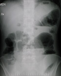
Large bowel obstruction

Upright abdominal X-ray of a person with a large bowel obstruction showing multiple air fluid levels and dilated loops of bowel LargeBowelObsUp2008.jpg
Upright abdominal X-ray of a person with a large bowel obstruction showing multiple air fluid levels and dilated loops of bowel

Causes of large bowel obstruction include: [22]

Outlet obstruction

Outlet obstruction is a sub-type of large bowel obstruction and refers to conditions affecting the anorectal region that obstruct defecation, specifically conditions of the pelvic floor and anal sphincters. Outlet obstruction can be classified into four groups. [23]

Treatment

Treatment of small and large bowel obstructions are initially similar and non-operative management is usually the initial management strategy as the majority of small bowel obstruction resolve spontaneously with non-operative management. [24] [25] Patients are be monitored by the surgical team for signs of improvement and resolution of the obstruction on imaging; if the obstruction does not clear then surgical management is required for the treatment of the causative lesion are required. [26] In malignant large bowel obstruction, endoscopically placed self-expanding metal stents may be used to temporarily relieve the obstruction as a bridge to surgery, [27] or as palliation. [28] Diagnosis of the type of bowel obstruction is normally conducted through initial plain radiograph of the abdomen, luminal contrast studies, computed tomography scan, or ultrasonography prior to determining the best type of treatment. [29]

Further research is needed to find out if parenteral nutrition is of benefit to people with an inoperable blockage of the bowel caused by advanced cancer. [30]

Small bowel obstruction

In the management of small bowel obstructions, a commonly quoted surgical aphorism is: "never let the sun rise or set on small-bowel obstruction" [31] because about 5.5% [31] of small bowel obstructions are ultimately fatal if treatment is delayed. Improvements in radiological imaging of small bowel obstructions allow for confident distinction between simple obstructions, that can be treated conservatively, and obstructions that are surgical emergencies (volvulus, closed-loop obstructions, ischemic bowel, incarcerated hernias, etc.). [2] Exam findings of bowel compromise requiring immediate surgery include: severe abdominal pain, signs of peritonitis such as rebound tenderness, elevated heart rate, fever, and elevated inflammatory markers on lab work, such as lactic acid. [32] [33]

A small flexible tube (nasogastric tube) may be inserted through the nose into the stomach to help decompress the dilated bowel. This tube is uncomfortable but relieves the abdominal cramps, distention, and vomiting. Intravenous therapy is utilized and the urine output may be monitored with a catheter in the bladder. [34] [35]

Most people with SBO are initially managed conservatively because in many cases, the bowel will open up. Some adhesions loosen up and the obstruction resolves. The patient is examined several times a day, and X-ray images are made to ensure he or she is not getting clinically worse. [36]

Conservative treatment involves insertion of a nasogastric tube, correction of dehydration and electrolyte abnormalities. Opioid pain relievers may be used for patients with severe pain but alternate pain relievers are preferred as opioids can decrease bowel motility. [37] Antiemetics may be administered if the patient is vomiting. Adhesive obstructions often settle without surgery. If the obstruction is complete surgery is usually required.

Most patients improve with conservative care in 2–5 days. When the obstruction is cancer, surgery is the only treatment. Those with bowel resection or lysis of adhesions usually stay in the hospital a few more days until they can eat and walk. [38]

Small bowel obstruction caused by Crohn's disease, peritoneal carcinomatosis, sclerosing peritonitis, radiation enteritis, and postpartum bowel obstruction are typically treated conservatively, i.e. without surgery.

Prognosis

The prognosis for non-ischemic cases of SBO is good with mortality rates of 3–5%, while prognosis for SBO with ischemia is fair with mortality rates as high as 30%. [39]

Cases of SBO related to cancer are more complicated and require additional intervention to address the malignancy, recurrence, and metastasis, and thus are associated with a more poor prognosis. [40] Surgical options in patients with malignant bowel obstruction need to be considered carefully as while it may provide relief of symptoms in the short term, there is a high risk of mortality and re-obstruction. [41]

All cases of abdominal surgical intervention are associated with increased risk of future small-bowel obstructions. Statistics from U.S. healthcare report 18.1% re-admittance rate within 30 days for patients who undergo SBO surgery. [42] More than 90% of patients also form adhesions after major abdominal surgery. [43] Common consequences of these adhesions include small-bowel obstruction, chronic abdominal pain, pelvic pain, and infertility. [43]

See also

Related Research Articles

<span class="mw-page-title-main">Appendicitis</span> Inflammation of the appendix

Appendicitis is inflammation of the appendix. Symptoms commonly include right lower abdominal pain, nausea, vomiting, and decreased appetite. However, approximately 40% of people do not have these typical symptoms. Severe complications of a ruptured appendix include widespread, painful inflammation of the inner lining of the abdominal wall and sepsis.

<span class="mw-page-title-main">Hernia</span> Abnormal exit of tissues or organs from the cavity they usually reside in

A hernia is the abnormal exit of tissue or an organ, such as the bowel, through the wall of the cavity in which it normally resides. The term is also used for the normal development of the intestinal tract, referring to the retraction of the intestine from the extra-embryonal navel coelom into the abdomen in the healthy embryo at about 7½ weeks.

<span class="mw-page-title-main">Hiatal hernia</span> Type of hernia

A hiatal hernia or hiatus hernia is a type of hernia in which abdominal organs slip through the diaphragm into the middle compartment of the chest. This may result in gastroesophageal reflux disease (GERD) or laryngopharyngeal reflux (LPR) with symptoms such as a taste of acid in the back of the mouth or heartburn. Other symptoms may include trouble swallowing and chest pains. Complications may include iron deficiency anemia, volvulus, or bowel obstruction.

<span class="mw-page-title-main">Cholecystitis</span> Inflammation of the gallbladder

Cholecystitis is inflammation of the gallbladder. Symptoms include right upper abdominal pain, pain in the right shoulder, nausea, vomiting, and occasionally fever. Often gallbladder attacks precede acute cholecystitis. The pain lasts longer in cholecystitis than in a typical gallbladder attack. Without appropriate treatment, recurrent episodes of cholecystitis are common. Complications of acute cholecystitis include gallstone pancreatitis, common bile duct stones, or inflammation of the common bile duct.

<span class="mw-page-title-main">Abdominal pain</span> Stomach aches

Abdominal pain, also known as a stomach ache, is a symptom associated with both non-serious and serious medical issues. Since the abdomen contains most of the body's vital organs, it can be an indicator of a wide variety of diseases. Given that, approaching the examination of a person and planning of a differential diagnosis is extremely important.

Colic in horses is defined as abdominal pain, but it is a clinical symptom rather than a diagnosis. The term colic can encompass all forms of gastrointestinal conditions which cause pain as well as other causes of abdominal pain not involving the gastrointestinal tract. What makes it tricky is that different causes can manifest with similar signs of distress in the animal. Recognizing and understanding these signs is pivotal, as timely action can spell the difference between a brief moment of discomfort and a life-threatening situation. The most common forms of colic are gastrointestinal in nature and are most often related to colonic disturbance. There are a variety of different causes of colic, some of which can prove fatal without surgical intervention. Colic surgery is usually an expensive procedure as it is major abdominal surgery, often with intensive aftercare. Among domesticated horses, colic is the leading cause of premature death. The incidence of colic in the general horse population has been estimated between 4 and 10 percent over the course of the average lifespan. Clinical signs of colic generally require treatment by a veterinarian. The conditions that cause colic can become life-threatening in a short period of time.

<span class="mw-page-title-main">Volvulus</span> Twisting of part of the intestine, causing a bowel obstruction

A volvulus is when a loop of intestine twists around itself and the mesentery that supports it, resulting in a bowel obstruction. Symptoms include abdominal pain, abdominal bloating, vomiting, constipation, and bloody stool. Onset of symptoms may be rapid or more gradual. The mesentery may become so tightly twisted that blood flow to part of the intestine is cut off, resulting in ischemic bowel. In this situation there may be fever or significant pain when the abdomen is touched.

Intestinal malrotation is a congenital anomaly of rotation of the midgut. It occurs during the first trimester as the fetal gut undergoes a complex series of growth and development. Malrotation can lead to a dangerous complication called volvulus, in which cases emergency surgery is indicated. Malrotation can refer to a spectrum of abnormal intestinal positioning, often including:

<span class="mw-page-title-main">Umbilical hernia</span> Medical condition

An umbilical hernia is a health condition where the abdominal wall behind the navel is damaged. It may cause the navel to bulge outwards—the bulge consisting of abdominal fat from the greater omentum or occasionally parts of the small intestine. The bulge can often be pressed back through the hole in the abdominal wall, and may "pop out" when coughing or otherwise acting to increase intra-abdominal pressure. Treatment is surgical, and surgery may be performed for cosmetic as well as health-related reasons.

<span class="mw-page-title-main">Pneumoperitoneum</span> Medical condition

Pneumoperitoneum is pneumatosis in the peritoneal cavity, a potential space within the abdominal cavity. The most common cause is a perforated abdominal organ, generally from a perforated peptic ulcer, although any part of the bowel may perforate from a benign ulcer, tumor or abdominal trauma. A perforated appendix seldom causes a pneumoperitoneum.

<span class="mw-page-title-main">Spigelian hernia</span> Surgical condition

A Spigelian is the type of ventral hernia where aponeurotic fascia pushes through a hole in the junction of the linea semilunaris and the arcuate line, creating a bulge. It appears in the lower quadrant of the abdomen between an area of dense fibrous tissue and abdominal wall muscles causing a.

Femoral hernias occur just below the inguinal ligament, when abdominal contents pass through a naturally occurring weakness in the abdominal wall called the femoral canal. Femoral hernias are a relatively uncommon type, accounting for only 3% of all hernias. While femoral hernias can occur in both males and females, almost all develop in women due to the increased width of the female pelvis. Femoral hernias are more common in adults than in children. Those that do occur in children are more likely to be associated with a connective tissue disorder or with conditions that increase intra-abdominal pressure. Seventy percent of pediatric cases of femoral hernias occur in infants under the age of one.

An acute abdomen refers to a sudden, severe abdominal pain. It is in many cases a medical emergency, requiring urgent and specific diagnosis. Several causes need immediate surgical treatment.

<span class="mw-page-title-main">Bowel resection</span> Surgical procedure in which a part of an intestine is removed

A bowel resection or enterectomy is a surgical procedure in which a part of an intestine (bowel) is removed, from either the small intestine or large intestine. Often the word enterectomy is reserved for the sense of small bowel resection, in distinction from colectomy, which covers the sense of large bowel resection. Bowel resection may be performed to treat gastrointestinal cancer, bowel ischemia, necrosis, or obstruction due to scar tissue, volvulus, and hernias. Some patients require ileostomy or colostomy after this procedure as alternative means of excretion. Complications of the procedure may include anastomotic leak or dehiscence, hernias, or adhesions causing partial or complete bowel obstruction. Depending on which part and how much of the intestines are removed, there may be digestive and metabolic challenges afterward, such as short bowel syndrome.

<span class="mw-page-title-main">Adhesion (medicine)</span> Medical condition

Adhesions are fibrous bands that form between tissues and organs, often as a result of injury during surgery. They may be thought of as internal scar tissue that connects tissues not normally connected.

<span class="mw-page-title-main">Bowel infarction</span> Injury to the intestine resulting from insufficient blood flow

Bowel infarction or gangrenous bowel represents an irreversible injury to the intestine resulting from insufficient blood flow. It is considered a medical emergency because it can quickly result in life-threatening infection and death. Any cause of bowel ischemia, the earlier reversible form of injury, may ultimately lead to infarction if uncorrected. The causes of bowel ischemia or infarction include primary vascular causes and other causes of bowel obstruction.

<span class="mw-page-title-main">Epiploic appendagitis</span> Medical condition

Epiploic appendagitis (EA) is an uncommon, benign, self-limiting inflammatory process of the epiploic appendices. Other, older terms for the process include appendicitis epiploica and appendagitis, but these terms are used less now in order to avoid confusion with acute appendicitis.

<span class="mw-page-title-main">Intestinal ischemia</span> Restriction of blood flow to the small intestine resulting in injury

Intestinal ischemia is a medical condition in which injury to the large or small intestine occurs due to not enough blood supply. It can come on suddenly, known as acute intestinal ischemia, or gradually, known as chronic intestinal ischemia. The acute form of the disease often presents with sudden severe abdominal pain and is associated with a high risk of death. The chronic form typically presents more gradually with abdominal pain after eating, unintentional weight loss, vomiting, and fear of eating.

<span class="mw-page-title-main">Obturator hernia</span> Medical condition

An obturator hernia is a rare type of hernia, encompassing 0.07-1% of all hernias, of the pelvic floor in which pelvic or abdominal contents protrudes through the obturator foramen. The obturator foramen is formed by a branch of the ischial as well as the pubic bone. The canal is typically 2-3 centimeters long and 1 centimeters wide, creating a space for pouches of pre-peritoneal fat.

Afferent loop syndrome is an uncommon side effect of gastric surgery. The afferent loop is made up of a segment of duodenum and/or proximal jejunum located upstream of a double-barrel gastrojejunostomy anastomosis. Abdominal pain and distension are signs of increased intraluminal pressure resulting from the accumulation of enteric secretions in the obstructed afferent loop.

References

  1. 1 2 3 4 5 6 7 8 9 10 11 12 Gore RM, Silvers RI, Thakrar KH, Wenzke DR, Mehta UK, Newmark GM, et al. (November 2015). "Bowel Obstruction". Radiologic Clinics of North America. 53 (6): 1225–40. doi:10.1016/j.rcl.2015.06.008. PMID   26526435.
  2. 1 2 3 4 5 6 7 8 9 10 11 Fitzgerald JE (2010). "Small Bowel Obstruction". Emergency Surgery. Oxford: Wiley-Blackwell. pp. 74–79. doi:10.1002/9781444315172.ch14. ISBN   978-1-4051-7025-3. Archived from the original on September 8, 2017.
  3. 1 2 Vos T, Allen C, Arora M, Barber RM, Bhutta ZA, Brown A, et al. (October 2016). "Global, regional, and national incidence, prevalence, and years lived with disability for 310 diseases and injuries, 1990-2015: a systematic analysis for the Global Burden of Disease Study 2015". Lancet. 388 (10053): 1545–1602. doi:10.1016/S0140-6736(16)31678-6. PMC   5055577 . PMID   27733282.
  4. 1 2 Wang H, Naghavi M, Allen C, Barber RM, Bhutta ZA, Carter A, et al. (October 2016). "Global, regional, and national life expectancy, all-cause mortality, and cause-specific mortality for 249 causes of death, 1980-2015: a systematic analysis for the Global Burden of Disease Study 2015". Lancet. 388 (10053): 1459–1544. doi:10.1016/s0140-6736(16)31012-1. PMC   5388903 . PMID   27733281.
  5. Adams JG (2012). Emergency Medicine: Clinical Essentials (Expert Consult -- Online). Elsevier Health Sciences. p. 331. ISBN   978-1-4557-3394-1. Archived from the original on September 8, 2017.
  6. 1 2 3 Ferri FF (2014). Ferri's Clinical Advisor 2015: 5 Books in 1. Elsevier Health Sciences. p. 1093. ISBN   978-0-323-08430-7. Archived from the original on September 8, 2017.
  7. Yeo CJ, McFadden DW, Pemberton JH, Peters JH, Matthews JB (2012). Shackelford's Surgery of the Alimentary Tract. Elsevier Health Sciences. p. 1851. ISBN   978-1-4557-3807-6. Archived from the original on September 8, 2017.
  8. "Large Bowel Obstruction". The Lecturio Medical Concept Library. Retrieved July 10, 2021.
  9. "Large Bowel Obstruction". The Lecturio Medical Concept Library. Retrieved July 10, 2021.
  10. Vercruysse G, Busch R, Dimcheff D, Al-Hawary M, Saad R, Seagull FJ, et al. (2021). Evaluation and Management of Mechanical Small Bowel Obstruction in Adults. Michigan Medicine Clinical Care Guidelines. Ann Arbor (MI): Michigan Medicine University of Michigan. PMID   34314126.
  11. Ferri F (July 12, 2023). Ferri's Clinical Advisor 2024 (1st ed.). Elsevier. pp. 829.e4–829.e6. ISBN   978-0-323-75576-4.
  12. "Large Bowel Obstruction". The Lecturio Medical Concept Library. Retrieved July 10, 2021.
  13. Jacobs SL, Rozenblit A, Ricci Z, Roberts J, Milikow D, Chernyak V, et al. (April 2007). "Small bowel faeces sign in patients without small bowel obstruction". Clinical Radiology. 62 (4): 353–7. doi:10.1016/j.crad.2006.11.007. PMID   17331829.
  14. Nguyen H, Loustaunau C, Facista A, Ramsey L, Hassounah N, Taylor H, et al. (July 2010). "Deficient Pms2, ERCC1, Ku86, CcOI in field defects during progression to colon cancer". Journal of Visualized Experiments (41). doi:10.3791/1931. PMC   3149991 . PMID   20689513.
  15. Singh A, Mansouri M (2018), Singh A (ed.), "Imaging of Bowel Obstruction", Emergency Radiology, Cham: Springer International Publishing, pp. 67–75, doi:10.1007/978-3-319-65397-6_5, ISBN   978-3-319-65396-9 , retrieved February 19, 2024
  16. Gottlieb M, Peksa GD, Pandurangadu AV, Nakitende D, Takhar S, Seethala RR (February 2018). "Utilization of ultrasound for the evaluation of small bowel obstruction: A systematic review and meta-analysis". The American Journal of Emergency Medicine. 36 (2): 234–242. doi:10.1016/j.ajem.2017.07.085. PMID   28797559. S2CID   24769945.
  17. Abbas S, Bissett IP, Parry BR (July 2007). "Oral water soluble contrast for the management of adhesive small bowel obstruction". The Cochrane Database of Systematic Reviews. 2010 (3): CD004651. doi:10.1002/14651858.CD004651.pub3. PMC   6465054 . PMID   17636770.
  18. 1 2 3 "UOTW #20 - Ultrasound of the Week". Ultrasound of the Week. October 1, 2014. Archived from the original on May 9, 2017. Retrieved May 27, 2017.
  19. "Small Bowel Obstruction". The Lecturio Medical Concept Library. Retrieved July 10, 2021.
  20. Segura-Sampedro JJ, Ashrafian H, Navarro-Sánchez A, Jenkins JT, Morales-Conde S, Martínez-Isla A (November 2015). "Small bowel obstruction due to laparoscopic barbed sutures: an unknown complication?". Revista Espanola de Enfermedades Digestivas. 107 (11): 677–80. doi: 10.17235/reed.2015.3863/2015 . hdl: 20.500.13003/12378 . PMID   26541657.
  21. ten Broek RP, Issa Y, van Santbrink EJ, Bouvy ND, Kruitwagen RF, Jeekel J, et al. (October 2013). "Burden of adhesions in abdominal and pelvic surgery: systematic review and met-analysis". BMJ. 347 (oct03 1): f5588. doi:10.1136/bmj.f5588. PMC   3789584 . PMID   24092941.
  22. "Intestinal obstruction and Ileus". MedlinePlus. Retrieved July 10, 2021.
  23. Zbar AP, Wexner SD (2010). Coloproctology. New York: Springer. p. 140. ISBN   978-1-84882-755-4.
  24. Vercruysse G, Busch R, Dimcheff D, Al-Hawary M, Saad R, Seagull FJ, et al. (2021). Evaluation and Management of Mechanical Small Bowel Obstruction in Adults. Michigan Medicine Clinical Care Guidelines. Ann Arbor (MI): Michigan Medicine University of Michigan. PMID   34314126.
  25. Ferri F (July 12, 2023). Ferri's Clinical Advisor 2024 (1st ed.). Elsevier. ISBN   978-0-323-75576-4.
  26. Bower KL, Lollar DI, Williams SL, Adkins FC, Luyimbazi DT, Bower CE (October 1, 2018). "Small Bowel Obstruction". Surgical Clinics of North America. Emergency General Surgery. 98 (5): 945–971. doi:10.1016/j.suc.2018.05.007. ISSN   0039-6109. PMID   30243455. S2CID   265759123.
  27. Young CJ, Suen MK, Young J, Solomon MJ (October 2011). "Stenting large bowel obstruction avoids a stoma: consecutive series of 100 patients". Colorectal Disease. 13 (10): 1138–41. doi:10.1111/j.1463-1318.2010.02432.x. PMID   20874797. S2CID   12724976.
  28. Mosler P, Mergener KD, Brandabur JJ, Schembre DB, Kozarek RA (February 2005). "Palliation of gastric outlet obstruction and proximal small bowel obstruction with self-expandable metal stents: a single center series". Journal of Clinical Gastroenterology. 39 (2): 124–8. PMID   15681907.
  29. Holzheimer RG (2001). Surgical Treatment. NCBI Bookshelf. ISBN   3-88603-714-2. Archived from the original on August 27, 2011.
  30. Sowerbutts AM, Lal S, Sremanakova J, Clamp A, Todd C, Jayson GC, et al. (August 2018). "Home parenteral nutrition for people with inoperable malignant bowel obstruction". The Cochrane Database of Systematic Reviews. 8 (8): CD012812. doi:10.1002/14651858.cd012812.pub2. PMC   6513201 . PMID   30095168.
  31. 1 2 Maglinte DD, Kelvin FM, Rowe MG, Bender GN, Rouch DM (January 2001). "Small-bowel obstruction: optimizing radiologic investigation and nonsurgical management". Radiology. 218 (1): 39–46. doi:10.1148/radiology.218.1.r01ja5439. PMID   11152777. Archived from the original on April 18, 2008. Retrieved June 6, 2008.
  32. Vercruysse G, Busch R, Dimcheff D, Al-Hawary M, Saad R, Seagull FJ, et al. (2021). Evaluation and Management of Mechanical Small Bowel Obstruction in Adults. Michigan Medicine Clinical Care Guidelines. Ann Arbor (MI): Michigan Medicine University of Michigan. PMID   34314126.
  33. Ferri F (July 12, 2023). Ferri's Clinical Advisor 2024 (1st ed.). Elsevier. ISBN   978-0-323-75576-4.
  34. Small Bowel Obstruction overview Archived February 12, 2010, at the Wayback Machine . Retrieved February 19, 2010.
  35. Vercruysse G, Busch R, Dimcheff D, Al-Hawary M, Saad R, Seagull FJ, et al. (2021). Evaluation and Management of Mechanical Small Bowel Obstruction in Adults. Michigan Medicine Clinical Care Guidelines. Ann Arbor (MI): Michigan Medicine University of Michigan. PMID   34314126.
  36. Small Bowel Obstruction: Treating Bowel Adhesions Non-Surgically Archived February 27, 2010, at the Wayback Machine . Clear Passage treatment center online portal Retrieved February 19, 2010
  37. Vercruysse G, Busch R, Dimcheff D, Al-Hawary M, Saad R, Seagull FJ, et al. (2021). Evaluation and Management of Mechanical Small Bowel Obstruction in Adults. Michigan Medicine Clinical Care Guidelines. Ann Arbor (MI): Michigan Medicine University of Michigan. PMID   34314126.
  38. Small Bowel Obstruction Archived July 5, 2010, at the Wayback Machine The Eastern Association for the Surgery of Trauma. February 19, 2010
  39. Kakoza R, Lieberman G (May 2006). Mechanical Small Bowel Obstruction (PDF). Archived from the original (PDF) on May 7, 2013. Retrieved October 9, 2012.
  40. "Small Bowel Obstruction". The Lecturio Medical Concept Library. Retrieved July 10, 2021.
  41. Song Y, Metzger DA, Bruce AN, Krouse RS, Roses RE, Fraker DL, et al. (January 2022). "Surgical Outcomes in Patients With Malignant Small Bowel Obstruction: A National Cohort Study". Annals of Surgery. 275 (1): e198–e205. doi:10.1097/SLA.0000000000003890. ISSN   0003-4932. PMID   32209901. S2CID   214643950.
  42. "Readmissions to U.S. Hospitals by Procedure" (PDF). Agency for Healthcare Research and Quality. April 2013. Archived (PDF) from the original on October 20, 2013. Retrieved August 27, 2013.
  43. 1 2 Liakakos T, Thomakos N, Fine PM, Dervenis C, Young RL (2001). "Peritoneal adhesions: etiology, pathophysiology, and clinical significance. Recent advances in prevention and management". Digestive Surgery. 18 (4): 260–73. doi:10.1159/000050149. PMID   11528133. S2CID   30816909.