Pneumoperitoneum

Last updated
Pneumoperitoneum
Pneumoperitoneum modification.jpg
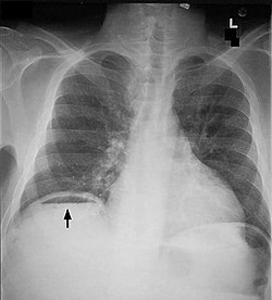
Frontal chest X-ray. The air bubble below the right hemidiaphragm (on the left of the image) is a pneumoperitoneum.
Specialty Gastroenterology   OOjs UI icon edit-ltr-progressive.svg

Pneumoperitoneum is pneumatosis (abnormal presence of air or other gas) in the peritoneal cavity, a potential space within the abdominal cavity. The most common cause is a perforated abdominal organ, generally from a perforated peptic ulcer, although any part of the bowel may perforate from a benign ulcer, tumor or abdominal trauma. A perforated appendix seldom causes a pneumoperitoneum.

Contents

Spontaneous pneumoperitoneum is a rare case that is not caused by an abdominal organ rupture. This is also called an idiopathic spontaneous pneumoperitoneum when the cause is not known.

In the mid-twentieth century, an "artificial" pneumoperitoneum was sometimes intentionally administered as a treatment for a hiatal hernia. This was achieved by insufflating the abdomen with carbon dioxide. The practice is currently used by surgical teams in order to aid in performing laparoscopic surgery.

Causes

Spontaneous pneumoperitoneum

A spontaneous pneumoperitoneum is a rare case that is not caused by an abdominal organ rupture. This is also called an idiopathic spontaneous pneumoperitoneum when the cause is not known. [16] Causes of a spontaneous pneumoperitoneum, with no peritonitis include a barotrauma due to mechanical ventilation, and a tracheal rupture following an emergency intubation. In the ventilation case, air had passed from the chest into the abdominal cavity through the diaphragm. In the tracheal rupture air had passed along the great vessels. [17]

Diagnosis

When present, pneumoperitoneum can often be seen on projectional radiography, but small amounts are often missed, and CT scan is nowadays regarded as a criterion standard in the assessment of a pneumoperitoneum. [18] CT can visualize quantities as small as 5 cm3 of air or gas.

Signs that can be seen on projectional radiography are shown below:

The double wall sign marks the presence of air on both sides of the intestine. [19] However, a false double wall sign can result from two loops of bowel being in contact with one another. [20] The sign is named after Leo George Rigler. [21] It is not the same as Rigler's triad.

The football sign is when the abdomen appears as a large oval radiolucency reminiscent of an American football on a supine projectional radiograph. [22] The football sign is most frequently seen in infants with spontaneous or iatrogenic gastric perforation causing pneumoperitoneum. It is also seen in bowel obstruction with secondary perforation, as in Hirschprung disease, midgut volvulus, meconium ileus and intestinal atresia. Iatrogenic causes like endoscopic perforation may also give football sign.[ citation needed ]

The Cupola sign is seen when air is accumulated under the central tendon of the diaphragm. [23]

Differential diagnosis

As differential diagnoses, a subphrenic abscess, bowel interposed between diaphragm and liver (Chilaiditi syndrome), and linear atelectasis at the base of the lungs can simulate free air under the diaphragm on a chest X-ray.[ citation needed ]

Treatment

Treatment depends on the cause of the condition.[ citation needed ]

Terminology

Pneumoperitoneum can be described as peritoneal emphysema, [25] just as pneumomediastinum can be called mediastinal emphysema, but pneumoperitoneum is the usual name.

See also

Related Research Articles

Peptic ulcer disease is a break in the inner lining of the stomach, the first part of the small intestine, or sometimes the lower esophagus. An ulcer in the stomach is called a gastric ulcer, while one in the first part of the intestines is a duodenal ulcer. The most common symptoms of a duodenal ulcer are waking at night with upper abdominal pain, and upper abdominal pain that improves with eating. With a gastric ulcer, the pain may worsen with eating. The pain is often described as a burning or dull ache. Other symptoms include belching, vomiting, weight loss, or poor appetite. About a third of older people with peptic ulcers have no symptoms. Complications may include bleeding, perforation, and blockage of the stomach. Bleeding occurs in as many as 15% of cases.

<span class="mw-page-title-main">Peritonitis</span> Medical condition

Peritonitis is inflammation of the localized or generalized peritoneum, the lining of the inner wall of the abdomen and cover of the abdominal organs. Symptoms may include severe pain, swelling of the abdomen, fever, or weight loss. One part or the entire abdomen may be tender. Complications may include shock and acute respiratory distress syndrome.

<span class="mw-page-title-main">Bowel obstruction</span> Medical condition

Bowel obstruction, also known as intestinal obstruction, is a mechanical or functional obstruction of the intestines which prevents the normal movement of the products of digestion. Either the small bowel or large bowel may be affected. Signs and symptoms include abdominal pain, vomiting, bloating and not passing gas. Mechanical obstruction is the cause of about 5 to 15% of cases of severe abdominal pain of sudden onset requiring admission to hospital.

<span class="mw-page-title-main">Abdominal pain</span> Stomach aches

Abdominal pain, also known as a stomach ache, is a symptom associated with both non-serious and serious medical issues. Since the abdomen contains most of the body's vital organs, it can be an indicator of a wide variety of diseases. Given that, approaching the examination of a person and planning of a differential diagnosis is extremely important.

<span class="mw-page-title-main">Meckel's diverticulum</span> Medical condition

A Meckel's diverticulum, a true congenital diverticulum, is a slight bulge in the small intestine present at birth and a vestigial remnant of the vitelline duct. It is the most common malformation of the gastrointestinal tract and is present in approximately 2% of the population, with males more frequently experiencing symptoms.

<span class="mw-page-title-main">Gastrointestinal disease</span> Medical condition

Gastrointestinal diseases refer to diseases involving the gastrointestinal tract, namely the esophagus, stomach, small intestine, large intestine and rectum, and the accessory organs of digestion, the liver, gallbladder, and pancreas.

<span class="mw-page-title-main">Diverticulum</span> Medical or biological term for an outpouching of a hollow (or a fluid-filled) structure in the body

In medicine or biology, a diverticulum is an outpouching of a hollow structure in the body. Depending upon which layers of the structure are involved, diverticula are described as being either true or false.

<span class="mw-page-title-main">Gastrointestinal perforation</span> Medical condition

Gastrointestinal perforation, also known as gastrointestinal rupture, is a hole in the wall of the gastrointestinal tract. The gastrointestinal tract is composed of hollow digestive organs leading from the mouth to the anus. Symptoms of gastrointestinal perforation commonly include severe abdominal pain, nausea, and vomiting. Complications include a painful inflammation of the inner lining of the abdominal wall and sepsis.

<span class="mw-page-title-main">Paracentesis</span> Form of body fluid sampling procedure

Paracentesis is a form of body fluid sampling procedure, generally referring to peritoneocentesis in which the peritoneal cavity is punctured by a needle to sample peritoneal fluid.

<span class="mw-page-title-main">Abdominal distension</span> Physical symptom

Abdominal distension occurs when substances, such as air (gas) or fluid, accumulate in the abdomen causing its expansion. It is typically a symptom of an underlying disease or dysfunction in the body, rather than an illness in its own right. People with this condition often describe it as "feeling bloated". Affected people often experience a sensation of fullness, abdominal pressure, and sometimes nausea, pain, or cramping. In the most extreme cases, upward pressure on the diaphragm and lungs can also cause shortness of breath. Through a variety of causes, bloating is most commonly due to buildup of gas in the stomach, small intestine, or colon. The pressure sensation is often relieved, or at least lessened, by belching or flatulence. Medications that settle gas in the stomach and intestines are also commonly used to treat the discomfort and lessen the abdominal distension.

Valentino's syndrome is pain presenting in the right lower quadrant of the abdomen caused by a duodenal ulcer with perforation through the retroperitoneum.

An acute abdomen refers to a sudden, severe abdominal pain. It is in many cases a medical emergency, requiring urgent and specific diagnosis. Several causes need immediate surgical treatment.

Hemoperitoneum is the presence of blood in the peritoneal cavity. The blood accumulates in the space between the inner lining of the abdominal wall and the internal abdominal organs. Hemoperitoneum is generally classified as a surgical emergency; in most cases, urgent laparotomy is needed to identify and control the source of the bleeding. In selected cases, careful observation may be permissible. The abdominal cavity is highly distensible and may easily hold greater than five liters of blood, or more than the entire circulating blood volume for an average-sized individual. Therefore, large-scale or rapid blood loss into the abdomen will reliably induce hemorrhagic shock and, if untreated, may rapidly lead to death.

<span class="mw-page-title-main">Bowel resection</span> Surgical procedure in which a part of an intestine is removed

A bowel resection or enterectomy is a surgical procedure in which a part of an intestine (bowel) is removed, from either the small intestine or large intestine. Often the word enterectomy is reserved for the sense of small bowel resection, in distinction from colectomy, which covers the sense of large bowel resection. Bowel resection may be performed to treat gastrointestinal cancer, bowel ischemia, necrosis, or obstruction due to scar tissue, volvulus, and hernias. Some patients require ileostomy or colostomy after this procedure as alternative means of excretion. Complications of the procedure may include anastomotic leak or dehiscence, hernias, or adhesions causing partial or complete bowel obstruction. Depending on which part and how much of the intestines are removed, there may be digestive and metabolic challenges afterward, such as short bowel syndrome.

<span class="mw-page-title-main">Gastric outlet obstruction</span> Medical condition

Gastric outlet obstruction (GOO) is a medical condition where there is an obstruction at the level of the pylorus, which is the outlet of the stomach. Individuals with gastric outlet obstruction will often have recurrent vomiting of food that has accumulated in the stomach, but which cannot pass into the small intestine due to the obstruction. The stomach often dilates to accommodate food intake and secretions. Causes of gastric outlet obstruction include both benign causes, as well as malignant causes, such as gastric cancer.

<span class="mw-page-title-main">Perforated ulcer</span> Medical condition

A perforated ulcer is a condition in which an untreated ulcer has burned through the mucosal wall in a segment of the gastrointestinal tract allowing gastric contents to leak into the abdominal cavity.

Abdominal guarding is the tensing of the abdominal wall muscles to guard inflamed organs within the abdomen from the pain of pressure upon them. The tensing is detected when the abdominal wall is pressed. Abdominal guarding is also known as 'défense musculaire'.

<span class="mw-page-title-main">Abdominal trauma</span> Medical condition

Abdominal trauma is an injury to the abdomen. Signs and symptoms include abdominal pain, tenderness, rigidity, and bruising of the external abdomen. Complications may include blood loss and infection.

<span class="mw-page-title-main">Diaphragmatic rupture</span> Tear in the thoracic diaphragm, usually caused by physical trauma

Diaphragmatic rupture is a tear of the diaphragm, the muscle across the bottom of the ribcage that plays a crucial role in breathing. Most commonly, acquired diaphragmatic tears result from physical trauma. Diaphragmatic rupture can result from blunt or penetrating trauma and occurs in about 0.5% of all people with trauma.

<span class="mw-page-title-main">Pneumatosis</span> Abnormal presence of air or other gas within tissues

Pneumatosis is the abnormal presence of air or other gas within tissues.

References

  1. "Peptic Ulcer Disease". The Lecturio Medical Concept Library. Retrieved 10 August 2021.
  2. "Large Bowel Obstruction". The Lecturio Medical Concept Library. Retrieved 10 August 2021.
  3. "Small Bowel Obstruction". The Lecturio Medical Concept Library. Retrieved 10 August 2021.
  4. "diverticulum | Definition of diverticulum in English by Lexico Dictionaries". Lexico Dictionaries | English. Archived from the original on July 17, 2019. Retrieved 2019-07-17.
  5. Stewart MG (2005). "Principles of ballistics and penetrating trauma". In Stewart MG (ed.). Head, Face, and Neck Trauma: Comprehensive Management. Thieme. pp. 188–94. ISBN   3-13-140331-4 . Retrieved 2008-06-12.
  6. "Megacolon". The Lecturio Medical Concept Library. Retrieved 10 August 2021.
  7. Necrotizing Enterocolitis Bugs, Drugs and Things That Go Bump in the Night
  8. "Colon Cancer Treatment (PDQ®)". NCI. May 12, 2014. Archived from the original on July 5, 2014. Retrieved June 29, 2014.
  9. Higgins PD, Davis KJ, Laine L (April 2004). "Systematic review: the epidemiology of ischaemic colitis". Alimentary Pharmacology & Therapeutics. 19 (7): 729–38. doi:10.1111/j.1365-2036.2004.01903.x. hdl: 2027.42/74164 . PMID   15043513. S2CID   9575677.
  10. Brandt LJ, Boley SJ (May 2000). "AGA technical review on intestinal ischemia. American Gastrointestinal Association". Gastroenterology. 118 (5): 954–68. doi:10.1016/S0016-5085(00)70183-1. PMID   10784596.
  11. American Gastroenterological Association (May 2000). "American Gastroenterological Association Medical Position Statement: guidelines on intestinal ischemia". Gastroenterology. 118 (5): 951–3. doi: 10.1016/S0016-5085(00)70182-X . PMID   10784595. Archived from the original on 2007-09-27.
  12. Imran M, Bhat R, Anijeet H (April 2011). "Pneumoperitoneum in peritoneal dialysis patients; one centre's experience". NDT Plus. 4 (2): 120–3. doi:10.1093/ndtplus/sfq208. PMC   4421564 . PMID   25984130.
  13. Kizer KW (May 1980). "Medical hazards of the water skiing douche". Annals of Emergency Medicine. 9 (5): 268–9. doi:10.1016/s0196-0644(80)80387-8. PMID   7369581. Many injuries can result from water skiing accidents, but the water skiing "douche" is unique to this sport. Although generally causing only discomfort, significant trauma can result from the forceful entry of water into the various body orifices.
  14. Jacobs VR, Mundhenke C, Maass N, Hilpert F, Jonat W (Oct 2000). "Sexual activity as cause for non-surgical pneumoperitoneum". JSLS. 4 (4): 297–300. PMC   3113190 . PMID   11051188.
  15. Cawich SO, Johnson PB, Williams E, Naraynsingh V (25 Sep 2013). "Non-surgical pneumoperitoneum after oro-genital intercourse". International Journal of Surgery Case Reports. 4 (11): 1048–51. doi:10.1016/j.ijscr.2013.08.022. PMC   3825970 . PMID   24121052.
  16. Pfister J, Riedtmann-Klee HJ (September 1993). "[Idiopathic spontaneous pneumoperitoneum. Case discussion based on two cases, assessment procedure and therapy and review of the literature]". Helvetica Chirurgica Acta. 60 (1–2): 49–56. PMID   8226082.
  17. Gutkin Z, Iellin A, Meged S, Sorkine P, Geller E (July 1992). "Spontaneous pneumoperitoneum without peritonitis". International Surgery. 77 (3): 219–23. PMID   1399374.
  18. Khan AN, Chandramohan M (2017-12-06). Coombs BD, Gay SD (eds.). "Pneumoperitoneum Imaging". Emedicine, Medscape. WebMD LLC.
  19. Harkin DW, Blake G (March 1999). "Small bowel obstruction in a young adult". Postgraduate Medical Journal. 75 (881): 173–5. doi:10.1136/pgmj.75.881.173. PMC   1741173 . PMID   10448501.
  20. de Lacey G, Bloomberg T, Wignall BK (July 1977). "Pneumoperitoneum: the misleading double wall sign". Clinical Radiology. 28 (4): 445–8. doi:10.1016/s0009-9260(77)80161-x. PMID   872511.
  21. Hirsch M, Cortés C (2009). "El signo de Leo Rigler: doble pared en neumoperitoneo" [The sign of Leo Rigler: double pared in neumoperitoneo.]. Revista chilena de radiología (in Spanish). 15 (3): 152–4. doi: 10.4067/S0717-93082009000300008 .
  22. Rampton JW (April 2004). "The football sign". Radiology. 231 (1): 81–2. doi:10.1148/radiol.2311011290. PMID   14990817.
  23. Sureka B, Bansal K, Arora A (2015). "Pneumoperitoneum: What to look for in a radiograph?". Journal of Family Medicine and Primary Care. 4 (3): 477–8. doi: 10.4103/2249-4863.161369 . PMC   4535122 . PMID   26288798.
  24. "UOTW #68 - Ultrasound of the Week". Ultrasound of the Week. 22 March 2016. Retrieved 27 May 2017.
  25. Marian K, Monika K, Bartosz K (March 2015). "Spontaneous pneumoperitoneum in a patient after ventilation therapy". Polski Przeglad Chirurgiczny. 86 (12): 601–3. doi:10.1515/pjs-2015-0008. PMID   25803061.