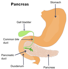
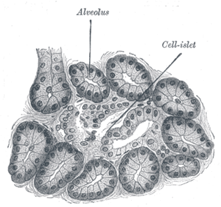
Acute pancreatitis

Last updated
Acute pancreatitis
Other namesAcute pancreatic necrosis [1]
3D Medical Animation Acute Pancreatitis.jpg
Still from 3D medical animation of acute pancreatitis
Specialty Gastroenterology, general surgery

Acute pancreatitis (AP) is a sudden inflammation of the pancreas. Causes, in order of frequency, include: a gallstone impacted in the common bile duct beyond the point where the pancreatic duct joins it; heavy alcohol use; systemic disease; trauma; and, in children, mumps. Acute pancreatitis may be a single event; it may be recurrent; or it may progress to chronic pancreatitis.

Contents

Mild cases are usually successfully treated with conservative measures: hospitalization, pain control, nothing by mouth, intravenous nutritional support, and intravenous fluid rehydration. Severe cases often require admission to an intensive care unit to monitor and manage complications of the disease. Complications are associated with a high mortality, even with optimal management. For people with this condition, the pancreas will begin to secrete active enzymes such as Trypsin, chymotrypsin and carboxypeptidase instead of their inactive forms. Damage to the pancreatic ducts can occur as a result of this.

Signs and symptoms

Common

Although these are common symptoms, frequently they are not all present; and epigastric pain may be the only symptom. [3]

Uncommon

The following are associated with severe disease:

Complications

Locoregional complications include pancreatic pseudocyst (most common, occurring in up to 25% of all cases, typically after 4–6 weeks) and phlegmon/abscess formation, splenic artery pseudoaneurysms, hemorrhage from erosions into splenic artery and vein, thrombosis of the splenic vein, superior mesenteric vein and portal veins (in descending order of frequency), duodenal obstruction, common bile duct obstruction, progression to chronic pancreatitis, pancreatic ascites, pleural effusion, sterile/infected pancreatic necrosis. [5]

Systemic complications include acute respiratory distress syndrome (ARDS), multiple organ dysfunction syndrome, disseminated intravascular coagulation (DIC), hypocalcemia (from fat saponification), hyperglycemia and insulin dependent diabetes mellitus (from pancreatic insulin-producing beta cell damage), malabsorption due to exocrine failure

Causes

Most common

Less common

Pathology

Anatomy of the pancreas Illu pancrease.svg
Anatomy of the pancreas

Pathogenesis

Acute pancreatitis occurs when there is abnormal activation of digestive enzymes within the pancreas. This occurs through inappropriate activation of inactive enzyme precursors called zymogens (or proenzymes) inside the pancreas, most notably trypsinogen. Normally, trypsinogen is converted to its active form (trypsin) in the first part of the small intestine (duodenum), where the enzyme assists in the digestion of proteins. During an episode of acute pancreatitis, trypsinogen comes into contact with lysosomal enzymes (specifically cathepsin), which activate trypsinogen to trypsin. The active form trypsin then leads to further activation of other molecules of trypsinogen. The activation of these digestive enzymes lead to inflammation, edema, vascular injury, and even cellular death. The death of pancreatic cells occurs via two main mechanisms: necrosis, which is less organized and more damaging, or apoptosis, which is more controlled. The balance between these two mechanisms of cellular death is mediated by caspases which regulate apoptosis and have important anti-necrosis functions during pancreatitis: preventing trypsinogen activation, preventing ATP depletion through inhibiting polyADP-ribose polymerase, and by inhibiting the inhibitors of apoptosis (IAPs). If, however, the caspases are depleted due to either chronic ethanol exposure or through a severe insult then necrosis can predominate.

Pathophysiology

The two types of acute pancreatitis are mild and severe, which are defined based on whether the predominant response to cell injury is inflammation (mild) or necrosis (severe). In mild pancreatitis, there is inflammation and edema of the pancreas. In severe pancreatitis, there is necrosis of the pancreas, and nearby organs may become injured.

As part of the initial injury there is an extensive inflammatory response due to pancreatic cells synthesizing and secreting inflammatory mediators: primarily TNF-alpha and IL-1. A hallmark of acute pancreatitis is a manifestation of the inflammatory response, namely the recruitment of neutrophils to the pancreas. The inflammatory response leads to the secondary manifestations of pancreatitis: hypovolemia from capillary permeability, acute respiratory distress syndrome, disseminated intravascular coagulations, renal failure, cardiovascular failure, and gastrointestinal hemorrhage.

Histopathology

The acute pancreatitis (acute hemorrhagic pancreatic necrosis) is characterized by acute inflammation and necrosis of pancreas parenchyma, focal enzymic necrosis of pancreatic fat and vessel necrosis (hemorrhage). These are produced by intrapancreatic activation of pancreatic enzymes. Lipase activation produces the necrosis of fat tissue in pancreatic interstitium and peripancreatic spaces as well as vessel damage. Necrotic fat cells appear as shadows, contours of cells, lacking the nucleus, pink, finely granular cytoplasm. It is possible to find calcium precipitates (hematoxylinophilic). Digestion of vascular walls results in thrombosis and hemorrhage. Inflammatory infiltrate is rich in neutrophils. Due to the pancreas lacking a capsule, the inflammation and necrosis can extend to include fascial layers in the immediate vicinity of the pancreas.

Diagnosis

Acute pancreatitis is diagnosed using clinical history and physical examination, based on the presence of at least 2 of 3 criteria: abdominal pain, elevated serum lipase or amylase, and abdominal imaging findings consistent with acute pancreatitis. [11] Additional blood studies are used to identify organ failure, offer prognostic information, and determine if fluid resuscitation is adequate and whether endoscopic retrograde cholangiopancreatography is necessary.

Differential diagnosis

The differential diagnosis includes: [12]

Biochemical

Regarding selection on these tests, two practice guidelines state:

"It is usually not necessary to measure both serum amylase and lipase. Serum lipase may be preferable because it remains normal in some nonpancreatic conditions that increase serum amylase including macroamylasemia, parotitis, and some carcinomas. In general, serum lipase is thought to be more sensitive and specific than serum amylase in the diagnosis of acute pancreatitis" [14]
"Although amylase is widely available and provides acceptable accuracy of diagnosis, where lipase is available it is preferred for the diagnosis of acute pancreatitis (recommendation grade A)" [15]

Most, but not all individual studies support the superiority of the lipase. [16] In one large study, there were no patients with pancreatitis who had an elevated amylase with a normal lipase. [17] Another study found that the amylase could add diagnostic value to the lipase, but only if the results of the two tests were combined with a discriminant function equation. [18]

While often quoted lipase levels of 3 or more times the upper-limit of normal is diagnostic of pancreatitis, there are also other differential diagnosis to be considered relating to this rise. [19]

Computed tomography

Axial CT in a patient with acute exudative pancreatitis showing extensive fluid collections surrounding the pancreas Akute exsudative Pankreatitis - CT axial.jpg
Axial CT in a patient with acute exudative pancreatitis showing extensive fluid collections surrounding the pancreas

Regarding the need for computed tomography, practice guidelines state:

CT is an important common initial assessment tool for acute pancreatitis. Imaging is indicated during the initial presentation if:

CT is recommended as a delayed assessment tool in the following situations:

Abdominal CT should not be performed before the first 12 hours of onset of symptoms as early CT (<12 hours) may result in equivocal or normal findings.

CT findings can be classified into the following categories for easy recall:

The principal value of CT imaging to the treating clinician is the capacity to identify devitalised areas of the pancreas which have become necrotic due to ischaemia. Pancreatic necrosis can be reliably identified by intravenous contrast-enhanced CT imaging, [20] and is of value if infection occurs and surgical or percutaneous debridement is indicated.

Magnetic resonance imaging

While computed tomography is considered the gold standard in diagnostic imaging for acute pancreatitis, [21] magnetic resonance imaging (MRI) has become increasingly valuable as a tool for the visualization of the pancreas, particularly of pancreatic fluid collections and necrotized debris. [22] Additional utility of MRI includes its indication for imaging of patients with an allergy to CT's contrast material, and an overall greater sensitivity to hemorrhage, vascular complications, pseudoaneurysms, and venous thrombosis. [23]

Another advantage of MRI is its utilization of magnetic resonance cholangiopancreatography (MRCP) sequences. MRCP provides useful information regarding the etiology of acute pancreatitis, i.e., the presence of tiny biliary stones (choledocholithiasis or cholelithiasis) and duct anomalies. [22] Clinical trials indicate that MRCP can be as effective a diagnostic tool for acute pancreatitis with biliary etiology as endoscopic retrograde cholangiopancreatography, but with the benefits of being less invasive and causing fewer complications. [24] [25]

Ultrasound

Abdominal ultrasonography of acute pancreatitis Ultrasonography of acute pancreatitis.jpg
Abdominal ultrasonography of acute pancreatitis

On abdominal ultrasonography, the finding of a hypoechoic and bulky pancreas is regarded as diagnostic of acute pancreatitis.[ citation needed ]

Treatment

Initial management of a patient with acute pancreatitis consists of supportive care with fluid resuscitation, pain control, nothing by mouth, and nutritional support.

Fluid replacement

Aggressive hydration at a rate of 5 to 10 mL/kg per hour of isotonic crystalloid solution (e.g., normal saline or lactated Ringer's solution) to all patients with acute pancreatitis, unless cardiovascular, renal, or other related comorbid factors preclude aggressive fluid replacement. In patients with severe volume depletion that manifests as hypotension and tachycardia, more rapid repletion with 20 mL/kg of intravenous fluid given over 30 minutes followed by 3 mL/kg/hour for 8 to 12 hours. [26] [27]

Fluid requirements should be reassessed at frequent intervals in the first six hours of admission and for the next 24 to 48 hours. The rate of fluid resuscitation should be adjusted based on clinical assessment, hematocrit and blood urea nitrogen (BUN) values.

In the initial stages (within the first 12 to 24 hours) of acute pancreatitis, fluid replacement has been associated with a reduction in morbidity and mortality. [28] [29] [30] [31]

Pain control

Abdominal pain is often the predominant symptom in patients with acute pancreatitis and should be treated with analgesics.

Opioids are safe and effective at providing pain control in patients with acute pancreatitis. [32] Adequate pain control requires the use of intravenous opiates, usually in the form of a patient-controlled analgesia pump. Hydromorphone or fentanyl (intravenous) may be used for pain relief in acute pancreatitis. Fentanyl is being increasingly used due to its better safety profile, especially in renal impairment. As with other opiates, fentanyl can depress respiratory function. It can be given both as a bolus as well as constant infusion. Meperidine has been historically favored over morphine because of the belief that morphine caused an increase in sphincter of Oddi pressure. However, no clinical studies suggest that morphine can aggravate or cause pancreatitis or cholecystitis. [33] In addition, meperidine has a short half-life and repeated doses can lead to accumulation of the metabolite normeperidine, which causes neuromuscular side effects and, rarely, seizures.

Bowel rest

In the management of acute pancreatitis, the treatment is to stop feeding the patient, giving them nothing by mouth, giving intravenous fluids to prevent dehydration, and sufficient pain control. As the pancreas is stimulated to secrete enzymes by the presence of food in the stomach, having no food pass through the system allows the pancreas to rest.[ citation needed ] Approximately 20% of patients have a relapse of pain during acute pancreatitis. [34] Approximately 75% of relapses occur within 48 hours of oral refeeding.[ citation needed ]

The incidence of relapse after oral refeeding may be reduced by post-pyloric enteral rather than parenteral feeding prior to oral refeeding. [34] IMRIE scoring is also useful.

Nutritional support

Recently, there has been a shift in the management paradigm from total parenteral nutrition (TPN) to early, post-pyloric enteral feeding (in which a feeding tube is endoscopically or radiographically introduced to the third portion of the duodenum). The advantage of enteral feeding is that it is more physiological, prevents gut mucosal atrophy, and is free from the side effects of TPN (such as fungemia). The additional advantages of post-pyloric feeding are the inverse relationship of pancreatic exocrine secretions and distance of nutrient delivery from the pylorus, as well as reduced risk of aspiration.

Disadvantages of a naso-enteric feeding tube include increased risk of sinusitis (especially if the tube remains in place greater than two weeks) and a still-present risk of accidentally intubating the trachea even in intubated patients (contrary to popular belief, the endotracheal tube cuff alone is not always sufficient to prevent NG tube entry into the trachea).

Oxygen

Oxygen may be provided in some patients (about 30%) if Pao2 levels fall below 70mm of Hg.

Antibiotics

Up to 20 percent of people with acute pancreatitis develop an infection outside the pancreas such as bloodstream infections, pneumonia, or urinary tract infections. [35] These infections are associated with an increase in mortality. [36] When an infection is suspected, antibiotics should be started while the source of the infection is being determined. However, if cultures are negative and no source of infection is identified, antibiotics should be discontinued.

Preventative antibiotics are not recommended in people with acute pancreatitis, regardless of the type (interstitial or necrotizing) or disease severity (mild, moderately severe, or severe) [11] [37]

Endoscopic retrograde cholangiopancreatography

In 30% of those with acute pancreatitis, no cause is identified. Endoscopic retrograde cholangiopancreatography (ERCP) with empirical biliary sphincterotomy has an equal chance of causing complications and treating the underlying cause, therefore, is not recommended for treating acute pancreatitis. [38] If a gallstone is detected, ERCP, performed within 24 to 72 hours of presentation with successful removal of the stone, is known to reduce morbidity and mortality. [39] The indications for early ERCP are:

The risks of ERCP are that it may worsen pancreatitis, it may introduce an infection to otherwise sterile pancreatitis, and bleeding.

Surgery

Surgery is indicated for (i) infected pancreatic necrosis and (ii) diagnostic uncertainty and (iii) complications. The most common cause of death in acute pancreatitis is secondary infection. Infection is diagnosed based on 2 criteria

Surgical options for infected necrosis include:

Other measures

Classification by severity: prognostic scoring systems

Acute pancreatitis patients recover in majority of cases. Some may develop abscess, pseudocyst or duodenal obstruction. In 5 percent cases, it may result in acute respiratory distress syndrome (ARDS), disseminated intravascular coagulation DIC. Acute pancreatitis can be further divided into mild and severe pancreatitis.

Mostly the Ranson Criteria are used to determine severity of acute pancreatitis. In severe pancreatitis serious amounts of necrosis determine the further clinical outcome. About 20% of the acute pancreatitis are severe with a mortality of about 20%. This is an important classification as severe pancreatitis will need intensive care therapy whereas mild pancreatitis can be treated on the common ward.

Necrosis will be followed by a systemic inflammatory response syndrome (SIRS) and will determine the immediate clinical course. The further clinical course is then determined by bacterial infection. SIRS is the cause of bacterial (Gram negative) translocation from the patients colon.

There are several ways to help distinguish between these two forms. One is the above-mentioned Ranson Score.

In predicting the prognosis, there are several scoring indices that have been used as predictors of survival. Two such scoring systems are the Ranson criteria and APACHE II (Acute Physiology and Chronic Health Evaluation) indices. Most, [42] [43] but not all [44] studies report that the Apache score may be more accurate. In the negative study of the APACHE-II, [44] the APACHE-II 24-hour score was used rather than the 48-hour score. In addition, all patients in the study received an ultrasound twice which may have influenced allocation of co-interventions. Regardless, only the APACHE-II can be fully calculated upon admission. As the APACHE-II is more cumbersome to calculate, presumably patients whose only laboratory abnormality is an elevated lipase or amylase do not need assessment with the APACHE-II; however, this approach is not studied. The APACHE-II score can be calculated at www.sfar.org.

Practice guidelines state:

2006: "The two tests that are most helpful at admission in distinguishing mild from severe acute pancreatitis are APACHE-II score and serum hematocrit. It is recommended that APACHE-II scores be generated during the first 3 days of hospitalization and thereafter as needed to help in this distinction. It is also recommended that serum hematocrit be obtained at admission, 12 h after admission, and 24 h after admission to help gauge adequacy of fluid resuscitation." [14]
2005: "Immediate assessment should include clinical evaluation, particularly of any cardiovascular, respiratory, and renal compromise, body mass index, chest x ray, and APACHE II score" [15]

Ranson score

The Ranson score is used to predict the severity of acute pancreatitis. They were introduced in 1974.

At admission

  • age in years > 55 years
  • white blood cell count > 16000 cells/mm3
  • blood glucose > 11.1 mmol/L (> 200 mg/dL)
  • serum AST > 250 IU/L
  • serum LDH > 350 IU/L

At 48 hours

  • Calcium (serum calcium < 2.0 mmol/L (< 8.0 mg/dL)
  • Hematocrit fall >10%
  • Oxygen (hypoxemia PO2 < 60 mmHg)
  • BUN increased by 1.8 or more mmol/L (5 or more mg/dL) after IV fluid hydration
  • Base deficit (negative base excess) > 4 mEq/L
  • Sequestration of fluids > 6 L

The criteria for point assignment is that a certain breakpoint be met at any time during that 48 hour period, so that in some situations it can be calculated shortly after admission. It is applicable to both gallstone and alcoholic pancreatitis.

Alternatively, pancreatitis can be diagnosed by meeting any of the following:[2]

Alternative Ranson score

Ranson's score of ≥ 8 Organ failure Substantial pancreatic necrosis (at least 30% glandular necrosis according to contrast-enhanced CT)

Interpretation If the score ≥ 3, severe pancreatitis likely. If the score < 3, severe pancreatitis is unlikely Or

Score 0 to 2 : 2% mortality Score 3 to 4 : 15% mortality Score 5 to 6 : 40% mortality Score 7 to 8 : 100% mortality

APACHE II score

"Acute Physiology And Chronic Health Evaluation" (APACHE II) score > 8 points predicts 11% to 18% mortality [14]

Balthazar score

Developed in the early 1990s by Emil J. Balthazar et al., [45] the Computed Tomography Severity Index (CTSI) is a grading system used to determine the severity of acute pancreatitis. The numerical CTSI has a maximum of ten points, and is the sum of the Balthazar grade points and pancreatic necrosis grade points:

Balthazar grade

Balthazar gradeAppearance on CTCT grade points
Grade ANormal CT0 points
Grade BFocal or diffuse enlargement of the pancreas1 point
Grade CPancreatic gland abnormalities and peripancreatic inflammation2 points
Grade DFluid collection in a single location3 points
Grade ETwo or more fluid collections and / or gas bubbles in or adjacent to pancreas4 points

Necrosis score

Necrosis percentagePoints
No necrosis0 points
0 to 30% necrosis2 points
30 to 50% necrosis4 points
Over 50% necrosis6 points

CTSI's staging of acute pancreatitis severity has been shown by a number of studies to provide more accurate assessment than APACHE II, Ranson, and C-reactive protein (CRP) level. [46] [47] [48] However, a few studies indicate that CTSI is not significantly associated with the prognosis of hospitalization in patients with pancreatic necrosis, nor is it an accurate predictor of AP severity. [49] [50]

Glasgow score

The Glasgow score is valid for both gallstone and alcohol induced pancreatitis, whereas the Ranson score is only for alcohol induced pancreatitis[ citation needed ]. If a patient scores 3 or more it indicates severe pancreatitis and the patient should be considered for transfer to ITU. It is scored through the mnemonic, PANCREAS:

BISAP score

Predicts mortality risk in pancreatitis with fewer variables than Ranson's criteria. Data should be taken from the first 24 hours of the patient's evaluation.

Patients with a score of zero had a mortality of less than one percent, whereas patients with a score of five had a mortality rate of 22 percent. In the validation cohort, the BISAP score had similar test performance characteristics for predicting mortality as the APACHE II score. [51] As is a problem with many of the other scoring systems, the BISAP has not been validated for predicting outcomes such as length of hospital stay, need for ICU care, or need for intervention.

Epidemiology

In the United States, the annual incidence is 18 cases of acute pancreatitis per 100,000 population, and it accounts for 220,000 hospitalizations in the US. [52] In a European cross-sectional study, incidence of acute pancreatitis increased from 12.4 to 15.9 per 100,000 annually from 1985 to 1995; however, mortality remained stable as a result of better outcomes. [53] Another study showed a lower incidence of 9.8 per 100,000 but a similar worsening trend (increasing from 4.9 in 1963–74) over time. [54]

In Western countries, the most common cause is alcohol, accounting for 65 percent of acute pancreatitis cases in the US, 20 percent of cases in Sweden, and 5 percent of those in the United Kingdom.[ citation needed ] In Eastern countries, gallstones are the most common cause of acute pancreatitis. The causes of acute pancreatitis also varies across age groups, with trauma and systemic disease (such as infection) being more common in children. Mumps is a more common cause in adolescents and young adults than in other age groups.

See also

Related Research Articles

<span class="mw-page-title-main">Pancreatitis</span> Inflammation of the pancreas

Pancreatitis is a condition characterized by inflammation of the pancreas. The pancreas is a large organ behind the stomach that produces digestive enzymes and a number of hormones. There are two main types: acute pancreatitis, and chronic pancreatitis.

<span class="mw-page-title-main">Abdominal pain</span> Stomach aches

Abdominal pain, also known as a stomach ache, Is a symptom associated with both non-serious and serious medical issues. Since the abdomen contains most of the body's vital organs, it can be an indicator of a wide variety of diseases. Given that, approaching the examination of a person and planning of a differential diagnosis is extremely important.

<span class="mw-page-title-main">Chronic pancreatitis</span> Medical condition

Chronic pancreatitis is a long-standing inflammation of the pancreas that alters the organ's normal structure and functions. It can present as episodes of acute inflammation in a previously injured pancreas, or as chronic damage with persistent pain or malabsorption. It is a disease process characterized by irreversible damage to the pancreas as distinct from reversible changes in acute pancreatitis. Tobacco smoke and alcohol misuse are two of the most frequently implicated causes, and the two risk factors are thought to have a synergistic effect with regards to the development of chronic pancreatitis. Chronic pancreatitis is a risk factor for the development of pancreatic cancer.

<span class="mw-page-title-main">Gastrointestinal disease</span> Medical condition

Gastrointestinal diseases refer to diseases involving the gastrointestinal tract, namely the esophagus, stomach, small intestine, large intestine and rectum, and the accessory organs of digestion, the liver, gallbladder, and pancreas.

<span class="mw-page-title-main">Pancreaticoduodenectomy</span> Major surgical procedure involving the pancreas, duodenum, and other organs

A pancreaticoduodenectomy, also known as a Whipple procedure, is a major surgical operation most often performed to remove cancerous tumours from the head of the pancreas. It is also used for the treatment of pancreatic or duodenal trauma, or chronic pancreatitis. Due to the shared blood supply of organs in the proximal gastrointestinal system, surgical removal of the head of the pancreas also necessitates removal of the duodenum, proximal jejunum, gallbladder, and, occasionally, part of the stomach.

<span class="mw-page-title-main">Pancreatic enzymes (medication)</span> Amylase, lipase, and protease mixture

Pancreatic enzymes, also known as pancreases or pancrelipase and pancreatin, are commercial mixtures of amylase, lipase, protease and lactase. They are used to treat malabsorption syndrome due to certain pancreatic problems. These pancreatic problems may be due to cystic fibrosis, surgical removal of the pancreas, long term pancreatitis, pancreatic cancer, or MODY 5, among others. The preparation is taken by mouth.

<span class="mw-page-title-main">Gastrointestinal perforation</span> Medical condition

Gastrointestinal perforation, also known as gastrointestinal rupture, is a hole in the wall of the gastrointestinal tract. The gastrointestinal tract is composed of hollow digestive organs leading from the mouth to the anus. Symptoms of gastrointestinal perforation commonly include severe abdominal pain, nausea, and vomiting. Complications include a painful inflammation of the inner lining of the abdominal wall and sepsis.

<span class="mw-page-title-main">Pancreatic pseudocyst</span> Medical condition

A pancreatic pseudocyst is a circumscribed collection of fluid rich in pancreatic enzymes, blood, and non-necrotic tissue, typically located in the lesser sac of the abdomen. Pancreatic pseudocysts are usually complications of pancreatitis, although in children they frequently occur following abdominal trauma. Pancreatic pseudocysts account for approximately 75% of all pancreatic masses.

<span class="mw-page-title-main">Pancreatic fistula</span> Medical condition

A pancreatic fistula is an abnormal communication between the pancreas and other organs due to leakage of pancreatic secretions from damaged pancreatic ducts. An external pancreatic fistula is one that communicates with the skin, and is also known as a pancreaticocutaneous fistula, whereas an internal pancreatic fistula communicates with other internal organs or spaces. Pancreatic fistulas can be caused by pancreatic disease, trauma, or surgery.

<span class="mw-page-title-main">Pancreas divisum</span> Congenital disorder of digestive system

Pancreatic divisum is a congenital anomaly in the anatomy of the ducts of the pancreas in which a single pancreatic duct is not formed, but rather remains as two distinct dorsal and ventral ducts. Most individuals with pancreas divisum remain without symptoms or complications. A minority of people with pancreatic divisum may develop episodes of abdominal pain, nausea or vomiting due to acute or chronic pancreatitis. The presence of pancreas divisum is usually identified with cross sectional diagnostic imaging, such as endoscopic retrograde cholangiopancreatography (ERCP) or magnetic resonance cholangiopancreatography (MRCP). In some cases, it may be detected intraoperatively. If no symptoms or complications are present, then treatment is not necessary. However, if there is recurrent pancreatitis, then a sphincterotomy of the minor papilla may be indicated.

<span class="mw-page-title-main">Exocrine pancreatic insufficiency</span> Human disease

Exocrine pancreatic insufficiency (EPI) is the inability to properly digest food due to a lack or reduction of digestive enzymes made by the pancreas. EPI can occur in humans and is prevalent in many conditions such as cystic fibrosis, Shwachman–Diamond syndrome, different types of pancreatitis, multiple types of diabetes mellitus, advanced renal disease, older adults, celiac disease, IBS-D, IBD, HIV, alcohol-related liver disease, Sjogren syndrome, tobacco use, and use of somatostatin analogues.

An acute abdomen refers to a sudden, severe abdominal pain. It is in many cases a medical emergency, requiring urgent and specific diagnosis. Several causes need immediate surgical treatment.

Pancreatic diseases are diseases that affect the pancreas, an organ in most vertebrates and in humans and other mammals located in the abdomen. The pancreas plays a role in the digestive and endocrine system, producing enzymes which aid the digestion process and the hormone insulin, which regulates blood sugar levels. The most common pancreatic disease is pancreatitis, an inflammation of the pancreas which could come in acute or chronic form. Other pancreatic diseases include diabetes mellitus, exocrine pancreatic insufficiency, cystic fibrosis, pseudocysts, cysts, congenital malformations, tumors including pancreatic cancer, and hemosuccus pancreaticus.

Autoimmune Pancreatitis (AIP) is an increasingly recognized type of chronic pancreatitis that can be difficult to distinguish from pancreatic carcinoma but which responds to treatment with corticosteroids, particularly prednisone. Although autoimmune pancreatitis is quite rare, it constitutes an important clinical problem for both patients and their clinicians: the disease commonly presents itself as a tumorous mass which is diagnostically indistinguishable from pancreatic cancer, a disease that is much more common in addition to being very dangerous. Hence, some patients undergo pancreatic surgery, which is associated to substantial mortality and morbidity, out of the fear by patients and clinicians to undertreat a malignancy. However, surgery is not a good treatment for this condition as AIP responds well to immunosuppressive treatment. There are two categories of AIP: Type 1 and Type 2, each with distinct clinical profiles.

The Ranson criteria form a clinical prediction rule for predicting the prognosis and mortality risk of acute pancreatitis. They were introduced in 1974 by the English-American pancreatic expert and surgeon Dr. John Ranson (1938–1995).

Ulinastatin, is a glycoprotein that is isolated from healthy human urine or synthetically produced and has molecular weight of 25 - 40kDa. It acts as a urinary trypsin inhibitor (UTI). Highly purified ulinastatin has been clinically used for the treatment of acute pancreatitis, chronic pancreatitis, Stevens–Johnson syndrome, burns, septic shock, and toxic epidermal necrolysis (TEN).

<span class="mw-page-title-main">Fat necrosis</span> Medical condition

Fat necrosis is a form of necrosis that is caused by the action of lipases on adipocytes.

<span class="mw-page-title-main">Sphincter of Oddi dysfunction</span> Medical condition

Sphincter of Oddi dysfunction refers to a group of functional disorders leading to abdominal pain due to dysfunction of the Sphincter of Oddi: functional biliary sphincter of Oddi and functional pancreatic sphincter of Oddi disorder. The sphincter of Oddi is a sphincter muscle, a circular band of muscle at the bottom of the biliary tree which controls the flow of pancreatic juices and bile into the second part of the duodenum. The pathogenesis of this condition is recognized to encompass stenosis or dyskinesia of the sphincter of Oddi ; consequently the terms biliary dyskinesia, papillary stenosis, and postcholecystectomy syndrome have all been used to describe this condition. Both stenosis and dyskinesia can obstruct flow through the sphincter of Oddi and can therefore cause retention of bile in the biliary tree and pancreatic juice in the pancreatic duct.

<span class="mw-page-title-main">Canine pancreatitis</span>

Canine pancreatitis is inflammation of the pancreas that can occur in two very different forms. Acute pancreatitis is sudden, while chronic pancreatitis is characterized by recurring or persistent form of pancreatic inflammation. Cases of both can be considered mild or severe.

Pancreatic abscess is a late complication of acute necrotizing pancreatitis, occurring more than 4 weeks after the initial attack. A pancreatic abscess is a collection of pus resulting from tissue necrosis, liquefaction, and infection. It is estimated that approximately 3% of the patients with acute pancreatitis will develop an abscess.

References

  1. Sommermeyer L (December 1935). "Acute Pancreatitis". American Journal of Nursing . 35 (12): 1157–1161. doi:10.2307/3412015. JSTOR   3412015.
  2. "Pancreatitis". Mayo Clinic. Retrieved 14 October 2020.
  3. "Symptoms & Causes of Pancreatitis". The National Institute of Diabetes and Digestive and Kidney Diseases. Retrieved 4 October 2020.
  4. Sriram Bhat M (2018-10-31). SRB's Clinical Methods in Surgery. JP Medical Ltd. pp. 488–. ISBN   978-93-5270-545-0.
  5. Bassi C, Falconi M, Butturini G, Pederzoli P (2001). "Early complications of severe acute pancreatitis". In Holzheimer RG, Mannick JA (eds.). Surgical Treatment: Evidence-Based and Problem-Oriented. Munich: Zuckschwerdt.
  6. Rawla P, Sunkara T, Thandra KC, Gaduputi V (December 2018). "Hypertriglyceridemia-induced pancreatitis: updated review of current treatment and preventive strategies". Clinical Journal of Gastroenterology. 11 (6): 441–448. doi:10.1007/s12328-018-0881-1. PMID   29923163. S2CID   49311482.
  7. 1 2 Rawla P, Bandaru SS, Vellipuram AR (June 2017). "Review of Infectious Etiology of Acute Pancreatitis". Gastroenterology Research. 10 (3): 153–158. doi:10.14740/gr858w. PMC   5505279 . PMID   28725301.
  8. Chung JW, Ryu SH, Jo JH, Park JY, Lee S, Park SW, Song SY, Chung JB (January 2013). "Clinical implications and risk factors of acute pancreatitis after cardiac valve surgery". Yonsei Medical Journal. 54 (1): 154–9. doi:10.3349/ymj.2013.54.1.154. PMC   3521256 . PMID   23225812.
  9. Hastier P, Buckley MJ, Peten EP, Demuth N, Dumas R, Demarquay JF, Caroli-Bosc FX, Delmont JP (November 2000). "A new source of drug-induced acute pancreatitis: codeine". The American Journal of Gastroenterology. 95 (11): 3295–8. doi:10.1111/j.1572-0241.2000.03213.x. PMID   11095359. S2CID   22261058.
  10. Moreno Escobosa MC, Amat López J, Cruz Granados S, Moya Quesada MC (2005). "Pancreatitis due to codeine". Allergologia et Immunopathologia. 33 (3): 175–7. doi:10.1157/13075703. PMID   15946633. S2CID   71532994.
  11. 1 2 Tenner S, Baillie J, DeWitt J, Vege SS (September 2013). "American College of Gastroenterology guideline: management of acute pancreatitis". The American Journal of Gastroenterology. 108 (9): 1400–15, 1416. doi: 10.1038/ajg.2013.218 . PMID   23896955. S2CID   12610145.
  12. Bailey & Love's/24th/1123
  13. Gumaste VV, Dave PB, Weissman D, Messer J (November 1991). "Lipase/amylase ratio. A new index that distinguishes acute episodes of alcoholic from nonalcoholic acute pancreatitis". Gastroenterology. 101 (5): 1361–6. doi: 10.1016/0016-5085(91)90089-4 . PMID   1718808.
  14. 1 2 3 Banks PA, Freeman ML, et al. (Practice Parameters Committee of the American College of Gastroenterology) (October 2006). "Practice guidelines in acute pancreatitis". The American Journal of Gastroenterology. 101 (10): 2379–400. doi: 10.1111/j.1572-0241.2006.00856.x . PMID   17032204. S2CID   1837007.
  15. 1 2 UK Working Party on Acute Pancreatitis (May 2005). "UK guidelines for the management of acute pancreatitis". Gut. 54 (Suppl 3): iii1–9. doi:10.1136/gut.2004.057026. PMC   1867800 . PMID   15831893.
  16. [ improper synthesis? ]In support of the superiority of the lipase:
    • Smith RC, Southwell-Keely J, Chesher D (June 2005). "Should serum pancreatic lipase replace serum amylase as a biomarker of acute pancreatitis?". ANZ Journal of Surgery. 75 (6): 399–404. doi:10.1111/j.1445-2197.2005.03391.x. PMID   15943725. S2CID   35768001.
    • Treacy J, Williams A, Bais R, Willson K, Worthley C, Reece J, Bessell J, Thomas D (October 2001). "Evaluation of amylase and lipase in the diagnosis of acute pancreatitis". ANZ Journal of Surgery. 71 (10): 577–82. doi:10.1046/j.1445-2197.2001.02220.x. PMID   11552931. S2CID   30880859.
    • Steinberg WM, Goldstein SS, Davis ND, Shamma'a J, Anderson K (May 1985). "Diagnostic assays in acute pancreatitis. A study of sensitivity and specificity". Annals of Internal Medicine. 102 (5): 576–80. doi:10.7326/0003-4819-102-5-576. PMID   2580467.
    • Lin XZ, Wang SS, Tsai YT, Lee SD, Shiesh SC, Pan HB, Su CH, Lin CY (February 1989). "Serum amylase, isoamylase, and lipase in the acute abdomen. Their diagnostic value for acute pancreatitis". Journal of Clinical Gastroenterology. 11 (1): 47–52. doi:10.1097/00004836-198902000-00011. PMID   2466075.
    • Keim V, Teich N, Fiedler F, Hartig W, Thiele G, Mössner J (January 1998). "A comparison of lipase and amylase in the diagnosis of acute pancreatitis in patients with abdominal pain". Pancreas. 16 (1): 45–9. doi:10.1097/00006676-199801000-00008. PMID   9436862. S2CID   21285537.
    Without support for the superiority of the lipase:
    • Ignjatović S, Majkić-Singh N, Mitrović M, Gvozdenović M (November 2000). "Biochemical evaluation of patients with acute pancreatitis". Clinical Chemistry and Laboratory Medicine. 38 (11): 1141–4. doi:10.1515/CCLM.2000.173. PMID   11156345. S2CID   34932274.
    • Sternby B, O'Brien JF, Zinsmeister AR, DiMagno EP (December 1996). "What is the best biochemical test to diagnose acute pancreatitis? A prospective clinical study". Mayo Clinic Proceedings. 71 (12): 1138–44. doi:10.4065/71.12.1138. PMID   8945483.
  17. Smith RC, Southwell-Keely J, Chesher D (June 2005). "Should serum pancreatic lipase replace serum amylase as a biomarker of acute pancreatitis?". ANZ Journal of Surgery. 75 (6): 399–404. doi:10.1111/j.1445-2197.2005.03391.x. PMID   15943725. S2CID   35768001.
  18. Corsetti JP, Cox C, Schulz TJ, Arvan DA (December 1993). "Combined serum amylase and lipase determinations for diagnosis of suspected acute pancreatitis". Clinical Chemistry. 39 (12): 2495–9. doi: 10.1093/clinchem/39.12.2495 . PMID   7504593.
  19. Hameed AM, Lam VW, Pleass HC (February 2015). "Significant elevations of serum lipase not caused by pancreatitis: a systematic review". HPB. 17 (2): 99–112. doi:10.1111/hpb.12277. PMC   4299384 . PMID   24888393.
  20. Larvin M, Chalmers AG, McMahon MJ (June 1990). "Dynamic contrast enhanced computed tomography: a precise technique for identifying and localising pancreatic necrosis". BMJ. 300 (6737): 1425–8. doi:10.1136/bmj.300.6737.1425. PMC   1663140 . PMID   2379000.
  21. Arvanitakis M, Koustiani G, Gantzarou A, Grollios G, Tsitouridis I, Haritandi-Kouridou A, Dimitriadis A, Arvanitakis C (May 2007). "Staging of severity and prognosis of acute pancreatitis by computed tomography and magnetic resonance imaging-a comparative study". Digestive and Liver Disease. 39 (5): 473–82. doi:10.1016/j.dld.2007.01.015. PMID   17363349.
  22. 1 2 Scaglione M, Casciani E, Pinto A, Andreoli C, De Vargas M, Gualdi GF (October 2008). "Imaging assessment of acute pancreatitis: a review". Seminars in Ultrasound, CT and MRI. 29 (5): 322–40. doi:10.1053/j.sult.2008.06.009. PMID   18853839.
  23. Miller FH, Keppke AL, Dalal K, Ly JN, Kamler VA, Sica GT (December 2004). "MRI of pancreatitis and its complications: part 1, acute pancreatitis". AJR. American Journal of Roentgenology. 183 (6): 1637–44. doi:10.2214/ajr.183.6.01831637. PMID   15547203.
  24. Testoni PA, Mariani A, Curioni S, Zanello A, Masci E (June 2008). "MRCP-secretin test-guided management of idiopathic recurrent pancreatitis: long-term outcomes". Gastrointestinal Endoscopy. 67 (7): 1028–34. doi:10.1016/j.gie.2007.09.007. PMID   18179795.
  25. Khalid A, Peterson M, Slivka A (August 2003). "Secretin-stimulated magnetic resonance pancreaticogram to assess pancreatic duct outflow obstruction in evaluation of idiopathic acute recurrent pancreatitis: a pilot study". Digestive Diseases and Sciences. 48 (8): 1475–81. doi:10.1023/A:1024747319606. PMID   12924639. S2CID   3066587.
  26. Gardner TB, Vege SS, Pearson RK, Chari ST (October 2008). "Fluid resuscitation in acute pancreatitis". Clinical Gastroenterology and Hepatology. 6 (10): 1070–6. doi: 10.1016/j.cgh.2008.05.005 . PMID   18619920.
  27. Haydock MD, Mittal A, Wilms HR, Phillips A, Petrov MS, Windsor JA (February 2013). "Fluid therapy in acute pancreatitis: anybody's guess". Annals of Surgery. 257 (2): 182–8. doi:10.1097/SLA.0b013e31827773ff. PMID   23207241. S2CID   46381021.
  28. Working Group IAP/APA Acute Pancreatitis Guidelines (2013). "IAP/APA evidence-based guidelines for the management of acute pancreatitis". Pancreatology. 13 (4 Suppl 2): e1–15. doi: 10.1016/j.pan.2013.07.063 . hdl: 1854/LU-8582111 . PMID   24054878.
  29. Talukdar R, Swaroop Vege S (April 2011). "Early management of severe acute pancreatitis". Current Gastroenterology Reports. 13 (2): 123–30. doi: 10.1007/s11894-010-0174-4 . PMID   21243452. S2CID   38955726.
  30. Trikudanathan G, Navaneethan U, Vege SS (August 2012). "Current controversies in fluid resuscitation in acute pancreatitis: a systematic review". Pancreas. 41 (6): 827–34. doi:10.1097/MPA.0b013e31824c1598. PMID   22781906. S2CID   1864635.
  31. Gardner TB, Vege SS, Chari ST, Petersen BT, Topazian MD, Clain JE, Pearson RK, Levy MJ, Sarr MG (2009). "Faster rate of initial fluid resuscitation in severe acute pancreatitis diminishes in-hospital mortality". Pancreatology. 9 (6): 770–6. doi:10.1159/000210022. PMID   20110744. S2CID   5614093.
  32. Basurto Ona X, Rigau Comas D, Urrútia G (July 2013). "Opioids for acute pancreatitis pain". The Cochrane Database of Systematic Reviews. 7 (7): CD009179. doi:10.1002/14651858.CD009179.pub2. PMID   23888429.
  33. Helm JF, Venu RP, Geenen JE, Hogan WJ, Dodds WJ, Toouli J, Arndorfer RC (October 1988). "Effects of morphine on the human sphincter of Oddi". Gut. 29 (10): 1402–7. doi:10.1136/gut.29.10.1402. PMC   1434014 . PMID   3197985.
  34. 1 2 Petrov MS, van Santvoort HC, Besselink MG, Cirkel GA, Brink MA, Gooszen HG (September 2007). "Oral refeeding after onset of acute pancreatitis: a review of literature". The American Journal of Gastroenterology. 102 (9): 2079–84, quiz 2085. doi:10.1111/j.1572-0241.2007.01357.x. hdl: 1874/26559 . PMID   17573797. S2CID   11581054.
  35. Besselink MG, van Santvoort HC, Boermeester MA, Nieuwenhuijs VB, van Goor H, Dejong CH, Schaapherder AF, Gooszen HG (March 2009). "Timing and impact of infections in acute pancreatitis". The British Journal of Surgery. 96 (3): 267–73. doi: 10.1002/bjs.6447 . PMID   19125434. S2CID   2226746.
  36. Wu BU, Johannes RS, Kurtz S, Banks PA (September 2008). "The impact of hospital-acquired infection on outcome in acute pancreatitis". Gastroenterology. 135 (3): 816–20. doi:10.1053/j.gastro.2008.05.053. PMC   2570951 . PMID   18616944.
  37. Jafri NS, Mahid SS, Idstein SR, Hornung CA, Galandiuk S (June 2009). "Antibiotic prophylaxis is not protective in severe acute pancreatitis: a systematic review and meta-analysis". American Journal of Surgery. 197 (6): 806–13. doi:10.1016/j.amjsurg.2008.08.016. PMID   19217608.
  38. Canlas KR, Branch MS (December 2007). "Role of endoscopic retrograde cholangiopancreatography in acute pancreatitis". World Journal of Gastroenterology. 13 (47): 6314–20. doi: 10.3748/wjg.v13.i47.6314 . PMC   4205448 . PMID   18081218.
  39. Apostolakos MJ, Papadakos PJ (2001). The Intensive Care Manual. McGraw-Hill Professional. ISBN   978-0-07-006696-0.
  40. DeCherney AH, Lauren N (2003). Current Obstetric & Gynecologic Diagnosis & Treatment. McGraw-Hill Professional. ISBN   978-0-8385-1401-6.
  41. Peitzman AB, Schwab CW, Yealy DM, Fabian TC (2007). The Trauma Manual: Trauma and Acute Care Surgery. Lippincott Williams & Wilkins. ISBN   978-0-7817-6275-5.
  42. Larvin M, McMahon MJ (July 1989). "APACHE-II score for assessment and monitoring of acute pancreatitis". Lancet. 2 (8656): 201–5. doi:10.1016/S0140-6736(89)90381-4. PMID   2568529. S2CID   26047869.
  43. Yeung YP, Lam BY, Yip AW (May 2006). "APACHE system is better than Ranson system in the prediction of severity of acute pancreatitis". Hepatobiliary & Pancreatic Diseases International. 5 (2): 294–9. PMID   16698595. Archived from the original on 2006-10-26.
  44. 1 2 Chatzicostas C, Roussomoustakaki M, Vlachonikolis IG, Notas G, Mouzas I, Samonakis D, Kouroumalis EA (November 2002). "Comparison of Ranson, APACHE II and APACHE III scoring systems in acute pancreatitis". Pancreas. 25 (4): 331–5. doi:10.1097/00006676-200211000-00002. PMID   12409825. S2CID   27166241. (comment=this study used an Apache cutoff of >=10)
  45. Balthazar EJ, Robinson DL, Megibow AJ, Ranson JH (February 1990). "Acute pancreatitis: value of CT in establishing prognosis". Radiology. 174 (2): 331–6. doi:10.1148/radiology.174.2.2296641. PMID   2296641.
  46. Knoepfli AS, Kinkel K, Berney T, Morel P, Becker CD, Poletti PA (2007). "Prospective study of 310 patients: can early CT predict the severity of acute pancreatitis?" (PDF). Abdominal Imaging. 32 (1): 111–5. doi:10.1007/s00261-006-9034-y. PMID   16944038. S2CID   20809378.
  47. Leung TK, Lee CM, Lin SY, Chen HC, Wang HJ, Shen LK, Chen YY (October 2005). "Balthazar computed tomography severity index is superior to Ranson criteria and APACHE II scoring system in predicting acute pancreatitis outcome". World Journal of Gastroenterology. 11 (38): 6049–52. doi: 10.3748/wjg.v11.i38.6049 . PMC   4436733 . PMID   16273623.
  48. Vriens PW, van de Linde P, Slotema ET, Warmerdam PE, Breslau PJ (October 2005). "Computed tomography severity index is an early prognostic tool for acute pancreatitis". Journal of the American College of Surgeons. 201 (4): 497–502. doi:10.1016/j.jamcollsurg.2005.06.269. PMID   16183486.
  49. Triantopoulou C, Lytras D, Maniatis P, Chrysovergis D, Manes K, Siafas I, Papailiou J, Dervenis C (October 2007). "Computed tomography versus Acute Physiology and Chronic Health Evaluation II score in predicting severity of acute pancreatitis: a prospective, comparative study with statistical evaluation". Pancreas. 35 (3): 238–42. doi:10.1097/MPA.0b013e3180619662. PMID   17895844. S2CID   24245362.
  50. Mortelé KJ, Mergo PJ, Taylor HM, Wiesner W, Cantisani V, Ernst MD, Kalantari BN, Ros PR (October 2004). "Peripancreatic vascular abnormalities complicating acute pancreatitis: contrast-enhanced helical CT findings". European Journal of Radiology. 52 (1): 67–72. doi:10.1016/j.ejrad.2003.10.006. PMID   15380848.
  51. Papachristou GI, Muddana V, Yadav D, O'Connell M, Sanders MK, Slivka A, Whitcomb DC (February 2010). "Comparison of BISAP, Ranson's, APACHE-II, and CTSI scores in predicting organ failure, complications, and mortality in acute pancreatitis". The American Journal of Gastroenterology. 105 (2): 435–41, quiz 442. doi:10.1038/ajg.2009.622. PMID   19861954. S2CID   41655611.
  52. Whitcomb DC (May 2006). "Clinical practice. Acute pancreatitis". The New England Journal of Medicine. 354 (20): 2142–50. doi:10.1056/NEJMcp054958. PMID   16707751.
  53. Eland IA, Sturkenboom MJ, Wilson JH, Stricker BH (October 2000). "Incidence and mortality of acute pancreatitis between 1985 and 1995". Scandinavian Journal of Gastroenterology. 35 (10): 1110–6. doi:10.1080/003655200451261. PMID   11099067. S2CID   218906363.
  54. Goldacre MJ, Roberts SE (June 2004). "Hospital admission for acute pancreatitis in an English population, 1963–98: database study of incidence and mortality". BMJ. 328 (7454): 1466–9. doi:10.1136/bmj.328.7454.1466. PMC   428514 . PMID   15205290.