Portal hypertensive gastropathy

Last updated
Portal hypertensive gastropathy
PHGastro.jpg
Image of portal hypertensive gastropathy seen on endoscopy of the stomach. The normally smooth mucosa of the stomach has developed a mosaic like appearance, that resembles snake-skin.
Specialty Gastroenterology

Portal hypertensive gastropathy refers to changes in the mucosa of the stomach in patients with portal hypertension; by far the most common cause of this is cirrhosis of the liver. These changes in the mucosa include friability of the mucosa and the presence of ectatic blood vessels at the surface. Patients with portal hypertensive gastropathy may experience bleeding from the stomach, which may uncommonly manifest itself in vomiting blood or melena; however, portal hypertension may cause several other more common sources of upper gastrointestinal bleeding, such as esophageal varices and gastric varices. On endoscopic evaluation of the stomach, this condition shows a characteristic mosaic or "snake-skin" appearance to the mucosa of the stomach.

Contents

Signs and symptoms

Most patients with portal hypertensive gastropathy have either a stable or improving course in the appearance of the gastropathy on endoscopy. However, according to retrospective data, roughly one in seven patients with portal hypertensive gastropathy will develop bleeding (either acute or chronic) attributable to the gastropathy. [1] Patients with chronic bleeding will usually present with anemia.

The diagnosis of portal hypertensive gastropathy is usually made on endoscopy. The usual appearance of portal hypertensive gastropathy on endoscopy is a mosaic-like or reticular pattern in the mucosa. Red spots may or may not be present. The pattern is usually seen throughout the stomach. [2] A similar pattern can be seen with a related condition called gastric antral vascular ectasia (GAVE), or watermelon stomach. However, in GAVE, the ectatic blood vessels are more commonly found in the antrum or lower part of the stomach. [2]

Pathogenesis

Several studies have found that patients with portal hypertension develop increased blood flow to the stomach. [3] The physiological findings that correlate with worsening portal hypertensive gastropathy include an increased portal venous pressure gradient and decreased hepatic blood flow. [4] Biopsies of the stomach in patients with portal hypertensive gastropathy show ectatic (or dilated) blood vessels, evidence of bleeding by means of red blood cells in the lamina propria, and edema in the stomach wall. [2]

Treatment

Argon plasma coagulation, seen here to treat gastric antral vascular ectasia, has also been used to treat bleeding from portal hypertensive gastropathy Gastric antral vascular ectasia (before and after).png
Argon plasma coagulation, seen here to treat gastric antral vascular ectasia, has also been used to treat bleeding from portal hypertensive gastropathy

Medications

Several treatment options have been developed for portal hypertensive gastropathy. The first is the use of beta-blockers, which reduce portal pressures. Non-selective beta blockers (such as propranolol and nadolol) have been used to decrease the pressure of the portal vein in patients with esophageal varices, and have been shown to regress portal hypertensive gastropathy that has been worsened by medical treatment of varices. [5] Propranolol has also been evaluated in patients with chronic cirrhosis and portal hypertensive gastropathy. [6] Other medications that primarily treat bleeding, including anti-fibrinolytic medications such as tranexamic acid have also been used in case reports of patients with portal hypertensive gastropathy. [7] These medications work by stabilizing deposits of fibrin at sites that ordinarily would bleed.

Finally, octreotide, an analogue of somatostatin that leads to vasoconstriction of the portal circulation, can be used for active bleeding due to portal hypertensive gastropathy. [8] Sucralfate, a coating medication has also been used, but evidence is from animal models. [9]

Procedural

Portal hypertensive gastropathy can also be treated with endoscopic treatment delivered through a fibre-optic camera into the stomach. Argon plasma coagulation and electrocautery have both been used to stop bleeding from ectatic vessels, and to attempt to obliterate the vessels, but have limited utility if the disease is diffuse. [8] [10]

Transjugular intrahepatic portosystemic shunt procedures, or TIPS involve decompressing the portal vein by shunting a portal venule to a lower pressure systemic venule, under guidance with fluoroscopy. Since it treats the root cause of portal hypertension gastropathy, it has been putatively used for the condition. The literature reports suggest both regression of portal hypertensive gastropathy on endoscopic images and improvement in bleeding after TIPS. [11]

Finally, cryotherapy involves the use of pressurized carbon dioxide administered through the endoscope to freeze and destroy tissue in a focal area. It is being studied for the treatment of portal hypertensive gastropathy. [12]

Similar conditions

Congestion of the mucosa in other parts of the gastrointestinal tract can also be seen in portal hypertension. When the condition involves the colon, it is termed portal hypertensive colopathy. [13]

Related Research Articles

<span class="mw-page-title-main">Upper gastrointestinal bleeding</span> Medical condition

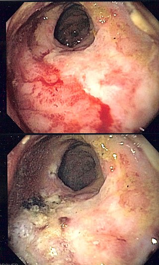
Upper gastrointestinal bleeding is gastrointestinal bleeding (hemorrhage) in the upper gastrointestinal tract, commonly defined as bleeding arising from the esophagus, stomach, or duodenum. Blood may be observed in vomit or in altered form as black stool. Depending on the amount of the blood loss, symptoms may include shock.

Hematemesis is the vomiting of blood. It can be confused with hemoptysis or epistaxis (nosebleed), which are more common. The source is generally the upper gastrointestinal tract, typically above the suspensory muscle of duodenum. It may be caused by ulcers, tumors of the stomach or esophagus, varices, prolonged and vigorous retching, gastroenteritis, ingested blood, or certain drugs.

Coffee ground vomitus refers to a particular appearance of vomit. Within organic heme molecules of red blood cells is the element iron, which oxidizes following exposure to gastric acid. This reaction causes the vomitus to look like ground coffee.

<span class="mw-page-title-main">Angiodysplasia</span> Medical condition

In medicine (gastroenterology), angiodysplasia is a small vascular malformation of the gut. It is a common cause of otherwise unexplained gastrointestinal bleeding and anemia. Lesions are often multiple, and frequently involve the cecum or ascending colon, although they can occur at other places. Treatment may be with colonoscopic interventions, angiography and embolization, medication, or occasionally surgery.

<span class="mw-page-title-main">Esophagogastroduodenoscopy</span> Diagnostic endoscopic procedure

Esophagogastroduodenoscopy (EGD) or oesophagogastroduodenoscopy (OGD), also called by various other names, is a diagnostic endoscopic procedure that visualizes the upper part of the gastrointestinal tract down to the duodenum. It is considered a minimally invasive procedure since it does not require an incision into one of the major body cavities and does not require any significant recovery after the procedure. However, a sore throat is common.

<span class="mw-page-title-main">Esophageal varices</span> Medical condition

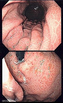
Esophageal varices are extremely dilated sub-mucosal veins in the lower third of the esophagus. They are most often a consequence of portal hypertension, commonly due to cirrhosis. People with esophageal varices have a strong tendency to develop severe bleeding which left untreated can be fatal. Esophageal varices are typically diagnosed through an esophagogastroduodenoscopy.

<span class="mw-page-title-main">Portal hypertension</span> Abnormally increased portal venous pressure

Portal hypertension is defined as increased portal venous pressure, with a hepatic venous pressure gradient greater than 5 mmHg. Normal portal pressure is 1–4 mmHg; clinically insignificant portal hypertension is present at portal pressures 5–9 mmHg; clinically significant portal hypertension is present at portal pressures greater than 10 mmHg. The portal vein and its branches supply most of the blood and nutrients from the intestine to the liver.

<span class="mw-page-title-main">Dieulafoy's lesion</span> Medical condition

Dieulafoy's lesion is a medical condition characterized by a large tortuous artery most commonly in the stomach wall (submucosal) that erodes and bleeds. It can present in any part of the gastrointestinal tract. It can cause gastric hemorrhage but is relatively uncommon. It is thought to cause less than 5% of all gastrointestinal bleeds in adults. It was named after French surgeon Paul Georges Dieulafoy, who described this condition in his paper "Exulceratio simplex: Leçons 1-3" in 1898. It is also called "caliber-persistent artery" or "aneurysm" of gastric vessels. However, unlike most other aneurysms, these are thought to be developmental malformations rather than degenerative changes.

<span class="mw-page-title-main">Gastrointestinal bleeding</span> Medical condition

Gastrointestinal bleeding, also called gastrointestinal hemorrhage (GIB), is all forms of bleeding in the gastrointestinal tract, from the mouth to the rectum. When there is significant blood loss over a short time, symptoms may include vomiting red blood, vomiting black blood, bloody stool, or black stool. Small amounts of bleeding over a long time may cause iron-deficiency anemia resulting in feeling tired or heart-related chest pain. Other symptoms may include abdominal pain, shortness of breath, pale skin, or passing out. Sometimes in those with small amounts of bleeding no symptoms may be present.

<span class="mw-page-title-main">Gastric varices</span> Medical condition

Gastric varices are dilated submucosal veins in the lining of the stomach, which can be a life-threatening cause of bleeding in the upper gastrointestinal tract. They are most commonly found in patients with portal hypertension, or elevated pressure in the portal vein system, which may be a complication of cirrhosis. Gastric varices may also be found in patients with thrombosis of the splenic vein, into which the short gastric veins that drain the fundus of the stomach flow. The latter may be a complication of acute pancreatitis, pancreatic cancer, or other abdominal tumours, as well as hepatitis C. Gastric varices and associated bleeding are a potential complication of schistosomiasis resulting from portal hypertension.

<span class="mw-page-title-main">Gastrointestinal disease</span> Medical condition

Gastrointestinal diseases refer to diseases involving the gastrointestinal tract, namely the esophagus, stomach, small intestine, large intestine and rectum, and the accessory organs of digestion, the liver, gallbladder, and pancreas.

<span class="mw-page-title-main">Transjugular intrahepatic portosystemic shunt</span> Artificial channel within the liver

Transjugular intrahepatic portosystemic shunt is an artificial channel within the liver that establishes communication between the inflow portal vein and the outflow hepatic vein. It is used to treat portal hypertension which frequently leads to intestinal bleeding, life-threatening esophageal bleeding and the buildup of fluid within the abdomen (ascites).

<span class="mw-page-title-main">Radiation proctitis</span> Medical condition

Radiation proctitis or radiation proctopathy is a condition characterized by damage to the rectum after exposure to x-rays or other ionizing radiation as a part of radiation therapy. Radiation proctopathy may occur as acute inflammation called "acute radiation proctitis" or with chronic changes characterized by radiation associated vascular ectasiae (RAVE) and chronic radiation proctopathy. Radiation proctitis most commonly occurs after pelvic radiation treatment for cancers such as cervical cancer, prostate cancer, bladder cancer, and rectal cancer. RAVE and chronic radiation proctopathy involves the lower intestine, primarily the sigmoid colon and the rectum, and was previously called chronic radiation proctitis, pelvic radiation disease and radiation enteropathy.

<span class="mw-page-title-main">Gastric antral vascular ectasia</span> Medical condition of the stomach

Gastric antral vascular ectasia (GAVE) is an uncommon cause of chronic gastrointestinal bleeding or iron deficiency anemia. The condition is associated with dilated small blood vessels in the pyloric antrum, which is a distal part of the stomach. The dilated vessels result in intestinal bleeding. It is also called watermelon stomach because streaky long red areas that are present in the stomach may resemble the markings on watermelon.

<span class="mw-page-title-main">Distal splenorenal shunt procedure</span> Medical procedure

In medicine, a distal splenorenal shunt procedure (DSRS), also splenorenal shunt procedure and Warren shunt, is a surgical procedure in which the distal splenic vein is attached to the left renal vein. It is used to treat portal hypertension and its main complication. It was developed by W. Dean Warren.

<span class="mw-page-title-main">Ménétrier's disease</span> Medical condition

Ménétrier disease is a rare, acquired, premalignant disease of the stomach characterized by massive gastric folds, excessive mucous production with resultant protein loss, and little or no acid production. The disorder is associated with excessive secretion of transforming growth factor alpha (TGF-α). It is named after a French physician Pierre Eugène Ménétrier, 1859–1935.

<span class="mw-page-title-main">Endoclip</span>

An endoclip is a metallic mechanical device used in endoscopy in order to close two mucosal surfaces without the need for surgery and suturing. Its function is similar to a suture in gross surgical applications, as it is used to join together two disjointed surfaces, but, can be applied through the channel of an endoscope under direct visualization. Endoclips have found use in treating gastrointestinal bleeding, in preventing bleeding after therapeutic procedures such as polypectomy, and in closing gastrointestinal perforations. Many forms of endoclips exist of different shapes and sizes, including two and three prong devices, which can be administered using single use and reloadable systems, and may or may not open and close to facilitate placement.

A stress ulcer is a single or multiple mucosal defect usually caused by physiological stress which can become complicated by upper gastrointestinal bleeding. These ulcers can be caused by shock, sepsis, trauma or other conditions and are found in patients with chronic illnesses. These ulcers are a significant issue in patients in critical and intensive care.

<span class="mw-page-title-main">Butyl cyanoacrylate</span> Chemical compound

n-Butyl cyanoacrylate, a cyanoacrylate ester, is a butyl ester of 2-cyano-2-propenoic acid. It is a colorless liquid with a sharp, irritating odor. It is insoluble in water. Its chief use is as the main component of medical cyanoacrylate glues. It can be encountered under various trade names, e.g. Cutseal, MediBond, MediCryl, PeriAcryl, GluStitch, Xoin, Gesika, VetGlu, Vetbond, LiquiVet, Indermil, LiquiBand, Histoacryl, IFABond, CutisSeal and others. The generic international nonproprietary name (INN) for NBCA is enbucrilate.

Therapeutic endoscopy is the medical term for an endoscopic procedure during which treatment is carried out via the endoscope. This contrasts with diagnostic endoscopy, where the aim of the procedure is purely to visualize a part of the gastrointestinal, respiratory or urinary tract in order to aid diagnosis. In practice, a procedure which starts as a diagnostic endoscopy may become a therapeutic endoscopy depending on the findings, such as in cases of upper gastrointestinal bleeding, or the finding of polyps during colonoscopy.

References

  1. Primignani M, Carpinelli L, Preatoni P, et al. (2000). "Natural history of portal hypertensive gastropathy in patients with liver cirrhosis. The New Italian Endoscopic Club for the study and treatment of esophageal varices (NIEC)". Gastroenterology. 119 (1): 181–7. doi: 10.1053/gast.2000.8555 . PMID   10889167.
  2. 1 2 3 Thuluvath PJ, Yoo HY (2002). "Portal Hypertensive gastropathy". Am. J. Gastroenterol. 97 (12): 2973–8. doi:10.1111/j.1572-0241.2002.07094.x. PMID   12492178. S2CID   29397451.
  3. Iwao T, Toyonaga A, Sumino M, et al. (1992). "Portal hypertensive gastropathy in patients with cirrhosis". Gastroenterology. 102 (6): 2060–5. doi:10.1016/0016-5085(92)90332-S. PMID   1587424.
  4. Gupta R, Sawant P, Parameshwar RV, Lele VR, Kulhalli PM, Mahajani SS (1998). "Gastric mucosal blood flow and hepatic perfusion index in patients with portal hypertensive gastropathy". J. Gastroenterol. Hepatol. 13 (9): 921–6. doi:10.1111/j.1440-1746.1998.tb00762.x. PMID   9794191. S2CID   26843163.
  5. Lo GH, Lai KH, Cheng JS, et al. (2001). "The effects of endoscopic variceal ligation and propranolol on portal hypertensive gastropathy: a prospective, controlled trial". Gastrointest. Endosc. 53 (6): 579–84. doi:10.1067/mge.2001.114062. PMID   11323582.
  6. Pérez-Ayuso RM, Piqué JM, Bosch J, et al. (1991). "Propranolol in prevention of recurrent bleeding from severe portal hypertensive gastropathy in cirrhosis". Lancet. 337 (8755): 1431–4. doi:10.1016/0140-6736(91)93125-S. PMID   1675316. S2CID   205038296.
  7. McCormick PA, Ooi H, Crosbie O (1998). "Tranexamic acid for severe bleeding gastric antral vascular ectasia in cirrhosis". Gut. 42 (5): 750–2. doi:10.1136/gut.42.5.750. PMC   1727106 . PMID   9659175.
  8. 1 2 Garcia N, Sanyal AJ (2001). "Portal Hypertensive Gastropathy and Gastric Antral Vascular Ectasia". Current Treatment Options in Gastroenterology. 4 (2): 163–171. doi:10.1007/s11938-001-0028-0. PMID   11469974. S2CID   179379.
  9. Geoffroy P, Duchateau A, Thiéfin G, Zeitoun P (1987). "Effects of propranolol and sucralfate on ethanol-induced gastric mucosal damage in chronic portal hypertensive rats". J. Hepatol. 5 (2): 162–6. doi:10.1016/S0168-8278(87)80568-8. PMID   3693860.
  10. Sato T, Yamazaki K, Toyota J, et al. (2005). "Efficacy of argon plasma coagulation for gastric antral vascular ectasia associated with chronic liver disease". Hepatol. Res. 32 (2): 121–6. doi:10.1016/j.hepres.2005.04.004. PMID   15967712.
  11. Urata J, Yamashita Y, Tsuchigame T, et al. (1998). "The effects of transjugular intrahepatic portosystemic shunt on portal hypertensive gastropathy". J. Gastroenterol. Hepatol. 13 (10): 1061–7. doi:10.1111/j.1440-1746.1998.tb00571.x. PMID   9835325. S2CID   21021556.
  12. Clarke JO, Thuluvath PJ (2007). "Endoscopic frontiers in the field of hepatology". Minerva Gastroenterologica e Dietologica. 53 (1): 101–9. PMID   17415347.
  13. Bini EJ, Lascarides CE, Micale PL, Weinshel EH (2000). "Mucosal abnormalities of the colon in patients with portal hypertension: an endoscopic study". Gastrointest. Endosc. 52 (4): 511–6. doi:10.1067/mge.2000.108478. PMID   11023569.