Adenomyomatosis

Last updated
Adenomyomatosis
Rokitansky-Aschoff sinus in a case of chronic cholecystitis.JPG
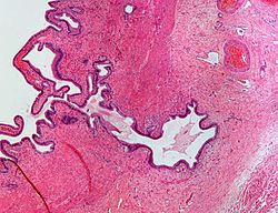
Micrograph showing Rokitansky–Aschoff sinus. H&E stain.

Adenomyomatosis is a benign condition characterized by hyperplastic changes of unknown cause involving the wall of the gallbladder. [1]

Contents

Pathophysiology

Adenomyomatosis of the gallbladder as seen on ultrasound [2]
Non-contrast abdominal ultrasound and contrast-enhanced ultrasound (CEUS) of adenomyomatosis of the gallbladder:
a The fundus of the gallbladder wall was thickened and the GB wall was obscure.
b The intramural echogenic foci were detected by high frequency transducer.
c CEUS--arterial phase (22 s) --heterogeneous hyper-enhancement and wall was intact.
d CEUS--venous phase (34 s) the anechoic spaces were more clear. Ultrasound of gallbladder adenomyosis.jpg
Non-contrast abdominal ultrasound and contrast-enhanced ultrasound (CEUS) of adenomyomatosis of the gallbladder:
a The fundus of the gallbladder wall was thickened and the GB wall was obscure.
b The intramural echogenic foci were detected by high frequency transducer.
c CEUS—arterial phase (22 s) —heterogeneous hyper-enhancement and wall was intact.
d CEUS—venous phase (34 s) the anechoic spaces were more clear.

Rokitansky–Aschoff sinuses

Rokitansky–Aschoff sinuses are pseudodiverticula or pockets in the wall of the gallbladder. They may be microscopic or macroscopic. Histologically, they are outpouchings of gallbladder mucosa into the gallbladder muscle layer and subserosal tissue as a result of hyperplasia and herniation of epithelial cells through the fibromuscular layer of the gallbladder wall. [4]

Rokitansky–Aschoff sinuses are not of themselves considered abnormal but they can be associated with cholecystitis. [5]

They form as a result of increased pressure in the gallbladder and recurrent damage to the wall of the gallbladder. [6]

Associations

Black pigment gallstones can form in Rokitansky–Aschoff sinuses of the gallbladder after the fourth to fifth decades of life in absence of the typical risk factors for bilirubin supersaturation of bile. [4] Hence, they are associated with gallstones (cholelithiasis). Cases of gall bladder cancer have also been reported to arise from Rokitansky–Aschoff sinuses. [7]

Diagnosis

Abdominal ultrasound has low accuracy in differentiating gall bladder adenomyomatosis from cancer and is operator dependent. However, it is used as the exam of the first-line due to its wide availability. Ultrasound findings may show thickened gall bladder wall, tiny anechoic spaces (Rokitansky–Aschoff sinuses or RAS), and twinkling artifact (or comet-tail reverberation). Comet tail reverberation, which is due to reflections from cholesterol crystals, is a highly specific sign for adenomyomatosis. [8]

On CT scan, it may show rosary sign, showing mucosal epithelium with intramural diverticula. [8]

Magnetic resonance imaging also plays an important role in the diagnosis of Rokitansky–Aschoff sinuses. [9] In fat-suppression MRI, RAS present with small, rounded, high signal intensity foci, called “pearl necklace sign”. [8]

Eponym

Rokitansky–Aschoff sinuses are named after Carl Freiherr von Rokitansky (1804–1878), a pathologist in Vienna, Austria and Ludwig Aschoff (1866–1942), a pathologist in Bonn, Germany. [10] [11]

See also

Related Research Articles

<span class="mw-page-title-main">Gallbladder</span> Organ in humans and other vertebrates

In vertebrates, the gallbladder, also known as the cholecyst, is a small hollow organ where bile is stored and concentrated before it is released into the small intestine. In humans, the pear-shaped gallbladder lies beneath the liver, although the structure and position of the gallbladder can vary significantly among animal species. It receives bile, produced by the liver, via the common hepatic duct, and stores it. The bile is then released via the common bile duct into the duodenum, where the bile helps in the digestion of fats.

<span class="mw-page-title-main">Bile duct</span> Type of organ

A bile duct is any of a number of long tube-like structures that carry bile, and is present in most vertebrates. The bile duct is separated into three main parts: the fundus (superior), the body (middle), and the neck (inferior).

<span class="mw-page-title-main">Gallstone</span> Disease where stones form in the gallbladder

A gallstone is a stone formed within the gallbladder from precipitated bile components. The term cholelithiasis may refer to the presence of gallstones or to any disease caused by gallstones, and choledocholithiasis refers to the presence of migrated gallstones within bile ducts.

<span class="mw-page-title-main">Cholecystitis</span> Inflammation of the gallbladder

Cholecystitis is inflammation of the gallbladder. Symptoms include right upper abdominal pain, pain in the right shoulder, nausea, vomiting, and occasionally fever. Often gallbladder attacks precede acute cholecystitis. The pain lasts longer in cholecystitis than in a typical gallbladder attack. Without appropriate treatment, recurrent episodes of cholecystitis are common. Complications of acute cholecystitis include gallstone pancreatitis, common bile duct stones, or inflammation of the common bile duct.

<span class="mw-page-title-main">Diverticulum</span> Medical or biological term for an outpouching of a hollow (or a fluid-filled) structure in the body

In medicine or biology, a diverticulum is an outpouching of a hollow structure in the body. Depending upon which layers of the structure are involved, diverticula are described as being either true or false.

Organomegaly is the abnormal enlargement of organs. For example, cardiomegaly is enlargement of the heart. Visceromegaly is the enlargement of abdominal organs. Examples of visceromegaly are enlarged liver (hepatomegaly), spleen (splenomegaly), stomach, kidneys, and pancreas.

<span class="mw-page-title-main">Gallbladder cancer</span> Medical condition

Gallbladder cancer is a relatively uncommon cancer, with an incidence of fewer than 2 cases per 100,000 people per year in the United States. It is particularly common in central and South America, central and eastern Europe, Japan and northern India; it is also common in certain ethnic groups e.g. Native American Indians and Hispanics. If it is diagnosed early enough, it can be cured by removing the gallbladder, part of the liver and associated lymph nodes. Most often it is found after symptoms such as abdominal pain, jaundice and vomiting occur, and it has spread to other organs such as the liver.

<span class="mw-page-title-main">Porcelain gallbladder</span> Medical condition

Porcelain gallbladder is a calcification of the gallbladder believed to be brought on by excessive gallstones, although the exact cause is not clear. As with gallstone disease in general, this condition occurs mostly in overweight female patients of middle age. It is a morphological variant of chronic cholecystitis. Inflammatory scarring of the wall, combined with dystrophic calcification within the wall transforms the gallbladder into a porcelain-like vessel. Removal of the gallbladder (cholecystectomy) is the recommended treatment.

Adenomyoma is a tumor (-oma) including components derived from glands (adeno-) and muscle (-my-). It is a type of complex and mixed tumor, and several variants have been described in the medical literature. Uterine adenomyoma, the localized form of uterine adenomyosis, is a tumor composed of endometrial gland tissue and smooth muscle in the myometrium. Adenomyomas containing endometrial glands are also found outside of the uterus, most commonly on the uterine adnexa but can also develop at distant sites outside of the pelvis. Gallbladder adenomyoma, the localized form of adenomyomatosis, is a polypoid tumor in the gallbladder composed of hyperplastic mucosal epithelium and muscularis propria.

<span class="mw-page-title-main">Biliary colic</span> Medical condition in which gallstones cause acute pain

Biliary colic, also known as symptomatic cholelithiasis, a gallbladder attack or gallstone attack, is when a colic occurs due to a gallstone temporarily blocking the cystic duct. Typically, the pain is in the right upper part of the abdomen, and can be severe. Pain usually lasts from 15 minutes to a few hours. Often, it occurs after eating a heavy meal, or during the night. Repeated attacks are common. Cholecystokinin - a gastrointestinal hormone - plays a role in the colic, as following the consumption of fatty meals, the hormone triggers the gallbladder to contract, which may expel stones into the duct and temporarily block it until being successfully passed.

<span class="mw-page-title-main">Gallbladder disease</span> Medical condition

Gallbladder diseases are diseases involving the gallbladder and is closely linked to biliary disease, with the most common cause being gallstones (cholelithiasis).

<span class="mw-page-title-main">Abdominal ultrasonography</span> Type of medical scan

Abdominal ultrasonography is a form of medical ultrasonography to visualise abdominal anatomical structures. It uses transmission and reflection of ultrasound waves to visualise internal organs through the abdominal wall. For this reason, the procedure is also called a transabdominal ultrasound, in contrast to endoscopic ultrasound, the latter combining ultrasound with endoscopy through visualize internal structures from within hollow organs.

<span class="mw-page-title-main">Cholesterolosis of gallbladder</span> Medical condition

In surgical pathology, strawberry gallbladder, more formally cholesterolosis of the gallbladder and gallbladder cholesterolosis, is a change in the gallbladder wall due to excess cholesterol.

Stereotactic biopsy, also known as stereotactic core biopsy, is a biopsy procedure that uses a computer and imaging performed in at least two planes to localize a target lesion in three-dimensional space and guide the removal of tissue for examination by a pathologist under a microscope. Stereotactic core biopsy makes use of the underlying principle of parallax to determine the depth or "Z-dimension" of the target lesion.

<span class="mw-page-title-main">Gallbladder polyp</span> Medical condition

Gallbladder polyps are growths or lesions resembling growths in the wall of the gallbladder. True polyps are abnormal accumulations of mucous membrane tissue that would normally be shed by the body.

Ioglycamic acid is a pharmaceutical drug that was used as an iodinated contrast medium for X-ray imaging of the gall bladder.

<span class="mw-page-title-main">Renal cyst</span> Medical condition

A renal cyst is a fluid collection in or on the kidney. There are several types based on the Bosniak classification. The majority are benign, simple cysts that can be monitored and not intervened upon. However, some are cancerous or are suspicious for cancer and are commonly removed in a surgical procedure called nephrectomy.

<span class="mw-page-title-main">Ground-glass opacity</span> Radiologic sign on radiographs and computed tomography scans

Ground-glass opacity (GGO) is a finding seen on chest x-ray (radiograph) or computed tomography (CT) imaging of the lungs. It is typically defined as an area of hazy opacification (x-ray) or increased attenuation (CT) due to air displacement by fluid, airway collapse, fibrosis, or a neoplastic process. When a substance other than air fills an area of the lung it increases that area's density. On both x-ray and CT, this appears more grey or hazy as opposed to the normally dark-appearing lungs. Although it can sometimes be seen in normal lungs, common pathologic causes include infections, interstitial lung disease, and pulmonary edema.

<span class="mw-page-title-main">Biliary pseudolithiasis</span> Medical condition

Biliary pseudolithiasis is an unusual complication of ceftriaxone where the drug complexes with calcium and mimics gallstones. It is reversed when ceftriaxone administration is stopped. It was first described in 1988 by Schaad et al. as "reversible ceftriaxone-associated biliary pseudolithiasis". Ceftriaxone has been frequently associated with biliary sludge or biliary pseudolithiasis in subsequent reports. Ceftriaxone is excreted primarily through the urine, but also through the bile, up to 40% of its excretion, with concentrations in the bile 20-150 times higher than in the serum. It forms a calcium salt in the gallbladder, which can exceed its solubility and create precipitates that resemble gallstones on ultrasonography. The incidence of pseudolithiasis in children treated with ceftriaxone is up to 25%, but most patients are asymptomatic. Risk factors for biliary pseudolithiasis include age greater than 24 months, gram-negative sepsis, high doses of ceftriaxone, hypercalcemia, surgery, and decreased bile flow/increased ceftriaxone excretion in bile. Conservative management with serial ultrasounds is recommended until the "stones" completely resolve. If associated with ceftriaxone, it resolves on average about 2 weeks after the ceftriaxone is stopped.

<span class="mw-page-title-main">Quadrilateral space syndrome</span> Medical condition

Quadrilateral space syndrome is a rotator cuff denervation syndrome in which the axillary nerve is compressed at the quadrilateral space of the rotator cuff.

References

  1. Ram MD, Midha D (August 1975). "Adenomyomatosis of the gallbladder". Surgery. 78 (2): 224–9. PMID   1154265.
  2. "UOTW#79 - Ultrasound of the Week". Ultrasound of the Week. 16 April 2017. Retrieved 27 May 2017.
  3. Tang S, Huang L, Wang Y, Wang Y (August 2015). "Contrast-enhanced ultrasonography diagnosis of fundal localized type of gallbladder adenomyomatosis". BMC Gastroenterology. 15 (1): 99. doi: 10.1186/s12876-015-0326-y . PMC   4524444 . PMID   26239485.
  4. 1 2 Cariati A, Cetta F (2002). "Rokitansky-Aschoff sinuses of the gallbladder are associated with black pigment gallstone formation: a scanning electron microscopy study". Ultrastructural Pathology. 27 (4): 265–70. doi:10.1080/01913120309913. PMID   12907372. S2CID   25219550.
  5. van Breda Vriesman AC, Engelbrecht MR, Smithuis RH, Puylaert JB (February 2007). "Diffuse gallbladder wall thickening: differential diagnosis". AJR. American Journal of Roentgenology. 188 (2): 495–501. doi:10.2214/AJR.05.1712. PMID   17242260.
  6. Stunell H, Buckley O, Geoghegan T, O'Brien J, Ward E, Torreggiani W (April 2008). "Imaging of adenomyomatosis of the gall bladder". Journal of Medical Imaging and Radiation Oncology. 52 (2): 109–17. doi:10.1111/j.1440-1673.2008.01926.x. PMID   18373800. S2CID   42685012.
  7. Matsumoto T, Shimada K (November 2009). "A case of gallbladder cancer arising from the Rokitansky-Aschoff sinus". Japanese Journal of Clinical Oncology. 39 (11): 776. doi: 10.1093/jjco/hyp149 . PMID   19884193.
  8. 1 2 3 Pang L, Zhang Y, Wang Y, Kong J (2018). "Pathogenesis of gallbladder adenomyomatosis and its relationship with early-stage gallbladder carcinoma: an overview". Brazilian Journal of Medical and Biological Research. 51 (6): e7411. doi:10.1590/1414-431x20187411. PMC   6002143 . PMID   29791592.
  9. Yoshimitsu K, Honda H, Jimi M, Kuroiwa T, Hanada K, Irie H, et al. (June 1999). "MR diagnosis of adenomyomatosis of the gallbladder and differentiation from gallbladder carcinoma: importance of showing Rokitansky-Aschoff sinuses". AJR. American Journal of Roentgenology. 172 (6): 1535–40. doi:10.2214/ajr.172.6.10350285. PMID   10350285.
  10. synd/983 at Who Named It?
  11. Kanne JP, Rohrmann CA, Lichtenstein JE (2005). "Eponyms in radiology of the digestive tract: historical perspectives and imaging appearances. Part 2. Liver, biliary system, pancreas, peritoneum, and systemic disease". Radiographics. 26 (2): 465–80. doi: 10.1148/rg.262055130 . PMID   16549610.