Sphincter of Oddi dysfunction

Last updated
Sphincter of Oddi dysfunction
Hepato-biliary.jpg
A diagram of the biliary tree showing the sphincter of Oddi. Note that part of the gall bladder has been removed, and the liver has been lifted off the tree.
Specialty Gastroenterology, general surgery

Sphincter of Oddi dysfunction refers to a group of functional disorders leading to abdominal pain due to dysfunction of the Sphincter of Oddi: functional biliary sphincter of Oddi and functional pancreatic sphincter of Oddi disorder. The sphincter of Oddi is a sphincter muscle, a circular band of muscle at the bottom of the biliary tree which controls the flow of pancreatic juices and bile into the second part of the duodenum. The pathogenesis of this condition is recognized to encompass stenosis or dyskinesia of the sphincter of Oddi (especially after cholecystectomy); consequently the terms biliary dyskinesia, papillary stenosis, and postcholecystectomy syndrome have all been used to describe this condition. Both stenosis and dyskinesia can obstruct flow through the sphincter of Oddi and can therefore cause retention of bile in the biliary tree and pancreatic juice in the pancreatic duct.

Contents

Individuals with sphincter of Oddi dysfunction present with abdominal pain resembling that of structural or inflammatory disorders of the gallbladder, biliary tree or pancreas. Among other characteristics, the pain is typically in the upper part of the abdomen or in the right upper quadrant of the abdomen, lasts 30 minutes or longer, and is not associated with a structural abnormality that could lead to these symptoms. The disorder is classified into two subtypes: functional biliary sphincter of Oddi disorder, where there is no disturbance in pancreatic enzyme measurements, such as amylase and lipase; and, functional pancreatic sphincter of Oddi disorder, where pancreatic enzyme measurements are elevated.

Attacks can be precipitated by opioid analgesics, [1] particularly in patients having undergone a cholecystectomy or bariatric surgery.

Classification

Functional disorders of the gallbladder, bile duct and pancreas have been defined and classified by the Rome criteria for functional gastrointestinal disorders. [2] The criteria outline three variants of functional disorders of the gallbladder, bile duct and pancreas, termed functional gallbladder disorder, functional biliary sphincter of Oddi disorder and functional pancreatic sphincter of Oddi disorder. All of the following criteria need to be met for as part of the definition of a functional disorder of the gallbladder: [2]

Functional gallbladder disorder

Individuals are classified as having a functional gallbladder disorder if the above criteria are met, if the gallbladder is present, and if the testing of liver enzymes, conjugated bilirubin, and pancreatic enzymes (amylase and lipase) are normal. [2]

Functional biliary sphincter of Oddi disorder

If all of the above criteria are met, individuals are classified as having a functional biliary sphincter of Oddi disorder, if the testing of pancreatic enzymes (amylase and lipase) is normal. [2]

The old Milwaukee classification of biliary sphincter of Oddi dysfunction (SOD) used to divide the condition into three subtypes: [3] but it is no longer in use. Type I included patients with biliary-type abdominal pain, with all of altered liver enzymes on blood testing, dilated biliary ducts on ultrasound or ERCP, and delayed bile clearance on HIDA scan. Type II included patients with biliary-type abdominal pain associated with at least one of the following: altered liver enzymes on blood testing, dilated biliary ducts on imaging tests, and delayed bile clearance on HIDA scan. Biliary-type pain in the absence of any sign of biliary or pancreatic alteration was the so-called Type III biliary SOD. The hypothesis that this pain could be linked to SOD has been studied in a large trial published in JAMA (2014) [4] where patients were randomized to sphincterotomy or sham surgery. Contrary to expectations, patients who were not subjected to sphincterotomy fared better. Overall, the investigators found that only 23% of the patients who underwent sphincterotomy improved versus 37% of the control group. The EPISOD trial has substantiated the ineffectiveness of endoscopic sphincterotomy in patients with these symptoms and SOD Type III is no longer considered to be a clinical entity.

Characteristics

Sphincter of Oddi dysfunction may be suggested by pain which seems to come from a biliary origin, which may or may not be associated with transient increases of liver or pancreatic enzymes. Common bile duct dilation and episodes of pancreatitis are also signs. [5]

Pathophysiology

Two mechanisms are involved in the development of sphincter of Oddi dysfunction, either or both of which may be contributory to the condition: stenosis, or narrowing of the sphincter of Oddi (also termed papillary stenosis), and dyskinesia, or alteration in the function of the sphincter of Oddi (also termed biliary dyskinesia). [5] Individuals with stenosis of the sphincter of Oddi typically have an elevated baseline pressure of the sphincter of Oddi, due to an anatomical problem that leads to narrowing of the sphincter, such as recurrent passage of gallstones through the ampulla of Vater, trauma to the sphincter from procedures such as endoscopic retrograde cholangiopancreatography or biliary surgery, or infections of the common bile duct. In contrast, dyskinesia of the sphincter of Oddi is a purely functional disorder, wherein there is intermittent obstruction of the bile duct due to inappropriate spasms. The reasons for dyskinesia of the sphincter of Oddi are not completely understood, but believed to be due to alteration in local gut hormones and peptides, such as cholecystokinin, which act on the sphincter or to altered neuronal control of the sphincter. [6]

Diagnosis

For diagnosis, measures of liver biochemistry and pancreatic enzymes are performed. Along with ruling out structural abnormalities, normally by performing an abdominal ultrasound and endoscopic retrograde cholangiopancreatography (ERCP). Measurements of bile transit when performing ERCP are taken to help evaluate different treatment options. [5] Sphincter of Oddi dysfunction is best diagnosed using manometry-an internal test done to measure the pressures within surrounding ducts to determine whether or not the muscle is functioning normally.

Treatment

Medication (to prevent spasms) or sphincterotomy (surgical procedure to cut the muscle) are the standard treatments for sphincter of Oddi dysfunction. One or the other may be better based on the classification of the condition.

See also

Related Research Articles

Pancreatitis Inflammation of the pancreas

Pancreatitis is a condition characterized by inflammation of the pancreas. The pancreas is a large organ behind the stomach that produces digestive enzymes and a number of hormones. There are two main types: acute pancreatitis, and chronic pancreatitis. Signs and symptoms of pancreatitis include pain in the upper abdomen, nausea and vomiting. The pain often goes into the back and is usually severe. In acute pancreatitis, a fever may occur, and symptoms typically resolve in a few days. In chronic pancreatitis weight loss, fatty stool, and diarrhea may occur. Complications may include infection, bleeding, diabetes mellitus, or problems with other organs.

Gallbladder Organ in humans and other vertebrates

In vertebrates, the gallbladder, also known as the cholecyst, is a small hollow organ where bile is stored and concentrated before it is released into the small intestine. In humans, the pear-shaped gallbladder lies beneath the liver, although the structure and position of the gallbladder can vary significantly among animal species. It receives and stores bile, produced by the liver, via the common hepatic duct, and releases it via the common bile duct into the duodenum, where the bile helps in the digestion of fats.

Gallstone Disease where stones form in the gallbladder

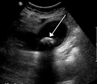
A gallstone is a stone formed within the gallbladder from precipitated bile components. The term cholelithiasis may refer to the presence of gallstones or to any disease caused by gallstones, and choledocholithiasis refers to the presence of migrated gallstones within bile ducts.

Cholecystitis Medical condition

Cholecystitis is inflammation of the gallbladder. Symptoms include right upper abdominal pain, pain in the right shoulder, nausea, vomiting, and occasionally fever. Often gallbladder attacks precede acute cholecystitis. The pain lasts longer in cholecystitis than in a typical gallbladder attack. Without appropriate treatment, recurrent episodes of cholecystitis are common. Complications of acute cholecystitis include gallstone pancreatitis, common bile duct stones, or inflammation of the common bile duct.

Cholecystectomy Surgical removal of the gallbladder

Cholecystectomy is the surgical removal of the gallbladder. Cholecystectomy is a common treatment of symptomatic gallstones and other gallbladder conditions. In 2011, cholecystectomy was the eighth most common operating room procedure performed in hospitals in the United States. Cholecystectomy can be performed either laparoscopically, or via an open surgical technique.

Endoscopic retrograde cholangiopancreatography Use of endoscopy and fluoroscopy to treat and diagnose digestive issues.

Endoscopic retrograde cholangiopancreatography (ERCP) is a technique that combines the use of endoscopy and fluoroscopy to diagnose and treat certain problems of the biliary or pancreatic ductal systems. It is primarily performed by highly skilled and specialty trained gastroenterologists. Through the endoscope, the physician can see the inside of the stomach and duodenum, and inject a contrast medium into the ducts in the biliary tree and pancreas so they can be seen on radiographs.

Common bile duct Gastrointestinal duct

The common bile duct, sometimes abbreviated as CBD, is a duct in the gastrointestinal tract of organisms that have a gallbladder. It is formed by the confluence of the common hepatic duct and cystic duct and terminates by uniting with pancreatic duct, forming the ampulla of Vater. The flow of bile from the ampulla of Vater into the duodenum is under the control of the sphincter of Oddi.

Gastrointestinal disease Medical condition

Gastrointestinal diseases refer to diseases involving the gastrointestinal tract, namely the oesophagus, stomach, small intestine, large intestine and rectum, and the accessory organs of digestion, the liver, gallbladder, and pancreas.

Common bile duct stone Medical condition

Common bile duct stone, also known as choledocholithiasis, is the presence of gallstones in the common bile duct (CBD). This condition can cause jaundice and liver cell damage. Treatments include choledocholithotomy and endoscopic retrograde cholangiopancreatography (ERCP).

Pancreatic divisum is a congenital anomaly in the anatomy of the ducts of the pancreas in which a single pancreatic duct is not formed, but rather remains as two distinct dorsal and ventral ducts. Most individuals with pancreas divisum remain without symptoms or complications. A minority of people with pancreatic divisum may develop episodes of abdominal pain, nausea or vomiting due to acute or chronic pancreatitis. The presence of pancreas divisum is usually identified with cross sectional diagnostic imaging, such as MRI or computed tomography (CT) imaging. In some cases, endoscopic retrograde cholangiopancreatography (ERCP) is performed, revealing the diagnosis of pancreas divisum. If no symptoms or complications are present, then treatment is not necessary. However, if there is recurrent pancreatitis, then a sphincterotomy of the minor papilla may be indicated.

Ascending cholangitis Medical condition

Ascending cholangitis, also known as acute cholangitis or simply cholangitis, is inflammation of the bile duct (cholangitis), usually caused by bacteria ascending from its junction with the duodenum. It tends to occur if the bile duct is already partially obstructed by gallstones.

Postcholecystectomy syndrome (PCS) describes the presence of abdominal symptoms after a cholecystectomy.

Biliary colic Medical condition in which gallstones cause acute pain

Biliary colic, also known as symptomatic cholelithiasis, a gallbladder attack or gallstone attack, is when a colic occurs due to a gallstone temporarily blocking the cystic duct. Typically, the pain is in the right upper part of the abdomen, and can be severe. Pain usually lasts from 15 minutes to a few hours. Often, it occurs after eating a heavy meal, or during the night. Repeated attacks are common.

Gallbladder disease Medical condition

Gallbladder diseases are diseases involving the gallbladder and is closely linked to biliary disease, with the most common cause being gallstones (cholelithiasis).

Abdominal ultrasonography

Abdominal ultrasonography is a form of medical ultrasonography to visualise abdominal anatomical structures. It uses transmission and reflection of ultrasound waves to visualise internal organs through the abdominal wall. For this reason, the procedure is also called a transabdominal ultrasound, in contrast to endoscopic ultrasound, the latter combining ultrasound with endoscopy through visualize internal structures from within hollow organs.

Biliary dyskinesia is a disorder of some component of biliary part of the digestive system in which bile cannot physically move in the proper direction through the tubular biliary tract. It most commonly involves abnormal biliary tract peristalsis muscular coordination within the gallbladder in response to dietary stimulation of that organ to squirt the liquid bile through the common bile duct into the duodenum. Ineffective peristaltic contraction of that structure produces postprandial right upper abdominal pain (cholecystodynia) and almost no other problem. When the dyskinesia is localized at the biliary outlet into the duodenum just as increased tonus of that outlet sphincter of Oddi, the backed-up bile can cause pancreatic injury with abdominal pain more toward the upper left side. In general, biliary dyskinesia is the disturbance in the coordination of peristaltic contraction of the biliary ducts, and/or reduction in the speed of emptying of the biliary tree into the duodenum.

Peter B. Cotton is a British Gastroenterologist best known for his advancement in digestive disease, pioneering and naming the ERCP procedure and creating the Digestive Disease Center at the Medical University of South Carolina.

Biloma is collection of bile within the abdominal cavity. It happens when there is a bile leak, for example after surgery for removing the gallbladder, with an incidence of 0.3–2%. Other causes are biliary surgery, liver biopsy, abdominal trauma, and, rarely, spontaneous perforation.

Choledochoduodenostomy

Choledochoduodenostomy (CDD) is a surgical procedure to create an anastomosis, a surgical connection, between the common bile duct (CBD) and an alternative portion of the duodenum. In healthy individuals, the CBD meets the pancreatic duct at the ampulla of Vater, which drains via the major duodenal papilla to the second part of duodenum. In cases of benign conditions such as narrowing of the distal CBD or recurrent CBD stones, performing a CDD provides the diseased patient with CBD drainage and decompression. A side-to-side anastomosis is usually performed.

Biliary endoscopic sphincterotomy Use of endoscopy and fluoroscopy to treat and diagnose digestive issues.

Biliary endoscopic sphincterotomy is a procedure where the sphincter of Oddi and the segment of the common bile duct where it enters the duodenum are cannulated and then cut with a sphincterotome, a device that includes a wire which cuts with an electric current (electrocautery).

References

  1. https://web.archive.org/web/20080731060352/http://www.anzca.edu.au/fpm/events/fpm-events/1-CBell.pdf%5B%5D
  2. 1 2 3 4 Rome Foundation. "Rome III Diagnostic Criteria for Functional Gastrointestinal Disorders" (PDF). Rome Foundation. Retrieved March 16, 2014.
  3. Prajapati, D. N.; Hogan, W. J. (2003). "Sphincter of Oddi dysfunction and other functional biliary disorders: Evaluation and treatment". Gastroenterology Clinics of North America. 32 (2): 601–18. doi:10.1016/s0889-8553(03)00025-6. PMID   12858608.
  4. Cotton PB, Durkalski V, Romagnuolo J, Pauls Q, Fogel E, Tarnasky P, Aliperti G, Freeman M, Kozarek R, Jamidar P, Wilcox M, Serrano J, Brawman-Mintzer O, Elta G, Mauldin P, Thornhill A, Hawes R, Wood-Williams A, Orrell K, Drossman D, Robuck P (2014). "Effect of endoscopic sphincterotomy for suspected sphincter of Oddi dysfunction on pain-related disability following cholecystectomy: the EPISOD randomized clinical trial". JAMA. 311 (20): 2101–2109. doi:10.1001/jama.2014.5220. PMC   4428324 . PMID   24867013.{{cite journal}}: CS1 maint: multiple names: authors list (link)
  5. 1 2 3 Corazziari, E (2003). "Sphincter of Oddi dysfunction". Digestive and Liver Disease. 35 Suppl 3: S26–9. doi:10.1016/s1590-8658(03)00090-2. PMID   12974506.
  6. Toouli, J (2009). "Sphincter of Oddi: Function, dysfunction, and its management". Journal of Gastroenterology and Hepatology. 24 Suppl 3: S57–62. doi:10.1111/j.1440-1746.2009.06072.x. PMID   19799700. S2CID   205465037.