Pancreatic pseudocyst

Last updated
Pancreatic pseudocyst
Pancreaticpseudocyst.png
A pancreatic pseudocyst as seen on CT
Specialty Gastroenterology   OOjs UI icon edit-ltr-progressive.svg
Symptoms Abdominal pain, bloating, nausea, vomiting and lack of appetite [1]
Complications Infection, hemorrhage, obstruction
CausesPancreatitis (chronic), Pancreatic neoplasm [2]
Diagnostic method Cyst fluid analysis [3]
Differential diagnosis Intraductal papillary mucinous neoplasm
Treatment Cystogastrostomy [4]

A pancreatic pseudocyst is a circumscribed collection of fluid rich in pancreatic enzymes, blood, and non-necrotic tissue, typically located in the lesser sac of the abdomen. Pancreatic pseudocysts are usually complications of pancreatitis, [5] although in children they frequently occur following abdominal trauma. Pancreatic pseudocysts account for approximately 75% of all pancreatic masses. [6]
Contents

Signs and symptoms

Signs and symptoms of pancreatic pseudocyst include abdominal pain, bloating, nausea, vomiting and lack of appetite. [1]

Complications

Complications of pancreatic pseudocysts include infection, hemorrhage, obstruction and rupture. For obstruction, it can cause compression in the GI tract from the stomach to colon, compression in urinary system, biliary system, and arteriovenous system.[ medical citation needed ]

Causes

Pancreatic pseudocyst can occur due to a variety of reasons, among them pancreatitis (chronic), pancreatic neoplasm and/or pancreatic trauma. [2]

Pathophysiology

Pancreatic pseudocysts are sometimes called false cysts because they do not have an epithelial lining. The wall of the pseudocyst is vascular and fibrotic, encapsulated in the area around the pancreas. Pancreatitis or abdominal trauma can cause its formation. [7] Treatment usually depends on the mechanism that brought about the pseudocyst. Pseudocysts take up to 6 weeks to completely form. [8]

Diagnosis

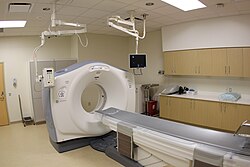
CT scan UPMCEast CTscan.jpg
CT scan

Diagnosis of pancreatic pseudocyst can be based on cyst fluid analysis: [3]

The most useful imaging tools are:

Treatment

TPN formula Tpn 3bag.jpg
TPN formula

Pancreatic pseudocyst treatment should be aimed at avoiding any complication (1 in 10 cases become infected). They also tend to rupture, and have shown that larger cysts have a higher likelihood to become more symptomatic, even needing surgery. [12] If no signs of infection are present, initial treatment may include conservative measures such as bowel rest (NPO), parenteral nutrition (TPN), and observation. If symptoms do not improve, then endoscopic drainage may be necessary. The majority of pseudocysts can be treated endoscopically; surgical intervention is rarely necessary. [13]

In the event of surgery:

See also

Related Research Articles

<span class="mw-page-title-main">Gastroenterology</span> Branch of medicine focused on the digestive system and its disorders

Gastroenterology is the branch of medicine focused on the digestive system and its disorders. The digestive system consists of the gastrointestinal tract, sometimes referred to as the GI tract, which includes the esophagus, stomach, small intestine and large intestine as well as the accessory organs of digestion which include the pancreas, gallbladder, and liver. The digestive system functions to move material through the GI tract via peristalsis, break down that material via digestion, absorb nutrients for use throughout the body, and remove waste from the body via defecation. Physicians who specialize in the medical specialty of gastroenterology are called gastroenterologists or sometimes GI doctors. Some of the most common conditions managed by gastroenterologists include gastroesophageal reflux disease, gastrointestinal bleeding, irritable bowel syndrome, inflammatory bowel disease (IBD) which includes Crohn's disease and ulcerative colitis, peptic ulcer disease, gallbladder and biliary tract disease, hepatitis, pancreatitis, colitis, colon polyps and cancer, nutritional problems, and many more.

<span class="mw-page-title-main">Pancreatitis</span> Inflammation of the pancreas

Pancreatitis is a condition characterized by inflammation of the pancreas. The pancreas is a large organ behind the stomach that produces digestive enzymes and a number of hormones. There are two main types: acute pancreatitis, and chronic pancreatitis.

<span class="mw-page-title-main">Bile duct</span> Type of organ

A bile duct is any of a number of long tube-like structures that carry bile, and is present in most vertebrates.

<span class="mw-page-title-main">Pancreatic cancer</span> Type of endocrine gland cancer

Pancreatic cancer arises when cells in the pancreas, a glandular organ behind the stomach, begin to multiply out of control and form a mass. These cancerous cells have the ability to invade other parts of the body. A number of types of pancreatic cancer are known.

<span class="mw-page-title-main">Acute pancreatitis</span> Medical condition

Acute pancreatitis (AP) is a sudden inflammation of the pancreas. Causes, in order of frequency, include: a gallstone impacted in the common bile duct beyond the point where the pancreatic duct joins it; heavy alcohol use; systemic disease; trauma; and, in minors, mumps. Acute pancreatitis may be a single event; it may be recurrent; or it may progress to chronic pancreatitis.

<span class="mw-page-title-main">Chronic pancreatitis</span> Medical condition

Chronic pancreatitis is a long-standing inflammation of the pancreas that alters the organ's normal structure and functions. It can present as episodes of acute inflammation in a previously injured pancreas, or as chronic damage with persistent pain or malabsorption. It is a disease process characterized by irreversible damage to the pancreas as distinct from reversible changes in acute pancreatitis.

<span class="mw-page-title-main">Gastrointestinal disease</span> Medical condition

Gastrointestinal diseases refer to diseases involving the gastrointestinal tract, namely the esophagus, stomach, small intestine, large intestine and rectum, and the accessory organs of digestion, the liver, gallbladder, and pancreas.

<span class="mw-page-title-main">Pancreatic duct</span> Duct associated with the human pancreas

The pancreatic duct, or duct of Wirsung, is a duct joining the pancreas to the common bile duct. This supplies it with pancreatic juice from the exocrine pancreas, which aids in digestion.

<span class="mw-page-title-main">Pseudocyst</span> Medical condition

Pseudocysts are like cysts, but lack epithelial or endothelial cells. Initial management consists of general supportive care. Symptoms and complications caused by pseudocysts require surgery. Computed tomography (CT) scans are used for initial imaging of cysts, and endoscopic ultrasounds are used in differentiating between cysts and pseudocysts. Endoscopic drainage is a popular and effective method of treating pseudocysts.

<span class="mw-page-title-main">Pancreatic fistula</span> Medical condition

A pancreatic fistula is an abnormal communication between the pancreas and other organs due to leakage of pancreatic secretions from damaged pancreatic ducts. An external pancreatic fistula is one that communicates with the skin, and is also known as a pancreaticocutaneous fistula, whereas an internal pancreatic fistula communicates with other internal organs or spaces. Pancreatic fistulas can be caused by pancreatic disease, trauma, or surgery.

<span class="mw-page-title-main">Pancreas divisum</span> Congenital disorder of digestive system

Pancreatic divisum is a congenital anomaly in the anatomy of the ducts of the pancreas in which a single pancreatic duct is not formed, but rather remains as two distinct dorsal and ventral ducts. Most individuals with pancreas divisum remain without symptoms or complications. A minority of people with pancreatic divisum may develop episodes of abdominal pain, nausea or vomiting due to acute or chronic pancreatitis. The presence of pancreas divisum is usually identified with cross sectional diagnostic imaging, such as endoscopic retrograde cholangiopancreatography (ERCP) or magnetic resonance cholangiopancreatography (MRCP). In some cases, it may be detected intraoperatively. If no symptoms or complications are present, then treatment is not necessary. However, if there is recurrent pancreatitis, then a sphincterotomy of the minor papilla may be indicated.

<span class="mw-page-title-main">Exocrine pancreatic insufficiency</span> Human disease

Exocrine pancreatic insufficiency (EPI) is the inability to properly digest food due to a lack or reduction of digestive enzymes made by the pancreas. EPI can occur in humans and is prevalent in many conditions such as cystic fibrosis, Shwachman–Diamond syndrome, different types of pancreatitis, multiple types of diabetes mellitus, advanced renal disease, older adults, celiac disease, IBS-D, IBD, HIV, alcohol-related liver disease, Sjogren syndrome, tobacco use, and use of somatostatin analogues.

Pancreatic diseases are diseases that affect the pancreas, an organ in most vertebrates and in humans and other mammals located in the abdomen. The pancreas plays a role in the digestive and endocrine system, producing enzymes which aid the digestion process and the hormone insulin, which regulates blood sugar levels. The most common pancreatic disease is pancreatitis, an inflammation of the pancreas which could come in acute or chronic form. Other pancreatic diseases include diabetes mellitus, exocrine pancreatic insufficiency, cystic fibrosis, pseudocysts, cysts, congenital malformations, tumors including pancreatic cancer, and hemosuccus pancreaticus.

<span class="mw-page-title-main">Hereditary pancreatitis</span> Medical condition

Hereditary pancreatitis (HP) is an inflammation of the pancreas due to genetic causes. It was first described in 1952 by Comfort and Steinberg but it was not until 1996 that Whitcomb et al isolated the first responsible mutation in the trypsinogen gene (PRSS1) on the long arm of chromosome seven (7q35).

<span class="mw-page-title-main">Hemosuccus pancreaticus</span> Medical condition

Hemosuccus pancreaticus is a rare cause of hemorrhage in the gastrointestinal tract. It is caused by a bleeding source in the pancreas, pancreatic duct, or structures adjacent to the pancreas, such as the splenic artery, that bleed into the pancreatic duct, which is connected with the bowel at the duodenum, the first part of the small intestine. Patients with hemosuccus may develop symptoms of gastrointestinal hemorrhage, such as blood in the stools, maroon stools, or melena, which is a dark, tarry stool caused by digestion of red blood cells. They may also develop abdominal pain. It is associated with pancreatitis, pancreatic cancer and aneurysms of the splenic artery. Hemosuccus may be identified with endoscopy (esophagogastroduodenoscopy), where fresh blood may be seen from the pancreatic duct. Alternatively, angiography may be used to inject the celiac axis to determine the blood vessel that is bleeding. This may also be used to treat hemosuccus, as embolization of the end vessel may terminate the hemorrhage. However, a distal pancreatectomy—surgery to remove of the tail of the pancreas—may be required to stop the hemorrhage.

Markus Wolfgang Büchler is a German surgeon and university full professor. He specialises in gastrointestinal, hepatobiliary and transplant surgery, and is especially known for pioneering operations on the pancreas.

Pancreatic abscess is a late complication of acute necrotizing pancreatitis, occurring more than 4 weeks after the initial attack. A pancreatic abscess is a collection of pus resulting from tissue necrosis, liquefaction, and infection. It is estimated that approximately 3% of the patients with acute pancreatitis will develop an abscess.

David B. Adams is an American physician who is Professor of Surgery, Chief, Division of Gastrointestinal and Laparoscopic Surgery and Co-Director of the Digestive Disease Center at the Medical University of South Carolina. Adams specializes in chronic pancreatitis surgeries. He has given over numerous presentations regarding his clinical interests and will host the Chronic Pancreatitis Symposium in 2014 on Kiawah Island.

Cystogastrostomy is a surgery to create an opening between a pancreatic pseudocyst and the stomach when the cyst is in a suitable position to be drained into the stomach. This conserves pancreatic juices that would otherwise be lost. This surgery is performed by a pancreatic surgeon to avoid a life-threatening rupture of the pancreatic pseudocyst.

Pancreatic mucinous cystic neoplasm (MCN) is a type of cystic lesion that occurs in the pancreas. Amongst individuals undergoing surgical resection of a pancreatic cyst, about 23 percent were mucinous cystic neoplasms. These lesions are benign, though there is a high rate of progression to cancer. As such, surgery should be pursued when feasible. The rate of malignancy present in MCN is about 10 percent. If resection is performed before invasive malignancy develops, prognosis is excellent. The extent of invasion is the single most important prognostic factor in predicting survival.

References

  1. 1 2 "Pancreatic pseudocyst: MedlinePlus Medical Encyclopedia". www.nlm.nih.gov. Retrieved 2015-08-10.
  2. 1 2 Atluri, Pavan (2005-01-01). The Surgical Review: An Integrated Basic and Clinical Science Study Guide. Lippincott Williams & Wilkins. ISBN   9780781756419.
  3. 1 2 "Pancreatic Pseudocysts: Practice Essentials, Background, Pathophysiology". 2018-10-30.{{cite journal}}: Cite journal requires |journal= (help)
  4. 1 2 (eds.), Edward H. Phillips ...; Rosenthal, Raul J. (1995). Operative strategies in laparoscopic surgery. Berlin [u.a.]: Springer. p. 136. ISBN   9783540592143 . Retrieved 26 November 2017.{{cite book}}: |last1= has generic name (help)
  5. Habashi S, Draganov PV (January 2009). "Pancreatic pseudocyst". World J. Gastroenterol. 15 (1): 38–47. doi: 10.3748/wjg.15.38 . PMC   2653285 . PMID   19115466.
  6. Beger, Hans G.; Buchler, Markus; Kozarek, Richard; Lerch, Markus; Neoptolemos, John P.; Warshaw, Andrew; Whitcomb, David; Shiratori, Keiko (2009-01-26). The Pancreas: An Integrated Textbook of Basic Science, Medicine, and Surgery. John Wiley & Sons. ISBN   9781444300130.
  7. Ignatavicius, Donna (2016). Medical surgical nursing. Elsevier. p. 1226. ISBN   978-1-4557-7255-1.
  8. Lillemoe, Keith (2013). Master techniques in Surgery. Lippincott Williams & Wilkins. p. 147. ISBN   978-1-60831-172-9.
  9. Aghdassi AA, Mayerle J, Kraft M, Sielenkämper AW, Heidecke CD, Lerch MM (2006). "Pancreatic pseudocysts - when and how to treat?". HPB (Oxford). 8 (6): 432–41. doi:10.1080/13651820600748012. PMC   2020756 . PMID   18333098.
  10. Aghdassi A, Mayerle J, Kraft M, Sielenkämper AW, Heidecke CD, Lerch MM (March 2008). "Diagnosis and treatment of pancreatic pseudocysts in chronic pancreatitis". Pancreas. 36 (2): 105–12. doi:10.1097/MPA.0b013e31815a8887. PMID   18376299. S2CID   1964674.
  11. Khanna, A. K.; Tiwary, Satyendra K.; Kumar, Puneet (2012). "Pancreatic Pseudocyst: Therapeutic Dilemma". International Journal of Inflammation. 2012: 1–7. doi: 10.1155/2012/279476 . PMC   3345229 . PMID   22577595.
  12. "Pancreatic pseudocyst Treatment". Medscape.com. eMedicine. Retrieved August 11, 2015.
  13. Elta, GH; Enestvedt, BK; Sauer, BG; Lennon, AM (April 2018). "ACG Clinical Guideline: Diagnosis and Management of Pancreatic Cysts". The American Journal of Gastroenterology. 113 (4): 464–479. doi:10.1038/ajg.2018.14. PMID   29485131. S2CID   3584079.
  14. Hughes, Steven (2015-03-26). Operative Techniques in Hepato-Pancreato-Biliary Surgery. Lippincott Williams & Wilkins. ISBN   9781496319067.
  15. Skandalakis, Lee J.; Skandalakis, John E. (2013-11-08). Surgical Anatomy and Technique: A Pocket Manual. Springer Science & Business Media. ISBN   9781461485636.
  16. Scott-Conner, Carol (2009). Scott-Conner & Dawson: Essential Operative Techniques and Anatomy. Lippincott Williams & Wilkins. p. 455. ISBN   978-1-4511-5172-5.

Further reading