Anal canal

Last updated
Anal canal
Anorectum-en.svg
Anatomy of the anus and rectum
Gray1079.png
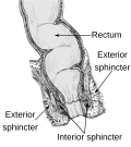
Coronal section through the anal canal. B. Cavity of urinary bladder V.D. Ductus deferens. S.V. Seminal vesicle. R. Second part of rectum. A.C. Anal canal. L.A. Levator ani. I.S. Sphincter ani internus. E.S. Sphincter ani externus.
Details
Precursor Hindgut, proctodeum
Artery superior rectal artery (above pectinate line) and inferior rectal artery (below line)
Vein superior rectal vein (above pectinate line) and inferior rectal vein (below line)
Nerve autonomic inferior hypogastric plexus (above pectinate line) and somatic inferior rectal nerves (below line)
Lymph Superficial inguinal lymph node (below pectinate line) and internal iliac lymph nodes (above line)
Identifiers
Latin canalis analis
MeSH D001003
TA98 A05.7.05.001
TA2 3009
FMA 15703
Anatomical terminology

The anal canal is the part that connects the rectum to the anus, located below the level of the pelvic diaphragm. [1] It is located within the anal triangle of the perineum, between the right and left ischioanal fossa. As the final functional segment of the bowel, it functions to regulate release of excrement by two muscular sphincter complexes. The anus is the aperture at the terminal portion of the anal canal.

Contents

Structure

In humans, the anal canal is approximately 2.5 to 4 cm (0.98 to 1.57 in) long, from the anorectal junction to the anus. [2] [3] [4] It is directed downwards and backwards. It is surrounded by inner involuntary and outer voluntary sphincters which keep the lumen closed in the form of an anteroposterior slit.

The canal is differentiated from the rectum by a transition along the internal surface from endodermal to skin-like ectodermal tissue.

The anal canal is traditionally divided into two segments, upper and lower, separated by the pectinate line (also known as the dentate line):

The anal verge refers to the distal end of the anal canal, a transitional zone between the epithelium of the anal canal and the perianal skin. It should not be confused with the pectinate line between the upper and lower zones within the anal canal.

The anal gland secretes lymphal discharge and built-up fecal matter from the colon lining. In some animals this gland expungement can be done routinely every 24–36 months to prevent infection and fistula formation.

Relations

Function

The external anal sphincter muscle is the voluntary muscle that surrounds and adheres to the anus at the lower margin of the anal canal. This muscle is in a state of tonic contraction, but during defecation, it relaxes to allow the release of feces.

Movement of the feces is also controlled by the involuntarily controlled internal anal sphincter, which is an extension of the circular muscle surrounding the anal canal. It relaxes to expel feces from the rectum and anal canal.

Additional images

See also

Related Research Articles

<span class="mw-page-title-main">Perineum</span> Region of the body including the perineal body and surrounding structures

The perineum in humans is the space between the anus and scrotum in the male, or between the anus and the vulva in the female. The perineum is the region of the body between the pubic symphysis and the coccyx, including the perineal body and surrounding structures. The perineal raphe is visible and pronounced to varying degrees. The perineum is an erogenous zone. This area is also known as the taint or chode in American slang.

<span class="mw-page-title-main">Pudendal nerve</span> Main nerve of the perineum

The pudendal nerve is the main nerve of the perineum. It is a mixed nerve and also conveys sympathetic autonomic fibers. It carries sensation from the external genitalia of both sexes and the skin around the anus and perineum, as well as the motor supply to various pelvic muscles, including the male or female external urethral sphincter and the external anal sphincter.

<span class="mw-page-title-main">Levator ani</span> Broad, thin muscle group, situated on either side of the pelvis

The levator ani is a broad, thin muscle group, situated on either side of the pelvis. It is formed from three muscle components: the pubococcygeus, the iliococcygeus, and the puborectalis.

Articles related to anatomy include:

A proctodeum is the back ectodermal part of an alimentary canal. It is created during embryogenesis by a folding of the outer body wall. It will form the lower part of the anal canal, below the pectinate line, which will be lined by stratified squamous non-keratinized and stratified squamous keratinized epithelium. The junction between them is Hilton's white line.

<span class="mw-page-title-main">External anal sphincter</span> Flat plane of skeletal muscle fibers

The external anal sphincter is an oval tube skeletal muscle fibers. Distally, it is adherent to the skin surrounding the margin of the anus. The sphincter exhibits a resting state of tonical contraction.

<span class="mw-page-title-main">Internal anal sphincter</span> Ring of smooth muscle that surrounds part of the anal canal

The internal anal sphincter, IAS, is a ring of smooth muscle that surrounds about 2.5–4.0 cm of the anal canal. It is about 5 mm thick, and is formed by an aggregation of the smooth (involuntary) circular muscle fibers of the rectum. it terminates distally about 6 mm from the anal orifice.

<span class="mw-page-title-main">Middle rectal artery</span>

The middle rectal artery is an artery in the pelvis that supplies blood to the rectum.

<span class="mw-page-title-main">Inferior rectal artery</span>

The inferior rectal artery is an artery that supplies blood to the lower third of the anal canal below the pectinate line.

<span class="mw-page-title-main">Ischioanal fossa</span> Wedge-shaped space located lateral to the anal canal and inferior to the pelvic diaphragm

The ischioanal fossa is the fat-filled wedge-shaped space located lateral to the anal canal and inferior to the pelvic diaphragm. It is somewhat prismatic in shape, with its base directed to the surface of the perineum and its apex at the line of meeting of the obturator and anal fasciae.

<span class="mw-page-title-main">Rectal venous plexus</span>

The rectal venous plexus is the venous plexus surrounding the rectum. It consists of an internal and an external rectal plexus. It is drained by the superior, middle, and inferior rectal veins. It forms a portosystemic (portocaval) anastomosis. This allows rectally administered medications to bypassing first pass metabolism.

<span class="mw-page-title-main">Superior rectal artery</span>

The superior rectal artery is an artery that descends into the pelvis to supply blood to the rectum.

<span class="mw-page-title-main">Inferior anal nerves</span>

The Inferior rectal nerves usually branch from the pudendal nerve but occasionally arises directly from the sacral plexus; they cross the ischiorectal fossa along with the inferior rectal artery and veins, toward the anal canal and the lower end of the rectum, and is distributed to the Sphincter ani externus and to the integument (skin) around the anus.

<span class="mw-page-title-main">Anal triangle</span> Posterior part of the perineum

The anal triangle is the posterior part of the perineum. It contains the anal canal.

<span class="mw-page-title-main">Pelvic fascia</span>

The pelvic fasciae are the fascia of the pelvis and can be divided into:

<span class="mw-page-title-main">Outline of human anatomy</span> Overview of and topical guide to human anatomy

The following outline is provided as an overview of and topical guide to human anatomy:

<span class="mw-page-title-main">Anocutaneous line</span>

The anocutaneous line, also called the Hilton white line or intersphincteric groove, is a boundary in the anal canal.

<span class="mw-page-title-main">Rectum</span> Final portion of the large intestine

The rectum is the final straight portion of the large intestine in humans and some other mammals, and the gut in others. The adult human rectum is about 12 centimetres (4.7 in) long, and begins at the rectosigmoid junction at the level of the third sacral vertebra or the sacral promontory depending upon what definition is used. Its diameter is similar to that of the sigmoid colon at its commencement, but it is dilated near its termination, forming the rectal ampulla. It terminates at the level of the anorectal ring or the dentate line, again depending upon which definition is used. In humans, the rectum is followed by the anal canal which is about 4 centimetres (1.6 in) long, before the gastrointestinal tract terminates at the anal verge. The word rectum comes from the Latin rectumintestinum, meaning straight intestine.

<span class="mw-page-title-main">Human anus</span> External opening of the rectum

In humans, the anus is the external opening of the rectum, located inside the intergluteal cleft and separated from the genitals by the perineum. Two sphincters control the exit of feces from the body during an act of defecation, which is the primary function of the anus. These are the internal anal sphincter and the external anal sphincter, which are circular muscles that normally maintain constriction of the orifice and which relaxes as required by normal physiological functioning. The inner sphincter is involuntary and the outer is voluntary. It is located behind the perineum which is located behind the vulva or scrotum.

Rectal discharge is intermittent or continuous expression of liquid from the anus. Normal rectal mucus is needed for proper excretion of waste. Otherwise, this is closely related to types of fecal incontinence but the term rectal discharge does not necessarily imply degrees of incontinence. Types of fecal incontinence that produce a liquid leakage could be thought of as a type of rectal discharge.

References

  1. Madoff, Robert D.; Melton-Meax, Genevieve B. (2020). "136. Diseases of the rectum and anus". In Goldman, Lee; Schafer, Andrew I. (eds.). Goldman-Cecil Medicine. Vol. 1 (26th ed.). Philadelphia: Elsevier. p. 933. ISBN   978-0-323-55087-1.
  2. "Anal canal".
  3. Anal+Canal at the U.S. National Library of Medicine Medical Subject Headings (MeSH)
  4. "Anal Canal - Location, Function and Pictures".