Buccopharyngeal fascia

Last updated
Buccopharyngeal fascia
Carotid sheath.PNG
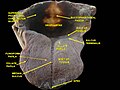
Carotid sheath outlined in red
Gray380.png
Muscles of the pharynx and cheek
Details
Identifiers
Latin fascia buccopharyngea
TA98 A04.1.04.010
A05.3.01.116
TA2 2211
FMA 55078
Anatomical terminology

The buccopharyngeal fascia is a fascia of the pharynx. [1] It represents the posterior portion of the pretracheal fascia [2] (visceral fascia). [3] It covers the superior pharyngeal constrictor muscles, and buccinator muscle. [4]

Contents

Structure

The buccopharyngeal fascia is a thin lamina given off from the pretracheal fascia.[ citation needed ] It is the portion of the pretracheal fascia situated posterior and lateral to the pharynx. It encloses the entire superior part of the alimentary canal. [3]

The buccopharyngeal fascia envelops the superior pharyngeal constrictor muscles. [4] [1] It extends anteriorly from the constrictor pharyngis superior [4] over the pterygomandibular raphe to cover the buccinator muscle [1] (though another source describes it as continuous with the fascia covering the buccinator muscle). [3]

Attachments

It is attached to the prevertebral fascia by loose connective tissue, with the retropharyngeal space found between them.[ citation needed ] It may also be attached to the alar fascia posteriorly at C3 and C6 levels. [5]

Relations

The thyroid gland wraps around the trachea and oesophagus anterior to the buccopharyngeal fascia, so that the lateral parts of the thyroid gland border it. [6]

The buccopharyngeal fascia runs parallel to the medial aspect of the carotid sheath.[ citation needed ]

Additional images

See also

Related Research Articles

<span class="mw-page-title-main">External carotid artery</span> Major artery of the head and neck

The external carotid artery is a major artery of the head and neck. It arises from the common carotid artery when it splits into the external and internal carotid artery. The external carotid artery supplies blood to the face, brain and neck.

<span class="mw-page-title-main">Endomysium</span> Connective tissue ensheathing individual muscle fibres

The endomysium, meaning within the muscle, is a wispy layer of areolar connective tissue that ensheaths each individual muscle fiber, or muscle cell. It also contains capillaries and nerves. It overlies the muscle fiber's cell membrane: the sarcolemma. Endomysium is the deepest and smallest component of muscle connective tissue. This thin layer helps provide an appropriate chemical environment for the exchange of calcium, sodium, and potassium, which is essential for the excitation and subsequent contraction of a muscle fiber.

<span class="mw-page-title-main">Facial artery</span>

The facial artery is a branch of the external carotid artery that supplies structures of the superficial face.

<span class="mw-page-title-main">Middle pharyngeal constrictor muscle</span> Muscle in the neck

The middle pharyngeal constrictor is a fan-shaped muscle located in the neck. It is one of three pharyngeal constrictor muscles. It is smaller than the inferior pharyngeal constrictor muscle.

<span class="mw-page-title-main">Inferior pharyngeal constrictor muscle</span> Skeletal muscle of the pharynx

The inferior pharyngeal constrictor muscle is a skeletal muscle of the neck. It is the thickest of the three outer pharyngeal muscles. It arises from the sides of the cricoid cartilage and the thyroid cartilage. It is supplied by the vagus nerve. It is active during swallowing, and partially during breathing and speech. It may be affected by Zenker's diverticulum.

<span class="mw-page-title-main">Superior pharyngeal constrictor muscle</span> Muscle

The superior pharyngeal constrictor muscle is a quadrilateral muscle of the pharynx. It is the uppermost and thinnest of the three pharyngeal constrictors.

<span class="mw-page-title-main">Stylopharyngeus muscle</span> Muscle

The stylopharyngeus muscle is a muscle in the head. It originates from the temporal styloid process. Some of its fibres insert onto the thyroid cartilage, while others end by intermingling with proximal structures. It is innervated by the glossopharyngeal nerve. It acts to elevate the larynx and pharynx, and dilate the pharynx, thus facilitating swallowing.

<span class="mw-page-title-main">Carotid sheath</span>

The carotid sheath is a condensation of the deep cervical fascia enveloping multiple vital neurovascular structures of the neck, including the common and internal carotid arteries, the internal jugular vein, the vagus nerve, and ansa cervicalis. The carotid sheath helps protects the structures contained therein.

<span class="mw-page-title-main">Inferior thyroid artery</span> Artery of the neck

The inferior thyroid artery is an artery in the neck. It arises from the thyrocervical trunk and passes upward, in front of the vertebral artery and longus colli muscle. It then turns medially behind the carotid sheath and its contents, and also behind the sympathetic trunk, the middle cervical ganglion resting upon the vessel.

<span class="mw-page-title-main">Ascending pharyngeal artery</span>

The ascending pharyngeal artery is an artery of the neck that supplies the pharynx.

<span class="mw-page-title-main">Superior laryngeal nerve</span> Branch of the vagus nerve

The superior laryngeal nerve is a branch of the vagus nerve. It arises from the middle of the inferior ganglion of vagus nerve and additionally also receives a sympathetic branch from the superior cervical ganglion.

<span class="mw-page-title-main">Ascending palatine artery</span>

The ascending palatine artery is an artery is a branch of the facial artery which ascends along the neck before spliting into two terminal branches; one branch supplies the soft palate, and the other supplies the palatine tonsil and pharyngotympanic tube.

<span class="mw-page-title-main">Retromandibular vein</span> Major face vein

The retromandibular vein is a major vein of the face. It is formed within the parotid gland by the confluence of the maxillary vein, and superficial temporal vein. It descends in the gland and splits into two branches upon emerging from the gland. Its anterior branch then joins the (anterior) facial vein forming the common facial vein, while its posterior branch joins the posterior auricular vein forming the external jugular vein.

<span class="mw-page-title-main">Deep cervical fascia</span>

The deep cervical fascia lies under cover of the platysma, and invests the muscles of the neck; it also forms sheaths for the carotid vessels, and for the structures situated in front of the vertebral column. Its attachment to the hyoid bone prevents the formation of a dewlap.

<span class="mw-page-title-main">Masseteric fascia</span>

The masseteric fascia and parotideomasseteric fascia are fascias of the head varyingly described depending upon the source consulted. They may or may not be described as one and the same structure.

<span class="mw-page-title-main">Carotid triangle</span>

The carotid triangle is a portion of the anterior triangle of the neck.

<span class="mw-page-title-main">Pterygomandibular raphe</span> Ligament formed from the buccopharyngeal fascia

The pterygomandibular raphe is a thin tendinous band of buccopharyngeal fascia. It is attached superiorly to the pterygoid hamulus of the medial pterygoid plate, and inferiorly to the posterior end of the mylohyoid line of the mandible. It gives attachment to the buccinator muscle, and the superior pharyngeal constrictor muscle (behind).

<span class="mw-page-title-main">Prevertebral fascia</span> Layer of deep cervical fascia that surrounds the vertebral column

The prevertebral fascia is the layer of deep cervical fascia that surrounds the vertebral column. It is the deepest layer of deep cervical fascia.

<span class="mw-page-title-main">Pretracheal fascia</span>

The pretracheal fascia is a layer of the deep cervical fascia at the front of the neck. It attaches to the hyoid bone above, and - extending down into the thorax - blends with the fibrous pericardium below. It encloses the thyroid gland and parathyroid glands, trachea, and esophagus. It extends medially in front of the carotid vessels. It assists in forming the carotid sheath.

The pharyngobasilar fascia is a fascia of the pharynx. It is situated between the mucous and muscular layers of the pharynx. It is formed as a thickening of the pharyngeal mucosa superior to the superior pharyngeal constrictor muscle. It attaches to the basilar part of occipital bone, the petrous part of the temporal bone, the medial pterygoid plate, and the pterygomandibular raphe. It diminishes in thickness inferiorly. Posteriorly, it is reinforced by the pharyngeal raphe. It reinforces the pharyngeal wall where muscle is deficient.

References

PD-icon.svgThis article incorporates text in the public domain from page 390 of the 20th edition of Gray's Anatomy (1918)

  1. 1 2 3 Standring, Susan (2020). Gray's Anatomy: The Anatomical Basis of Clinical Practice (42nd ed.). New York. p. 709. ISBN   978-0-7020-7707-4. OCLC   1201341621.{{cite book}}: CS1 maint: location missing publisher (link)
  2. Morton, David A. (2019). The Big Picture: Gross Anatomy. K. Bo Foreman, Kurt H. Albertine (2nd ed.). New York. p. 266. ISBN   978-1-259-86264-9. OCLC   1044772257.{{cite book}}: CS1 maint: location missing publisher (link)
  3. 1 2 3 Fehrenbach, Margaret J.; Herring, Susan W. (2017). Illustrated Anatomy of the Head and Neck (5th ed.). St. Louis: Elsevier. pp. 266–267. ISBN   978-0-323-39634-9.
  4. 1 2 3 Stecco, Carla; Hammer, Warren; Vleeming, Andry; De Caro, Raffaele (2015-01-01), Stecco, Carla; Hammer, Warren; Vleeming, Andry; De Caro, Raffaele (eds.), "4 - Fasciae of the Head and Neck", Functional Atlas of the Human Fascial System, Churchill Livingstone, pp. 103–139, ISBN   978-0-7020-4430-4 , retrieved 2021-01-12
  5. Yousem, David M. (2015-01-01), Yousem, David M. (ed.), "Fair Game", Head and Neck Imaging (Fourth Edition), Case Review Series, Philadelphia: W.B. Saunders, pp. 103–304, ISBN   978-1-4557-7629-0 , retrieved 2021-01-13
  6. Thompson, Stevan H.; Yeung, Alison Y. (2016-01-01), Hupp, James R.; Ferneini, Elie M. (eds.), "4 - Anatomy Relevant to Head, Neck, and Orofacial Infections", Head, Neck, and Orofacial Infections, St. Louis: Elsevier, pp. 60–93, doi:10.1016/b978-0-323-28945-0.00004-1, ISBN   978-0-323-28945-0 , retrieved 2021-01-13