Superior pharyngeal constrictor muscle

Last updated
Superior pharyngeal constrictor muscle
Musculusconstrictorpharyngissuperior.png
Muscles of the pharynx and cheek.
Gray1031.png
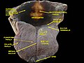
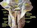
Muscles of the pharynx, viewed from behind, together with the associated vessels and nerves.
Details
Origin Medial pterygoid plate, pterygomandibular raphé, alveolar process
Insertion Pharyngeal raphe, pharyngeal tubercle
Artery Ascending pharyngeal artery and tonsillar branch of facial artery
Nerve Pharyngeal plexus of vagus nerve
Actions Swallowing
Identifiers
Latin musculus constrictor pharyngis superior
TA98 A05.3.01.103
TA2 2179
FMA 46621
Anatomical terms of muscle

The superior pharyngeal constrictor muscle is a quadrilateral muscle of the pharynx. It is the uppermost and thinnest of the three pharyngeal constrictors.[ citation needed ]

Contents

The muscle is divided into four parts according to its four distincts origins: a pterygopharyngeal, buccopharyngeal, mylopharyngeal, and a glossopharyngeal part. The muscle inserts onto the pharyngeal raphe, and pharyngeal spine. It is innervated by pharyngeal branch of the vagus nerve via the pharyngeal plexus. It acts to convey a bolus down towards the esophagus, facilitating swallowing.

Anatomy

The superior constrictor muscle is a quadrilateral, sheet-like muscle. It is thinner than the middle and inferior constrictor muscles. [1]

Origin

The sites of origin of the muscles collectively are the pterygoid hamulus (and occasionally the adjoining posterior margin of the medial pterygoid plate) anteriorly, (the posterior margin of) the pterygomandibular raphe, the posterior extremity of the mylohyoid line of mandible, and (negligibly) the side of the tongue. [1]

Four parts of the muscle are distinguished according to the origin:[ citation needed ]

Insertion

The muscle's fibres extend posterior-ward from its origin to form the midline pharyngeal raphe which then attaches onto the pharyngeal tubercle (of the basilar part of the occipital bone). [1]

Innervation

The superior pharyngeal constrictor muscle receives motor innervation from the pharyngeal plexus of vagus nerve. [1]

Vasculature

Arterial supply is provided primarily by the pharyngeal branch of the ascending pharyngeal artery, and the tonsilar branch of the facial artery. [1]

Relations

The interval between the upper border of the muscle and the base of the skull is closed by the pharyngeal aponeurosis, and is known as the sinus of Morgagni. [2]

There is an interval between superior pharyngeal constrictor and middle pharyngeal constrictor, this space contains glossopharyngeal nerve, lingual nerve and the stylopharyngeus muscle. [2]

The superior fibers arch beneath the levator veli palatini muscle and the Eustachian tube.[ citation needed ]

Actions/movements

Constriction of the muscle constricts the superior portion of the pharynx. [1]

Function

As soon as the bolus of food is received in the pharynx, the elevator muscles relax, the pharynx descends, and the constrictors contract upon the bolus, conveying it downward into the esophagus.[ citation needed ]

Additional images

Related Research Articles

Swallowing, also called deglutition or inglutition in scientific contexts, is the process in the body of a human or other animal that allows for a substance to pass from the mouth, to the pharynx, and into the esophagus, while shutting the epiglottis. Swallowing is an important part of eating and drinking. If the process fails and the material goes through the trachea, then choking or pulmonary aspiration can occur. In the human body the automatic temporary closing of the epiglottis is controlled by the swallowing reflex.

Articles related to anatomy include:

<span class="mw-page-title-main">Glossopharyngeal nerve</span> Cranial nerve IX, for the tongue and pharynx

The glossopharyngeal nerve, also known as the ninth cranial nerve, cranial nerve IX, or simply CN IX, is a cranial nerve that exits the brainstem from the sides of the upper medulla, just anterior to the vagus nerve. Being a mixed nerve (sensorimotor), it carries afferent sensory and efferent motor information. The motor division of the glossopharyngeal nerve is derived from the basal plate of the embryonic medulla oblongata, whereas the sensory division originates from the cranial neural crest.

<span class="mw-page-title-main">External carotid artery</span> Major artery of the head and neck

The external carotid artery is a major artery of the head and neck. It arises from the common carotid artery when it splits into the external and internal carotid artery. The external carotid artery supplies blood to the face, brain and neck.

<span class="mw-page-title-main">Styloglossus</span>

The styloglossus muscle is a bilaterally paired muscle of the tongue. It originates at the styloid process of the temporal bone. It inserts onto the side of the tongue. It acts to elevate and retract the tongue. It is innervated by the hypoglossal nerve.

<span class="mw-page-title-main">Middle pharyngeal constrictor muscle</span> Muscle in the neck

The middle pharyngeal constrictor is a fan-shaped muscle located in the neck. It is one of three pharyngeal constrictor muscles. It is smaller than the inferior pharyngeal constrictor muscle.

<span class="mw-page-title-main">Inferior pharyngeal constrictor muscle</span> Skeletal muscle of the pharynx

The inferior pharyngeal constrictor muscle is a skeletal muscle of the neck. It is the thickest of the three outer pharyngeal muscles. It arises from the sides of the cricoid cartilage and the thyroid cartilage. It is supplied by the vagus nerve. It is active during swallowing, and partially during breathing and speech. It may be affected by Zenker's diverticulum.

<span class="mw-page-title-main">Stylopharyngeus muscle</span> Muscle

The stylopharyngeus muscle is a muscle in the head. It originates from the temporal styloid process. Some of its fibres insert onto the thyroid cartilage, while others end by intermingling with proximal structures. It is innervated by the glossopharyngeal nerve. It acts to elevate the larynx and pharynx, and dilate the pharynx, thus facilitating swallowing.

<span class="mw-page-title-main">Mylohyoid line</span> Bony ridge on the inner surface of the body of the mandible

The mylohyoid line is a bony ridge on the internal surface of the mandible. It runs posterosuperiorly. It is the site of origin of the mylohyoid muscle, the superior pharyngeal constrictor muscle, and the pterygomandibular raphe.

<span class="mw-page-title-main">Superior laryngeal nerve</span> Branch of the vagus nerve

The superior laryngeal nerve is a branch of the vagus nerve. It arises from the middle of the inferior ganglion of vagus nerve and additionally also receives a sympathetic branch from the superior cervical ganglion.

<span class="mw-page-title-main">Deep cervical fascia</span>

The deep cervical fascia lies under cover of the platysma, and invests the muscles of the neck; it also forms sheaths for the carotid vessels, and for the structures situated in front of the vertebral column. Its attachment to the hyoid bone prevents the formation of a dewlap.

<span class="mw-page-title-main">Pharyngeal muscles</span>

The pharyngeal muscles are a group of muscles that form the pharynx, which is posterior to the oral cavity, determining the shape of its lumen, and affecting its sound properties as the primary resonating cavity.

<span class="mw-page-title-main">Carotid triangle</span>

The carotid triangle is a portion of the anterior triangle of the neck.

<span class="mw-page-title-main">Pterygomandibular raphe</span> Ligament formed from the buccopharyngeal fascia

The pterygomandibular raphe is a thin tendinous band of buccopharyngeal fascia. It is attached superiorly to the pterygoid hamulus of the medial pterygoid plate, and inferiorly to the posterior end of the mylohyoid line of the mandible. It gives attachment to the buccinator muscle, and the superior pharyngeal constrictor muscle (behind).

<span class="mw-page-title-main">Pharyngeal plexus of vagus nerve</span> Nerve fibers innervating most of the palate and pharynx

The pharyngeal plexus is a nerve plexus located upon the outer surface of the pharynx. It contains a motor component, a sensory component, and sympathetic component.

<span class="mw-page-title-main">Buccopharyngeal fascia</span>

The buccopharyngeal fascia is a fascia of the pharynx. It represents the posterior portion of the pretracheal fascia. It covers the superior pharyngeal constrictor muscles, and buccinator muscle.

The pharyngobasilar fascia is a fascia of the pharynx. It is situated between the mucous and muscular layers of the pharynx. It is formed as a thickening of the pharyngeal mucosa superior to the superior pharyngeal constrictor muscle. It attaches to the basilar part of occipital bone, the petrous part of the temporal bone, the medial pterygoid plate, and the pterygomandibular raphe. It diminishes in thickness inferiorly. Posteriorly, it is reinforced by the pharyngeal raphe. It reinforces the pharyngeal wall where muscle is deficient.

<span class="mw-page-title-main">Outline of human anatomy</span> Overview of and topical guide to human anatomy

The following outline is provided as an overview of and topical guide to human anatomy:

<span class="mw-page-title-main">Pharynx</span> Part of the throat that is behind the mouth and nasal cavity

The pharynx is the part of the throat behind the mouth and nasal cavity, and above the esophagus and trachea. It is found in vertebrates and invertebrates, though its structure varies across species. The pharynx carries food to the esophagus and air to the larynx. The flap of cartilage called the epiglottis stops food from entering the larynx.

<span class="mw-page-title-main">Parapharyngeal space</span>

The parapharyngeal space, is a potential space in the head and the neck. It has clinical importance in otolaryngology due to parapharyngeal space tumours and parapharyngeal abscess developing in this area. It is also a key anatomic landmark for localizing disease processes in the surrounding spaces of the neck; the direction of its displacement indirectly reflects the site of origin for masses or infection in adjacent areas, and consequently their appropriate differential diagnosis.

References

PD-icon.svgThis article incorporates text in the public domain from page 1143 of the 20th edition of Gray's Anatomy (1918)

  1. 1 2 3 4 5 6 Standring, Susan (2020). Gray's Anatomy: The Anatomical Basis of Clinical Practice (42th ed.). New York. p. 712. ISBN   978-0-7020-7707-4. OCLC   1201341621.{{cite book}}: CS1 maint: location missing publisher (link)
  2. 1 2 McMinn, R., 1994. Last's anatomy: Regional and applied (9th ed).