Related Research Articles
Swallowing, sometimes called deglutition in scientific contexts, is the process in the human or animal body that allows for a substance to pass from the mouth, to the pharynx, and into the esophagus, while shutting the epiglottis. Swallowing is an important part of eating and drinking. If the process fails and the material goes through the trachea, then choking or pulmonary aspiration can occur. In the human body the automatic temporary closing of the epiglottis is controlled by the swallowing reflex.
Articles related to anatomy include:

In anatomy, the Eustachian tube, also known as the auditory tube or pharyngotympanic tube, is a tube that links the nasopharynx to the middle ear, of which it is also a part. In adult humans, the Eustachian tube is approximately 35 mm (1.4 in) long and 3 mm (0.12 in) in diameter. It is named after the sixteenth-century Italian anatomist Bartolomeo Eustachi.

The glossopharyngeal nerve, also known as the ninth cranial nerve, cranial nerve IX, or simply CN IX, is a cranial nerve that exits the brainstem from the sides of the upper medulla, just anterior to the vagus nerve. Being a mixed nerve (sensorimotor), it carries afferent sensory and efferent motor information. The motor division of the glossopharyngeal nerve is derived from the basal plate of the embryonic medulla oblongata, whereas the sensory division originates from the cranial neural crest.

The soft palate is, in mammals, the soft tissue constituting the back of the roof of the mouth. The soft palate is part of the palate of the mouth; the other part is the hard palate. The soft palate is distinguished from the hard palate at the front of the mouth in that it does not contain bone.

The facial artery is a branch of the external carotid artery that supplies structures of the superficial face.

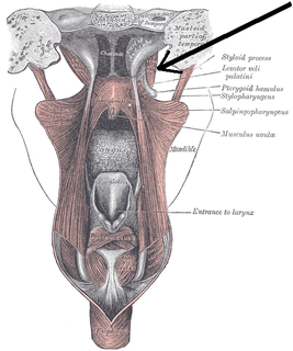
The levator veli palatini is the elevator muscle of the soft palate in the human body. It is supplied via the pharyngeal plexus. During swallowing, it contracts, elevating the soft palate to help prevent food from entering the nasopharynx.

The palatopharyngeusmuscle is a small muscle in the roof of the mouth.

The salpingopharyngeus muscle is a muscle of the pharynx. It arises from cartilage around the Eustachian tube, and inserts into the palatopharyngeus muscle by blending with its posterior fasciculus. It raises the pharynx and larynx during deglutition (swallowing) and laterally draws the pharyngeal walls up. It opens the pharyngeal orifice of the Eustachian tube during swallowing allowing for the equalization of pressure between it and the pharynx.

The superior pharyngeal constrictor muscle is a muscle in the pharynx. It is the highest located muscle of the three pharyngeal constrictors. The muscle is a quadrilateral muscle, thinner and paler than the inferior pharyngeal constrictor muscle and middle pharyngeal constrictor muscle.
The tensor veli palatini muscle is a broad, thin, ribbon-like muscle in the head that tenses the soft palate.
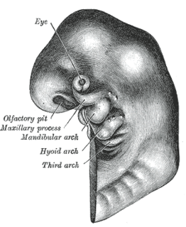
The pharyngeal arches, also known as visceral arches, are structures seen in the embryonic development of vertebrates that are recognisable precursors for many structures. In fish, the arches are known as the branchial arches, or gill arches.

The pterygoid processes of the sphenoid, one on either side, descend perpendicularly from the regions where the body and the greater wings of the sphenoid bone unite.
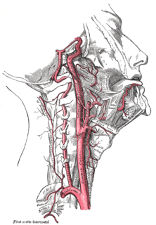
The ascending palatine artery is an artery in the head that branches off the facial artery and runs up the superior pharyngeal constrictor muscle.

The base of the cartilaginous portion of the Eustachian tube lies directly under the mucous membrane of the nasal part of the pharynx, where it forms an elevation or cushion, the torus tubarius or torus of the auditory tube, behind the pharyngeal orifice of the tube. The torus tubarius is very close to the tubal tonsil, which is sometimes also called the tonsil of (the) torus tubarius.
The pharyngeal branch of the vagus nerve, the principal motor nerve of the pharynx, arises from the upper part of the ganglion nodosum, and consists principally of filaments from the cranial portion of the accessory nerve.

The following outline is provided as an overview of and topical guide to human anatomy:

The pharynx is the part of the throat behind the mouth and nasal cavity, and above the oesophagus and trachea. It is found in vertebrates and invertebrates, though its structure varies across species. The pharynx carries food and air to the esophagus and larynx respectively. The flap of cartilage called the epiglottis stops food from entering the larynx.

The parapharyngeal space, is a potential space in the head and the neck. It has clinical importance in otolaryngology due to parapharyngeal space tumours and parapharyngeal abscess developing in this area. It is also a key anatomic landmark for localizing disease processes in the surrounding spaces of the neck; the direction of its displacement indirectly reflects the site of origin for masses or infection in adjacent areas, and consequently their appropriate differential diagnosis.
Guttural pouches are large, auditory-tube diverticula that contain between 300 and 600 ml of air. They are present in odd-toed mammals, some bats, hyraxes, and the American forest mouse. They are paired bilaterally just below the ears, behind the skull and connect to the nasopharynx.
References
- ↑ Gray's Anatomy 1918, Chapter: The Pharynx Archived 2012-01-21 at the Wayback Machine
| | This anatomy article is a stub. You can help Wikipedia by expanding it. |