Soft palate

Last updated
Soft palate
Tonsils diagram.jpg
Blausen 0872 UpperRespiratorySystem.png
Upper respiratory system, with soft palate labeled near center.
Details
Artery Lesser palatine arteries, ascending palatine artery
Nerve Pharyngeal branch of vagus nerve, medial pterygoid nerve, lesser palatine nerves, glossopharyngeal nerve [1]
Identifiers
Latin palatum molle, velum palatinum
MeSH D010160
TA98 A05.1.01.104
A05.2.01.003
TA2 2780
FMA 55021
Anatomical terminology

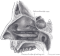
The soft palate (also known as the velum, palatal velum, or muscular palate) is, in mammals, the soft tissue constituting the back of the roof of the mouth. The soft palate is part of the palate of the mouth; the other part is the hard palate. The soft palate is distinguished from the hard palate at the front of the mouth in that it does not contain bone.

Contents

Structure

Muscles

Dissection of the muscles of the palate from behind. Gray1028.png
Dissection of the muscles of the palate from behind.

The five muscles of the soft palate play important roles in swallowing and breathing. The muscles are:

  1. Tensor veli palatini, which is involved in swallowing
  2. Palatoglossus, involved in swallowing
  3. Palatopharyngeus, involved in breathing
  4. Levator veli palatini, involved in swallowing
  5. Musculus uvulae, which moves the uvula

These muscles are innervated by the pharyngeal plexus via the vagus nerve, with the exception of the tensor veli palatini. The tensor veli palatini is innervated by the mandibular division of the trigeminal nerve (V3). [2]

Function

The soft palate is moveable, consisting of muscle fibers sheathed in mucous membrane. It is responsible for closing off the nasal passages during the act of swallowing, and also for closing off the airway. During sneezing, it protects the nasal passage by diverting a portion of the excreted substance to the mouth.

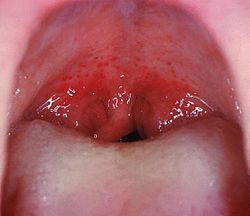
In humans, the uvula hangs from the end of the soft palate. Touching the uvula or the end of the soft palate evokes a strong gag reflex in most people.

Speech

A speech sound made with the middle part of the tongue (dorsum) touching the soft palate is known as a velar consonant.

It is possible for the soft palate to retract and elevate during speech to separate the oral cavity (mouth) from the nasal cavity in order to produce the oral speech sounds. If this separation is incomplete, air escapes through the nose, causing speech to be perceived as nasal.

Modelling

Within the microstructure of the soft palate lie a variety of variably-oriented fibers that create a nonuniform surface with a nonuniform density distribution. The tissue has been characterized as viscoelastic, nonlinear, and anisotropic in the direction of the fibers. Young modulus values range from 585 Pa at the posterior free edge of the soft palate to 1409 Pa where the soft palate attaches to the maxilla. [3] These properties are useful when quantifying the effects of corrective orthopedic devices such as the Hotz Plate on cleft lip.

Quantitative analyses have been done on bilateral and unilateral cleft palate to better understand geometric differences in cleft palate throughout the course of its development and correction. [4] Despite the difficulty in finding common, comparable landmarks between normal soft palates and cleft palates, analytical methods have been devised to assess differences in degree of curvature of the alveolar crest, two-dimensional and three-dimensional surface area, and slope of the alveolar crest.

Finite element analysis has demonstrated effective modeling of soft-palate extension and movement. It has also been an effective tool for evaluating the craniofacial effects of corrective orthopedic devices and cleft lip.

Clinical significance

Disease

Pathology of the soft palate includes mucosal lesions such as pemphigus vulgaris dsg - 3, herpangina and migratory stomatitis, [5] and muscular conditions such as the congenital cleft palate and cleft uvula.

Palatal petechiae. Streptococcal pharyngitis.jpg
Palatal petechiae.

Petechiae on the soft palate are mainly associated with streptococcal pharyngitis, [6] and as such it is an uncommon but highly specific finding. [7] 10 to 30 percent of palatal petechiae cases are estimated to be caused by suction, which can be habitual or secondary to fellatio. [8]

Additional images

See also

Related Research Articles

<span class="mw-page-title-main">Uvula</span> Fleshy appendage that hangs from the back of the palate

The uvula, also known as the palatine uvula, is a conic projection from the back edge of the middle of the soft palate, composed of connective tissue containing a number of racemose glands, and some muscular fibers. It also contains many serous glands, which produce thin saliva. It is only found in humans.

<span class="mw-page-title-main">Palate</span> Roof of the mouth

The palate is the roof of the mouth in humans and other mammals. It separates the oral cavity from the nasal cavity. A similar structure is found in crocodilians, but in most other tetrapods, the oral and nasal cavities are not truly separated. The palate is divided into two parts, the anterior, bony hard palate and the posterior, fleshy soft palate.

Swallowing, sometimes called deglutition in scientific contexts, is the process in the human or animal body that allows for a substance to pass from the mouth, to the pharynx, and into the esophagus, while shutting the epiglottis. Swallowing is an important part of eating and drinking. If the process fails and the material goes through the trachea, then choking or pulmonary aspiration can occur. In the human body the automatic temporary closing of the epiglottis is controlled by the swallowing reflex.

<span class="mw-page-title-main">Eustachian tube</span> Tube connecting middle ear to throat

In anatomy, the Eustachian tube, also called the auditory tube or pharyngotympanic tube, is a tube that links the nasopharynx to the middle ear, of which it is also a part. In adult humans, the Eustachian tube is approximately 35 mm (1.4 in) long and 3 mm (0.12 in) in diameter. It is named after the sixteenth-century Italian anatomist Bartolomeo Eustachi.

<span class="mw-page-title-main">Cleft lip and cleft palate</span> Medical condition

A cleft lip contains an opening in the upper lip that may extend into the nose. The opening may be on one side, both sides, or in the middle. A cleft palate occurs when the palate contains an opening into the nose. The term orofacial cleft refers to either condition or to both occurring together. These disorders can result in feeding problems, speech problems, hearing problems, and frequent ear infections. Less than half the time the condition is associated with other disorders.

<span class="mw-page-title-main">Nucleus ambiguus</span>

The nucleus ambiguus is a group of large motor neurons, situated deep in the medullary reticular formation named by Jacob Clarke. The nucleus ambiguus contains the cell bodies of neurons that innervate the muscles of the soft palate, pharynx, and larynx which are associated with speech and swallowing. As well as motor neurons, the nucleus ambiguus contains preganglionic parasympathetic neurons which innervate postganglionic parasympathetic neurons in the heart.

<span class="mw-page-title-main">Palatoglossus muscle</span> Muscle of the soft palate

The palatoglossal muscle is a muscle of the soft palate and an extrinsic muscle of the tongue. Its surface is covered by oral mucosa and forms the visible palatoglossal arch.

<span class="mw-page-title-main">Levator veli palatini</span> Muscle of the soft palate

The levator veli palatini is a muscle of the soft palate and pharynx. It is innervated by the vagus nerve via its pharyngeal plexus. During swallowing, it contracts, elevating the soft palate to help prevent food from entering the nasopharynx.

<span class="mw-page-title-main">Palatopharyngeus muscle</span>

The palatopharyngeusmuscle is a small muscle in the roof of the mouth.

<span class="mw-page-title-main">Tensor veli palatini muscle</span> Muscle of the soft palate

The tensor veli palatini muscle is a thin, triangular muscle of the head that tenses the soft palate and opens the Eustachian tube to equalise pressure in the middle ear.

<span class="mw-page-title-main">Pharyngeal arch</span> Embryonic precursor structures in vertebrates

The pharyngeal arches, also known as visceral arches, are structures seen in the embryonic development of vertebrates that are recognisable precursors for many structures. In fish, the arches are known as the branchial arches, or gill arches.

<span class="mw-page-title-main">Musculus uvulae</span> Human muscle

The musculus uvulae is a bilaterally muscle of the soft palate that acts to shorten the uvula when both muscles contract. It forms most of the mass of the uvula. It is innervated by the pharyngeal plexus of vagus nerve.

<span class="mw-page-title-main">Palatal obturator</span>

A palatal obturator is a prosthesis that totally occludes an opening such as an oronasal fistula. They are similar to dental retainers, but without the front wire. Palatal obturators are typically short-term prosthetics used to close defects of the hard/soft palate that may affect speech production or cause nasal regurgitation during feeding. Following surgery, there may remain a residual orinasal opening on the palate, alveolar ridge, or vestibule of the larynx. A palatal obturator may be used to compensate for hypernasality and to aid in speech therapy targeting correction of compensatory articulation caused by the cleft palate. In simpler terms, a palatal obturator covers any fistulas in the roof of the mouth that lead to the nasal cavity, providing the wearer with a plastic/acrylic, removable roof of the mouth, which aids in speech, eating, and proper air flow.

Velopharyngeal insufficiency is a disorder of structure that causes a failure of the velum to close against the posterior pharyngeal wall during speech in order to close off the nose during oral speech production. This is important because speech requires sound and airflow to be directed into the oral cavity (mouth) for the production of all speech sound with the exception of nasal sounds. If complete closure does not occur during speech, this can cause hypernasality and/or audible nasal emission during speech. In addition, there may be inadequate airflow to produce most consonants, making them sound weak or omitted.

Oral and maxillofacial pathology refers to the diseases of the mouth, jaws and related structures such as salivary glands, temporomandibular joints, facial muscles and perioral skin. The mouth is an important organ with many different functions. It is also prone to a variety of medical and dental disorders.

<span class="mw-page-title-main">Pharynx</span> Part of the throat that is behind the mouth and nasal cavity

The pharynx is the part of the throat behind the mouth and nasal cavity, and above the esophagus and trachea. It is found in vertebrates and invertebrates, though its structure varies across species. The pharynx carries food to the esophagus and air to the larynx. The flap of cartilage called the epiglottis stops food from entering the larynx.

Orofacial Myofunctional Disorders (OMD) are muscle disorders of the face, mouth, lips, or jaw due to chronic mouth breathing.

<span class="mw-page-title-main">Mouth assessment</span> Part of patient assessment performed on the mouth

A mouth assessment is performed as part of a patient's health assessment. The mouth is the beginning of the digestive system and a substantial part of the respiratory tract. Before an assessment of the mouth, patient is sometimes advised to remove any dentures. The assessment begins with a dental-health questionnaire, including questions about toothache, hoarseness, dysphagia(difficulty swallowing), altered taste or a frequent sore throat, current and previous tobacco use and alcohol consumption and any sores, lesions or bleeding of the gums.

<span class="mw-page-title-main">Hypernasal speech</span> Medical condition

Hypernasal speech is a disorder that causes abnormal resonance in a human's voice due to increased airflow through the nose during speech. It is caused by an open nasal cavity resulting from an incomplete closure of the soft palate and/or velopharyngeal sphincter. In normal speech, nasality is referred to as nasalization and is a linguistic category that can apply to vowels or consonants in a specific language. The primary underlying physical variable determining the degree of nasality in normal speech is the opening and closing of a velopharyngeal passage way between the oral vocal tract and the nasal vocal tract. In the normal vocal tract anatomy, this opening is controlled by lowering and raising the velum or soft palate, to open or close, respectively, the velopharyngeal passageway.

<span class="mw-page-title-main">Human mouth</span> Part of human anatomy

In human anatomy, the mouth is the first portion of the alimentary canal that receives food and produces saliva. The oral mucosa is the mucous membrane epithelium lining the inside of the mouth.

References

  1. Walker, H. Kenneth (1990). "Cranial Nerves IX and X: The Glossopharyngeal and Vagus Nerves". In Walker, H. Kenneth; Hall, W. Dallas; Hurst, J. Willis (eds.). Clinical Methods: The History, Physical, and Laboratory Examinations (3rd ed.). Butterworths. pp. 327–328. ISBN   9780409900774. Archived from the original on 2018-05-04. Retrieved 2017-09-12.
  2. Drake, Richard L.; Vogl, Wayne; Tibbitts, Adam W.M. Mitchell; illustrations by Richard; Richardson, Paul (2005). Gray's anatomy for students. Philadelphia: Elsevier/Churchill Livingstone. p. 1000. ISBN   978-0-8089-2306-0.
  3. Birch, M. J.; Srodon, P. D. (2009). "Biomechanical Properties of the Human Soft Palate". The Cleft Palate-Craniofacial Journal. 46 (3): 268–74. doi:10.1597/08-012.1. PMID   19642755. S2CID   24414466.
  4. Berkowitz, S; Krischer, J; Pruzansky, S (1974). "Quantitative analysis of cleft palate casts. A geometric study". The Cleft Palate Journal. 11: 134–61. PMID   4524356.
  5. Zadik, Yehuda; Drucker, Scott; Pallmon, Sarit (2011). "Migratory stomatitis (ectopic geographic tongue) on the floor of the mouth". Journal of the American Academy of Dermatology. 65 (2): 459–60. doi:10.1016/j.jaad.2010.04.016. PMID   21763590.
  6. Fact Sheet: Tonsillitis Archived 2011-11-26 at the Wayback Machine from American Academy of Otolaryngology. "Updated 1/11". Retrieved November 2011
  7. Brook, I; Dohar, J. E. (2006). "Management of group a beta-hemolytic streptococcal pharyngotonsillitis in children". The Journal of Family Practice. 55 (12): S1–11, quiz S12. PMID   17137534.
  8. Page 134 Archived 2018-05-04 at the Wayback Machine in: Michael Glick; Greenberg, Martin Harry; Burket, Lester W. (2003). Burket's oral medicine: diagnosis & treatment. Hamilton, Ont: BC Decker. ISBN   978-1-55009-186-1.