Gums

Last updated
Gums
Blausen 0863 ToothAnatomy 02.png
Cross-section of a tooth with gums labeled
Details
Identifiers
Latin gingiva
MeSH D005881
TA98 A05.1.01.108
A03.1.03.003
A03.1.03.004
TA2 2790
FMA 59762
Anatomical terminology

The gums or gingiva (PL: gingivae) consist of the mucosal tissue that lies over the mandible and maxilla inside the mouth. Gum health and disease can have an effect on general health. [1]

Contents

Structure

The gums are part of the soft tissue lining of the mouth. They surround the teeth and provide a seal around them. Unlike the soft tissue linings of the lips and cheeks, most of the gums are tightly bound to the underlying bone which helps resist the friction of food passing over them. Thus when healthy, it presents an effective barrier to the barrage of periodontal insults to deeper tissue. Healthy gums are usually coral pink in light skinned people, and may be naturally darker with melanin pigmentation.

Changes in color, particularly increased redness, together with swelling and an increased tendency to bleed, suggest an inflammation that is possibly due to the accumulation of bacterial plaque. Overall, the clinical appearance of the tissue reflects the underlying histology, both in health and disease. When gum tissue is not healthy, it can provide a gateway for periodontal disease to advance into the deeper tissue of the periodontium, leading to a poorer prognosis for long-term retention of the teeth. Both the type of periodontal therapy and homecare instructions given to patients by dental professionals and restorative care are based on the clinical conditions of the tissue. [2]

A diagram of the periodontium. The crown of the tooth is covered by enamel (A). Dentin (B). The root of the tooth is covered by cementum. C, alveolar bone. D, subepithelial connective tissue. E, oral epithelium. F, free gingival margin. G, gingival sulcus. H, principal gingival fibers. I, alveolar crest fibers of the periodontal ligament (PDL). J, horizontal fibers of the PDL. K, oblique fibers of the PDL. Periodontium.svg
A diagram of the periodontium. The crown of the tooth is covered by enamel (A). Dentin (B). The root of the tooth is covered by cementum. C, alveolar bone. D, subepithelial connective tissue. E, oral epithelium. F, free gingival margin. G, gingival sulcus. H, principal gingival fibers. I, alveolar crest fibers of the periodontal ligament (PDL). J, horizontal fibers of the PDL. K, oblique fibers of the PDL.

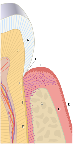
The gums are divided anatomically into marginal, attached and interdental areas.

Marginal gums

The marginal gum is the edge of the gums surrounding the teeth in collar-like fashion. In about half of individuals, it is demarcated from the adjacent, attached gums by a shallow linear depression, the free gingival groove. This slight depression on the outer surface of the gum does not correspond to the depth of the gingival sulcus but instead to the apical border of the junctional epithelium. This outer groove varies in depth according to the area of the oral cavity. The groove is very prominent on mandibular anteriors and premolars.

The marginal gum varies in width from 0.5 to 2.0 mm from the free gingival crest to the attached gingiva. The marginal gingiva follows the scalloped pattern established by the contour of the cementoenamel junction (CEJ) of the teeth. The marginal gingiva has a more translucent appearance than the attached gingiva, yet has a similar clinical appearance, including pinkness, dullness, and firmness. In contrast, the marginal gingiva lacks the presence of stippling, and the tissue is mobile or free from the underlying tooth surface, as can be demonstrated with a periodontal probe. The marginal gingiva is stabilized by the gingival fibers that have no bony support. The gingival margin, or free gingival crest, at the most superficial part of the marginal gingiva, is also easily seen clinically, and its location should be recorded on a patient's chart. [2]

Attached gum

The attached gums are continuous with the marginal gum. It is firm, resilient, and tightly bound to the underlying periosteum of alveolar bone. The facial aspect of the attached gum extends to the relatively loose and movable alveolar mucosa, from which it is demarcated by the mucogingival junction. Attached gum may present with surface stippling. The tissue when dried is dull, firm, and immobile, with varying amounts of stippling. The width of the attached gum varies according to its location. The width of the attached gum on the facial aspect differs in different areas of the mouth. It is generally greatest in the incisor region (3.5 to 4.5 mm in the maxilla and 3.3 to 3.9 mm in the mandible) and less in the posterior segments, with the least width in the first premolar area (1.9 mm in the maxilla and 1.8 mm in the mandible). However, certain levels of attached gum may be necessary for the stability of the underlying root of the tooth. [2]

Interdental gum

The interdental gum lies between the teeth. They occupy the gingival embrasure, which is the interproximal space beneath the area of tooth contact. The interdental papilla can be pyramidal or have a "col" shape. Attached gums are resistant to the forces of chewing and covered in keratin.

The col varies in depth and width, depending on the expanse of the contacting tooth surfaces. The epithelium covering the col consists of the marginal gum of the adjacent teeth, except that it is nonkeratinized. It is mainly present in the broad interdental gingiva of the posterior teeth, and generally is not present with those interproximal tissue associated with anterior teeth because the latter tissue is narrower. In the absence of contact between adjacent teeth, the attached gum extends uninterrupted from the facial to the lingual aspect. The col may be important in the formation of periodontal disease but is visible clinically only when teeth are extracted. [2]

Characteristics of healthy gums

Color

Gummy Smile.jpg
Natural "coral pink" gums without any pigmentation
Hyperpigmentation of the gum in a 22 year old non smoker female patient Hyperpigmentation of the gum.jpg
Hyperpigmentation of the gum in a 22 year old non smoker female patient

Healthy gums usually have a color that has been described as "coral pink". Other colours like red, white, and blue can signify inflammation (gingivitis) or pathology. Smoking or drug use can cause discoloring as well (such as "meth mouth"). Although described as the colour coral pink, variation in colour is possible. This can be the result of factors such as: thickness and degree of keratinization of the epithelium, blood flow to the gums, natural pigmentation of the skin, disease, and medications. [3]

Since the colour of the gums can vary, uniformity of colour is more important than the underlying color itself. Excess deposits of melanin can cause dark spots or patches on the gums (melanin gingival hyperpigmentation), especially at the base of the interdental papillae. Gum depigmentation (aka gum bleaching) is a procedure used in cosmetic dentistry to remove these discolorations.

Contour

Healthy gums have a smooth curved or scalloped appearance around each tooth. Healthy gums fill and fit each space between the teeth, unlike the swollen gum papilla seen in gingivitis or the empty interdental embrasure seen in periodontal disease. Healthy gums hold tight to each tooth in that the gum surface narrows to "knife-edge" thin at the free gingival margin. On the other hand, inflamed gums have a "puffy" or "rolled" margin.

Texture

Healthy gums have a firm texture that is resistant to movement, and the surface texture often exhibits surface stippling. Unhealthy gums, on the other hand, are often swollen and less firm. Healthy gums have an orange-peel like texture to it due to the stippling.

Reaction to disturbance

Healthy gums usually have no reaction to normal disturbance such as brushing or periodontal probing. Unhealthy gums, conversely, will show bleeding on probing (BOP) and/or purulent exudate.

Clinical significance

The gingival cavity microecosystem, fueled by food residues and saliva, can support the growth of many microorganisms, of which some can be injurious to health. Improper or insufficient oral hygiene can thus lead to many gum and periodontal disorders, including gingivitis or periodontitis, which are major causes for tooth failure. Recent studies have also shown that anabolic steroids are also closely associated with gingival enlargement requiring a gingivectomy for many cases. Gingival recession is when there is an apical movement of the gum margin away from the biting (occlusal) surface. [4] It may indicate an underlying inflammation such as periodontitis [5] or pyorrhea, [5] a pocket formation, dry mouth [5] or displacement of the marginal gums away from the tooth by mechanical (such as brushing), [5] chemical, or surgical means. [6] Gingival retraction, in turn, may expose the dental neck and leave it vulnerable to the action of external stimuli, and may cause root sensitivity. [5]

See also

Related Research Articles

<span class="mw-page-title-main">Periodontal disease</span> Medical condition

Periodontal disease, also known as gum disease, is a set of inflammatory conditions affecting the tissues surrounding the teeth. In its early stage, called gingivitis, the gums become swollen and red and may bleed. It is considered the main cause of tooth loss for adults worldwide. In its more serious form, called periodontitis, the gums can pull away from the tooth, bone can be lost, and the teeth may loosen or fall out. Bad breath may also occur.

<span class="mw-page-title-main">Necrotizing gingivitis</span> Non-contagious, painful bacterial infection of the gums

Necrotizing gingivitis (NG) is a common, non-contagious infection of the gums with sudden onset. The main features are painful, bleeding gums, and ulceration of inter-dental papillae. This disease, along with necrotizing periodontitis (NP) and necrotizing stomatitis, is classified as a necrotizing periodontal disease, one of the three general types of gum disease caused by inflammation of the gums (periodontitis).

Periodontology or periodontics is the specialty of dentistry that studies supporting structures of teeth, as well as diseases and conditions that affect them. The supporting tissues are known as the periodontium, which includes the gingiva (gums), alveolar bone, cementum, and the periodontal ligament. A periodontist is a dentist that specializes in the prevention, diagnosis and treatment of periodontal disease and in the placement of dental implants.

Dental plaque is a biofilm of microorganisms that grows on surfaces within the mouth. It is a sticky colorless deposit at first, but when it forms tartar, it is often brown or pale yellow. It is commonly found between the teeth, on the front of teeth, behind teeth, on chewing surfaces, along the gumline (supragingival), or below the gumline cervical margins (subgingival). Dental plaque is also known as microbial plaque, oral biofilm, dental biofilm, dental plaque biofilm or bacterial plaque biofilm. Bacterial plaque is one of the major causes for dental decay and gum disease.

<span class="mw-page-title-main">Veterinary dentistry</span> Branch of veterinary medicine

Veterinary dentistry is the field of dentistry applied to the care of animals. It is the art and science of prevention, diagnosis, and treatment of conditions, diseases, and disorders of the oral cavity, the maxillofacial region, and its associated structures as it relates to animals.

<span class="mw-page-title-main">Gingival recession</span>

Gingival recession, also known as gum recession and receding gums, is the exposure in the roots of the teeth caused by a loss of gum tissue and/or retraction of the gingival margin from the crown of the teeth. Gum recession is a common problem in adults over the age of 40, but it may also occur starting in adolescence, or around the age of 10. It may exist with or without concomitant decrease in crown-to-root ratio.

<span class="mw-page-title-main">Gingival sulcus</span> Space between tooth and gums

The gingival sulcus is an area of potential space between a tooth and the surrounding gingival tissue and is lined by sulcular epithelium. The depth of the sulcus is bounded by two entities: apically by the gingival fibers of the connective tissue attachment and coronally by the free gingival margin. A healthy sulcular depth is three millimeters or less, which is readily self-cleansable with a properly used toothbrush or the supplemental use of other oral hygiene aids.

<span class="mw-page-title-main">Gingival and periodontal pocket</span>

Gingival and periodontal pockets are dental terms indicating the presence of an abnormal depth of the gingival sulcus near the point at which the gingival tissue contacts the tooth.

Bleeding on probing (BoP) which is also known as bleeding gums or gingival bleeding is a term used by dentists and dental hygienists when referring to bleeding that is induced by gentle manipulation of the tissue at the depth of the gingival sulcus, or interface between the gingiva and a tooth. BoP is a sign of periodontal inflammation and indicates some sort of destruction and erosion to the lining of the sulcus or the ulceration of sulcular epithelium. The blood comes from lamina propria after the ulceration of the lining. BoP seems to be correlated with Periodontal Inflamed Surface Area (PISA).

<span class="mw-page-title-main">Scaling and root planing</span> Dental procedure

Scaling and root planing, also known as conventional periodontal therapy, non-surgical periodontal therapy or deep cleaning, is a procedure involving removal of dental plaque and calculus and then smoothing, or planing, of the (exposed) surfaces of the roots, removing cementum or dentine that is impregnated with calculus, toxins, or microorganisms, the agents that cause inflammation. It is a part of non-surgical periodontal therapy. This helps to establish a periodontium that is in remission of periodontal disease. Periodontal scalers and periodontal curettes are some of the tools involved.

A mucogingival junction is an anatomical feature found on the intraoral mucosa. The mucosa of the cheeks and floor of the mouth are freely moveable and fragile, whereas the mucosa around the teeth and on the palate are firm and keratinized. Where the two tissue types meet is known as a mucogingival junction.

The interdental papilla, also known as the interdental gingiva, is the part of the gums (gingiva) that exists coronal to the free gingival margin on the buccal and lingual surfaces of the teeth. The interdental papillae fill in the area between the teeth apical to their contact areas to prevent food impaction; they assume a conical shape for the anterior teeth and a blunted shape buccolingually for the posterior teeth.

<span class="mw-page-title-main">Periodontal scaler</span> Dental tool

Periodontal scalers are dental instruments used in the prophylactic and periodontal care of teeth, including scaling and root planing. The working ends come in a variety of shapes and sizes, but they are always narrow at the tip, so as to allow for access to narrow embrasure spaces between teeth. They differ from periodontal curettes, which possess a blunt tip.

<span class="mw-page-title-main">Gingivitis</span> Inflammation of the gums

Gingivitis is a non-destructive disease that causes inflammation of the gums. The most common form of gingivitis, and the most common form of periodontal disease overall, is in response to bacterial biofilms that is attached to tooth surfaces, termed plaque-induced gingivitis. Most forms of gingivitis are plaque-induced.

<span class="mw-page-title-main">Plasma cell gingivitis</span> Medical condition

Plasma cell gingivitis is a rare condition, appearing as generalized erythema (redness) and edema (swelling) of the attached gingiva, occasionally accompanied by cheilitis or glossitis. It is called plasma cell gingivitis where the gingiva (gums) are involved, plasma cell cheilitis, where the lips are involved, and other terms such as plasma cell orifacial mucositis, or plasma cell gingivostomatitis where several sites in the mouth are involved. On the lips, the condition appears as sharply outlined, infiltrated, dark red plaque with a lacquer-like glazing of the surface of the involved oral area.

In dentistry, numerous types of classification schemes have been developed to describe the teeth and gum tissue in a way that categorizes various defects. All of these classification schemes combine to provide the periodontal diagnosis of the aforementioned tissues in their various states of health and disease.

Clinical attachment loss (CAL) is the predominant clinical manifestation and determinant of periodontal disease.

Hereditary gingival fibromatosis (HGF), also known as idiopathic gingival hyperplasia, is a rare condition of gingival overgrowth. HGF is characterized as a benign, slowly progressive, nonhemorrhagic, fibrous enlargement of keratinized gingiva. It can cover teeth in various degrees, and can lead to aesthetic disfigurement. Fibrous enlargement is most common in areas of maxillary and mandibular tissues of both arches in the mouth. Phenotype and genotype frequency of HGF is 1:175,000 where males and females are equally affected but the cause is not entirely known. It mainly exists as an isolated abnormality but can also be associated with a multi-system syndrome.

Periodontal surgery is a form of dental surgery that prevents or corrects anatomical, traumatic, developmental, or plaque-induced defects in the bone, gingiva, or alveolar mucosa. The objectives of this surgery include accessibility of instruments to root surface, elimination of inflammation, creation of an oral environment for plaque control, periodontal diseases control, oral hygiene maintenance, maintain proper embrasure space, address gingiva-alveolar mucosa problems, and esthetic improvement. The surgical procedures include crown lengthening, frenectomy, and mucogingival flap surgery.

References

  1. Gum disease opens up the body to a host of infections April 6, 2016 Science News
  2. 1 2 3 4 Illustrated Dental Embryology, Histology, and Anatomy, Bath-Balogh and Fehrenbach, Elsevier, 2011, page 123
  3. Mosby's Medical Dictionary, 8th edition. 2009, Elsevier.
  4. Gingival Recession - Causes and treatment Archived 2010-09-17 at the Wayback Machine JADA, Vol 138. http://jada.ada.org. Oct 2007. American Dental Association
  5. 1 2 3 4 5 mexicodentaldirectory.com: dental sensitivity Retrieved on August 2010
  6. Mondofacto medical dictionary > gingival retraction Archived 2018-12-09 at the Wayback Machine 05 Mar 2000