
Human teeth function to mechanically break down items of food by cutting and crushing them in preparation for swallowing and digesting. As such, they are considered part of the human digestive system. Humans have four types of teeth: incisors, canines, premolars, and molars, which each have a specific function. The incisors cut the food, the canines tear the food and the molars and premolars crush the food. The roots of teeth are embedded in the maxilla or the mandible and are covered by gums. Teeth are made of multiple tissues of varying density and hardness.

In mammalian oral anatomy, the canine teeth, also called cuspids, dogteeth, eye teeth, vampire teeth, or fangs, are the relatively long, pointed teeth. In the context of the upper jaw, they are also known as fangs. They can appear more flattened, however, causing them to resemble incisors and leading them to be called incisiform. They developed and are used primarily for firmly holding food in order to tear it apart, and occasionally as weapons. They are often the largest teeth in a mammal's mouth. Individuals of most species that develop them normally have four, two in the upper jaw and two in the lower, separated within each jaw by incisors; humans and dogs are examples. In most species, canines are the anterior-most teeth in the maxillary bone. The four canines in humans are the two upper maxillary canines and the two lower mandibular canines. They are specially prominent in dogs (Canidae), hence the name.

Incisors are the front teeth present in most mammals. They are located in the premaxilla above and on the mandible below. Humans have a total of eight. Opossums have 18, whereas armadillos have none.
The premolars, also called premolar teeth, or bicuspids, are transitional teeth located between the canine and molar teeth. In humans, there are two premolars per quadrant in the permanent set of teeth, making eight premolars total in the mouth. They have at least two cusps. Premolars can be considered transitional teeth during chewing, or mastication. They have properties of both the canines, that lie anterior and molars that lie posterior, and so food can be transferred from the canines to the premolars and finally to the molars for grinding, instead of directly from the canines to the molars.

The maxillary central incisor is a human tooth in the front upper jaw, or maxilla, and is usually the most visible of all teeth in the mouth. It is located mesial to the maxillary lateral incisor. As with all incisors, their function is for shearing or cutting food during mastication (chewing). There is typically a single cusp on each tooth, called an incisal ridge or incisal edge. Formation of these teeth begins at 14 weeks in utero for the deciduous (baby) set and 3–4 months of age for the permanent set.

The maxillary lateral incisors are a pair of upper (maxillary) teeth that are located laterally from both maxillary central incisors of the mouth and medially from both maxillary canines. As with all incisors, their function is for shearing or cutting food during mastication, commonly known as chewing. There are generally no cusps on the teeth, but the rare condition known as talon cusps are most prevalent on the maxillary lateral incisors. The surface area of the tooth used in eating is called an incisal ridge or incisal edge. Though relatively the same, there are some minor differences between the deciduous (baby) maxillary lateral incisor and that of the permanent maxillary lateral incisor. The maxillary lateral incisors occlude in opposition to the mandibular lateral incisors.

The maxillary first premolar is one of two teeth located in the upper jaw, laterally from both the maxillary canines of the mouth but mesial from both maxillary second premolars. The function of this premolar is similar to that of canines in regard to tearing being the principal action during mastication, commonly known as chewing. There are two cusps on maxillary first premolars, and the buccal cusp is sharp enough to resemble the prehensile teeth found in carnivorous animals. There are no deciduous maxillary premolars. Around 10-11 years of age, the primary molars are shed and the permanent premolars erupt in their place. It takes about 3 years for the adult premolar and its root to fully calcify. Due to its long buccal root with narrow root canal and short palatal root with wide root canal, the upper 1st premolar is very prone to fracture during exodontia, hence, it is sometimes referred to some dentists as the "King of Fracture". In the universal system of notation, the permanent maxillary premolars are designated by a number. The right permanent maxillary first premolar is known as "5", and the left one is known as "12". In the Palmer notation, a number is used in conjunction with a symbol designating in which quadrant the tooth is found. For this tooth, the left and right first premolars would have the same number, "4", but the right one would have the symbol, "┘", underneath it, while the left one would have, "└". The international notation has a different numbering system than the previous two, and the right permanent maxillary first premolar is known as "14", and the left one is known as "24".
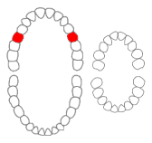
The maxillary second premolar is one of two teeth located in the upper maxilar, laterally from both the maxillary first premolars of the mouth but mesial from both maxillary first molars. The function of this premolar is similar to that of first molars in regard to grinding being the principal action during mastication, commonly known as chewing. There are two cusps on maxillary second premolars, but both of them are less sharp than those of the maxillary first premolars. There are no deciduous (baby) maxillary premolars. Instead, the teeth that precede the permanent maxillary premolars are the deciduous maxillary molars.

The maxillary first molar is the human tooth located laterally from both the maxillary second premolars of the mouth but mesial from both maxillary second molars.

The maxillary second molar is the tooth located distally from both the maxillary first molars of the mouth but mesial from both maxillary third molars. This is true only in permanent teeth. In deciduous (baby) teeth, the maxillary second molar is the last tooth in the mouth and does not have a third molar behind it. The function of this molar is similar to that of all molars in regard to grinding being the principal action during mastication, commonly known as chewing. There are usually four cusps on maxillary molars, two on the buccal and two palatal.

The mandibular central incisor is the tooth located on the jaw, adjacent to the midline of the face. It is mesial from both mandibular lateral incisors. As with all incisors, its function includes shearing or cutting food during mastication, commonly known as chewing. There are no cusps on the tooth. Instead, the surface area of the tooth used in eating is called an incisal ridge or incisal edge. Though the two are similar, there are some minor differences between the deciduous (baby) mandibular central incisor and that of the permanent mandibular central incisor. The mandibular central incisors are usually the first teeth to appear in the mouth, typically around the age of 6–8 months.

The mandibular lateral incisor is the tooth located distally from both mandibular central incisors of the mouth and mesially from both mandibular canines. As with all incisors, their function is for shearing or cutting food during mastication, commonly known as chewing. There are no cusps on the teeth. Instead, the surface area of the tooth used in eating is called an incisal ridge or incisal edge. Though relatively the same, there are some minor differences between the deciduous (baby) mandibular lateral incisor and that of the permanent mandibular lateral incisor.

The mandibular canine is the tooth located distally from both mandibular lateral incisors of the mouth but mesially from both mandibular first premolars. Both the maxillary and mandibular canines are called the "cornerstone" of the mouth because they are all located three teeth away from the midline, and separate the premolars from the incisors. The location of the canines reflect their dual function as they complement both the premolars and incisors during mastication, commonly known as chewing. Nonetheless, the most common action of the canines is tearing of food. The canine teeth are able to withstand the tremendous lateral pressures from chewing. There is a single cusp on canines, and they resemble the prehensile teeth found in carnivorous animals. Though relatively the same, there are some minor differences between the deciduous (baby) mandibular canine and that of the permanent mandibular canine.

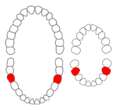
The mandibular first premolar is the tooth located laterally from both the mandibular canines of the mouth but mesial from both mandibular second premolars. The function of this premolar is similar to that of canines in regard to tearing being the principal action during mastication, commonly known as chewing. Mandibular first premolars have two cusps. The one large and sharp is located on the buccal side of the tooth. Since the lingual cusp is small and nonfunctional, the mandibular first premolar resembles a small canine. There are no deciduous (baby) mandibular premolars. Instead, the teeth that precede the permanent mandibular premolars are the deciduous mandibular molars.

The mandibular second premolar is the tooth located distally from both the mandibular first premolars of the mouth but mesial from both mandibular first molars. The function of this premolar is assist the mandibular first molar during mastication, commonly known as chewing. Mandibular second premolars have three cusps. There is one large cusp on the buccal side of the tooth. The lingual cusps are well developed and functional. Therefore, whereas the mandibular first premolar resembles a small canine, the mandibular second premolar is more alike to the first molar. There are no deciduous (baby) mandibular premolars. Instead, the teeth that precede the permanent mandibular premolars are the deciduous mandibular molars.
The mandibular first molar or six-year molar is the tooth located distally from both the mandibular second premolars of the mouth but mesial from both mandibular second molars. It is located on the mandibular (lower) arch of the mouth, and generally opposes the maxillary (upper) first molars and the maxillary 2nd premolar in normal class I occlusion. The function of this molar is similar to that of all molars in regard to grinding being the principal action during mastication, commonly known as chewing. There are usually five well-developed cusps on mandibular first molars: two on the buccal, two lingual, and one distal. The shape of the developmental and supplementary grooves, on the occlusal surface, are described as being M-shaped. There are great differences between the deciduous (baby) mandibular molars and those of the permanent mandibular molars, even though their function are similar. The permanent mandibular molars are not considered to have any teeth that precede it. Despite being named molars, the deciduous molars are followed by permanent premolars.

The mandibular second molar is the tooth located distally from both the mandibular first molars of the mouth but mesial from both mandibular third molars. This is true only in permanent teeth. The function of this molar is similar to that of all molars in regard to grinding being the principal action during mastication, commonly known as chewing. Though there is more variation between individuals than that of the first mandibular molar, there are usually four cusps on mandibular second molars: two on the buccal and two lingual. There are great differences between the deciduous (baby) mandibular molars and those of the permanent mandibular molars, even though their function is similar. The permanent mandibular molars are not considered to have any teeth that precede them. Despite being named molars, the deciduous molars are followed by permanent premolars.

FDI World Dental Federation notation is the world's most commonly used dental notation. It is designated by the International Organization for Standardization as standard ISO 3950 "Dentistry — Designation system for teeth and areas of the oral cavity".
Dental anatomy is a field of anatomy dedicated to the study of human tooth structures. The development, appearance, and classification of teeth fall within its purview. Tooth formation begins before birth, and the teeth's eventual morphology is dictated during this time. Dental anatomy is also a taxonomical science: it is concerned with the naming of teeth and the structures of which they are made, this information serving a practical purpose in dental treatment.
Serial extraction is the planned extraction of certain deciduous teeth and specific permanent teeth in an orderly sequence and predetermined pattern to guide the erupting permanent teeth into a more favorable position.
Brand, Richard W. and Isselhard, Donald E., 2003. "Anatomy of Orofacial Structures." 7th edition

