In mammalian oral anatomy, the canine teeth, also called cuspids, dog teeth, or fangs, eye teeth, vampire teeth, or vampire fangs, are the relatively long, pointed teeth. They can appear more flattened however, causing them to resemble incisors and leading them to be called incisiform. They developed and are used primarily for firmly holding food in order to tear it apart, and occasionally as weapons. They are often the largest teeth in a mammal's mouth. Individuals of most species that develop them normally have four, two in the upper jaw and two in the lower, separated within each jaw by incisors; humans and dogs are examples. In most species, canines are the anterior-most teeth in the maxillary bone.

The premolars, also called premolar teeth, or bicuspids, are transitional teeth located between the canine and molar teeth. In humans, there are two premolars per quadrant in the permanent set of teeth, making eight premolars total in the mouth. They have at least two cusps. Premolars can be considered transitional teeth during chewing, or mastication. They have properties of both the canines, that lay anterior and molars that lay posterior, and so food can be transferred from the canines to the premolars and finally to the molars for grinding, instead of directly from the canines to the molars.

The maxillary central incisor is a human tooth in the front upper jaw, or maxilla, and is usually the most visible of all teeth in the mouth. It is located mesial to the maxillary lateral incisor. As with all incisors, their function is for shearing or cutting food during mastication (chewing). There is typically a single cusp on each tooth, called an incisal ridge or incisal edge. Formation of these teeth begins at 14 weeks in utero for the deciduous (baby) set and 3–4 months of age for the permanent set.

The maxillary lateral incisors are a pair of upper (maxillary) teeth that are located laterally from both maxillary central incisors of the mouth and medially from both maxillary canines. As with all incisors, their function is for shearing or cutting food during mastication, commonly known as chewing. There are generally no cusps on the teeth, but the rare condition known as talon cusps are most prevalent on the maxillary lateral incisors. The surface area of the tooth used in eating is called an incisal ridge or incisal edge. Though relatively the same, there are some minor differences between the deciduous (baby) maxillary lateral incisor and that of the permanent maxillary lateral incisor. The maxillary lateral incisors occlude in opposition to the mandibular lateral incisors.

In human dentistry, the maxillary canine is the tooth located laterally from both maxillary lateral incisors of the mouth but mesial from both maxillary first premolars. Both the maxillary and mandibular canines are called the "cornerstone" of the mouth because they are all located three teeth away from the midline, and separate the premolars from the incisors. The location of the canines reflect their dual function as they complement both the premolars and incisors during mastication, commonly known as chewing. Nonetheless, the most common action of the canines is tearing of food. The canines often erupt in the upper gums several millimeters above the gum line. The canine teeth are able to withstand the tremendous lateral pressure caused by chewing. There is a single cusp on canines, and they resemble the prehensile teeth found in carnivorous animals such as the extinct Saber-toothed cat. Though relatively the same, there are some minor differences between the deciduous (baby) maxillary canine and that of the permanent maxillary canine.

The maxillary first premolar is one of two teeth located in the upper jaw, laterally from both the maxillary canines of the mouth but mesial from both maxillary second premolars. The function of this premolar is similar to that of canines in regard to tearing being the principal action during mastication, commonly known as chewing. There are two cusps on maxillary first premolars, and the buccal cusp is sharp enough to resemble the prehensile teeth found in carnivorous animals. There are no deciduous maxillary premolars. Around 10-11 years of age, the primary molars are shed and the permanent premolars erupt in their place. It takes about 3 years for the adult premolar and its root to fully calcify. Due to its long buccal root with narrow root canal and short palatal root with wide root canal, the upper 1st premolar is very prone to fracture during exodontia, hence, it is sometimes referred to some dentists as the "King of Fracture". In the universal system of notation, the permanent maxillary premolars are designated by a number. The right permanent maxillary first premolar is known as "5", and the left one is known as "12". In the Palmer notation, a number is used in conjunction with a symbol designating in which quadrant the tooth is found. For this tooth, the left and right first premolars would have the same number, "4", but the right one would have the symbol, "┘", underneath it, while the left one would have, "└". The international notation has a different numbering system than the previous two, and the right permanent maxillary first premolar is known as "14", and the left one is known as "24".
The maxillary second premolar is one of two teeth located in the upper jaw, laterally from both the maxillary first premolars of the mouth but mesial from both maxillary first molars. The function of this premolar is similar to that of first molars in regard to grinding being the principal action during mastication, commonly known as chewing. There are two cusps on maxillary second premolars, but both of them are less sharp than those of the maxillary first premolars. There are no deciduous (baby) maxillary premolars. Instead, the teeth that precede the permanent maxillary premolars are the deciduous maxillary molars.

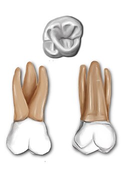
The maxillary second molar is the tooth located distally from both the maxillary first molars of the mouth but mesial from both maxillary third molars. This is true only in permanent teeth. In deciduous (baby) teeth, the maxillary second molar is the last tooth in the mouth and does not have a third molar behind it. The function of this molar is similar to that of all molars in regard to grinding being the principal action during mastication, commonly known as chewing. There are usually four cusps on maxillary molars, two on the buccal and two palatal.

The mandibular canine is the tooth located distally from both mandibular lateral incisors of the mouth but mesially from both mandibular first premolars. Both the maxillary and mandibular canines are called the "cornerstone" of the mouth because they are all located three teeth away from the midline, and separate the premolars from the incisors. The location of the canines reflect their dual function as they complement both the premolars and incisors during mastication, commonly known as chewing. Nonetheless, the most common action of the canines is tearing of food. The canine teeth are able to withstand the tremendous lateral pressures from chewing. There is a single cusp on canines, and they resemble the prehensile teeth found in carnivorous animals. Though relatively the same, there are some minor differences between the deciduous (baby) mandibular canine and that of the permanent mandibular canine.

The mandibular first premolar is the tooth located laterally from both the mandibular canines of the mouth but mesial from both mandibular second premolars. The function of this premolar is similar to that of canines in regard to tearing being the principal action during mastication, commonly known as chewing. Mandibular first premolars have two cusps. The one large and sharp is located on the buccal side of the tooth. Since the lingual cusp is small and nonfunctional, the mandibular first premolar resembles a small canine. There are no deciduous (baby) mandibular premolars. Instead, the teeth that precede the permanent mandibular premolars are the deciduous mandibular molars.

The mandibular second premolar is the tooth located distally from both the mandibular first premolars of the mouth but mesial from both mandibular first molars. The function of this premolar is assist the mandibular first molar during mastication, commonly known as chewing. Mandibular second premolars have three cusps. There is one large cusp on the buccal side of the tooth. The lingual cusps are well developed and functional. Therefore, whereas the mandibular first premolar resembles a small canine, the mandibular second premolar is more alike to the first molar. There are no deciduous (baby) mandibular premolars. Instead, the teeth that precede the permanent mandibular premolars are the deciduous mandibular molars.
The mandibular first molar or six-year molar is the tooth located distally from both the mandibular second premolars of the mouth but mesial from both mandibular second molars. It is located on the mandibular (lower) arch of the mouth, and generally opposes the maxillary (upper) first molars and the maxillary 2nd premolar in normal class I occlusion. The function of this molar is similar to that of all molars in regard to grinding being the principal action during mastication, commonly known as chewing. There are usually five well-developed cusps on mandibular first molars: two on the buccal, two lingual, and one distal. The shape of the developmental and supplementary grooves, on the occlusal surface, are describes as being 'M' shaped. There are great differences between the deciduous (baby) mandibular molars and those of the permanent mandibular molars, even though their function are similar. The permanent mandibular molars are not considered to have any teeth that precede it. Despite being named molars, the deciduous molars are followed by permanent premolars.

The mandibular second molar is the tooth located distally from both the mandibular first molars of the mouth but mesial from both mandibular third molars. This is true only in permanent teeth. The function of this molar is similar to that of all molars in regard to grinding being the principal action during mastication, commonly known as chewing. Though there is more variation between individuals to that of the first mandibular molar, there are usually four cusps on mandibular second molars: two on the buccal and two palatal. There are great differences between the deciduous (baby) mandibular molars and those of the permanent mandibular molars, even though their function are similar. The permanent mandibular molars are not considered to have any teeth that precede it. Despite being named molars, the deciduous molars are followed by permanent premolars.
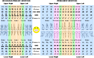
Dental professionals, in writing or speech, use several different dental notation systems for associating information with a specific tooth. The three most common systems are the ISO System, Universal Numbering System, and Palmer notation method. The ISO system is used worldwide, and the Universal is used widely in the United States. The ISO System can be easily adapted to computerized charting.
Dental anatomy is a field of anatomy dedicated to the study of human tooth structures. The development, appearance, and classification of teeth fall within its purview. Tooth formation begins before birth, and the teeth's eventual morphology is dictated during this time. Dental anatomy is also a taxonomical science: it is concerned with the naming of teeth and the structures of which they are made, this information serving a practical purpose in dental treatment.

A cusp is a pointed, projecting, or elevated feature. In animals, it is usually used to refer to raised points on the crowns of teeth. The concept is also used with regard to the valve between the right atrium and the right ventricle in the human heart. This valve is closed during ventricular contraction by the tricuspid valve, so named because it usually consists of three cusps or leaflets.
This is a list of definitions of commonly used terms of location and direction in dentistry. This set of terms provides orientation within the oral cavity, much as anatomical terms of location provide orientation throughout the body.
Dental pertains to the teeth, including dentistry. Topics related to the dentistry, the human mouth and teeth include:

The Lewis offset is a term for the portion of the central groove on a permanent mandibular first molar which lies between the two central pits. It was named for long time dental anatomy instructor Dr. Christopher S. Lewis, a Mercer Island, WA dentist.
Serial extraction is the planned extraction of certain deciduous teeth and specific permanent teeth in an orderly sequence and predetermined pattern to guide the erupting permanent teeth into a more favorable position.