Molar (tooth)

Last updated

Molar
Lower wisdom tooth.jpg
A lower wisdom tooth after extraction.
Gray997.png
Permanent teeth of right half of lower dental arch, seen from above: In this diagram, a healthy wisdom tooth (third, rearmost molar) is included
Details
Artery Posterior superior alveolar artery
Identifiers
Latin dentes molares
MeSH D008963
TA98 A05.1.03.007
TA2 910
FMA 55638
Anatomical terminology

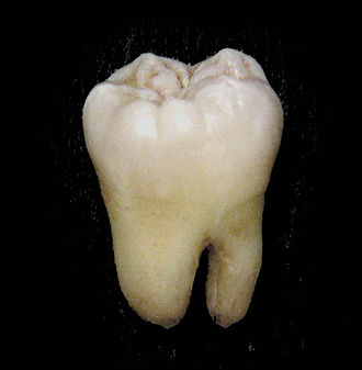
The molars or molar teeth are large, flat teeth at the back of the mouth. They are more developed in mammals. They are used primarily to grind food during chewing. The name molar derives from Latin, molaris dens, meaning "millstone tooth", from mola, millstone and dens, tooth. Molars show a great deal of diversity in size and shape across the mammal groups. The third molar of humans is sometimes vestigial.

Contents

Human anatomy

In humans, the molar teeth have either four or five cusps. Adult humans have 12 molars, in four groups of three at the back of the mouth. The third, rearmost molar in each group is called a wisdom tooth. It is the last tooth to appear, breaking through the front of the gum at about the age of 20, although this varies among individuals and populations, and in many cases the tooth is missing. [1]

The human mouth contains upper (maxillary) and lower (mandibular) molars. They are: maxillary first molar, maxillary second molar, maxillary third molar, mandibular first molar, mandibular second molar, and mandibular third molar.

Mammal evolution

In mammals, the crown of the molars and premolars is folded into a wide range of complex shapes. The basic elements of the crown are the more or less conical projections called cusps and the valleys that separate them. The cusps contain both dentine and enamel, whereas minor projections on the crown, called crenulations, are the result of different enamel thickness. Cusps are occasionally joined to form ridges and expanded to form crests. Cingula are often incomplete ridges that pass around the base of the crown. [2]

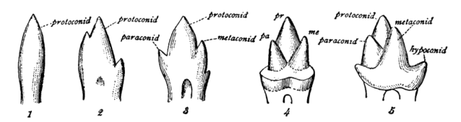
Mammalian, multicusped cheek teeth probably evolved from single-cusped teeth in synapsids, although the diversity of therapsid molar patterns and the complexity in the molars of the earliest mammals make determining how this happened impossible. According to the widely accepted "differentiation theory", additional cusps have arisen by budding or outgrowth from the crown, while the rivalling "concrescence theory" instead proposes that complex teeth evolved by the clustering of originally separate conical teeth. Therian mammals (placentals and marsupials) are generally agreed to have evolved from an ancestor with tribosphenic cheek teeth, with three main cusps arranged in a triangle. [2]

Comparison of cheek teeth in various taxa: 1, a single-cusped pelycosaur; 2, Dromatherium (a Triassic cynodont); 3, Microconodon (a Triassic eucynodont); 4, Spalacotherium (a Cretaceous "symmetrodont"); 5, Amphitherium (a Jurassic prototribosphenid mammal) Cambridge Natural History Mammalia Fig 039.png
Comparison of cheek teeth in various taxa: 1, a single-cusped pelycosaur; 2, Dromatherium (a Triassic cynodont); 3, Microconodon (a Triassic eucynodont); 4, Spalacotherium (a Cretaceous "symmetrodont"); 5, Amphitherium (a Jurassic prototribosphenid mammal)

Morphology

Image showing molar teeth and their typical arrangement in humans 3D Medical Animation Still Showing Types of Teeth.jpg
Image showing molar teeth and their typical arrangement in humans

Each major cusp on an upper molar is called a cone and is identified by a prefix dependent on its relative location on the tooth: proto-, para-, meta-, hypo-, and ento-. Suffixes are added to these names: -id is added to cusps on a lower molar (e.g., protoconid); -ule to a minor cusp (e.g., protoconulid). A shelf-like ridge left lower part of the crown (on an upper molar) is called a cingulum; the same feature on the lower molar a cingulid, and a minor cusp on these, for example, a cingular cuspule or conulid. [3]

Tribosphenic

A diagram of generalized tribosphenic molars with notable features labelled. Upper left molar in pink, lower left molar in blue. Tribosphenic molar diagram.png
A diagram of generalized tribosphenic molars with notable features labelled. Upper left molar in pink, lower left molar in blue.

The design that is considered one of the most important characteristics of therian mammals is called a tribosphenic molar. Among living mammals, the tribosphenic tooth is found in most insectivorous mammals as well as young platypuses, even though adults platypuses are toothless.

In tribosphenic teeth, the lower molar is divided into two regions: the three-cusped trigonid, or shearing end, and the talonid, or crushing heel. In modern tribosphenic molars, the trigonid is towards the front of the jaw and the talonid is towards the rear. The trigonid is defined by three large cusps: the protoconid is on the buccal/labial (cheek) side of the tooth, while the anterior paraconid and posterior metaconid are on the lingual (tongue) side.

Generalized tribosphenic left upper molar, showing the protocone, paracone, and metacone. Tribosphenic molar.svg
Generalized tribosphenic left upper molar, showing the protocone, paracone, and metacone.

Upper molars look like three-pointed mountain ranges, with their features mirrored from the lower molars. The protocone cusp is on the lingual side of the tooth, while the anterior paracone and posterior metacone are on the buccal side. The protocone of the upper molar and talonid basin of the lower molar mesh together as a crushing system similar to a mortar and pestle.

Tribosphenic molars were present in the direct ancestors of all three living mammal groups, but it was most likely not ancestral to mammals as a whole. Many paleontologists argue that it developed independently in monotremes (from australosphenidans), rather than being inherited from a common ancestor that they share with marsupials and placentals (from boreosphenidans); this idea still has some critics. [4] For example, the dentition of the Early Cretaceous monotreme Steropodon is similar to those of Peramus and dryolestoids, which suggests that monotremes are related to some pre-tribosphenic mammals, [5] but, on the other hand, the status of neither of these two groups is well-established.

Some Jurassic mammalia forms, such as docodonts and shuotheriids, have "reversed tribosphenic" molars, in which a talonid-like structure develops towards the front of the lower molar, rather than towards the rear. This variant is regarded as an example of convergent evolution. [6]

Quadrate

Pig tooth Pig tooth.jpg
Pig tooth

From the primitive tribosphenic tooth, molars have diversified into several unique morphologies. In many groups, a fourth cusp, the hypocone (hypoconid), subsequently evolved (see below). Quadrate (also called quadritubercular or euthemorphic) molars have a hypocone, an additional fourth cusp on the lingual (tongue) side of the upper molar, located posterior to the protocone. Quadrate molars appeared early in mammal evolution and are present in many species, including hedgehogs, raccoons, and many primates, including humans. [7] There may be a fifth cusp.

In many mammals, additional smaller cusps called conules appear between the larger cusps. They are named after their locations, e.g. a paraconule is located between a paracone and a metacone, a hypoconulid is located between a hypoconid and an entoconid. [7]

Bunodont

Upper and lower dentition of a chimpanzee Chimpjaw.png
Upper and lower dentition of a chimpanzee

In bunodont molars, the cusps are low and rounded hills rather than sharp peaks. They are most common among omnivores such as pigs, bears, and humans. [7] Bunodont molars are effective crushing devices and often basically quadrate in shape. [8]

Hypsodont

Hypsodont dentition is characterized by high-crowned teeth and enamel that extends far past the gum line, which provides extra material for wear and tear. [9] Some examples of animals with hypsodont dentition are cattle and horses, all animals that feed on gritty, fibrous material. Hypsodont molars can continue to grow throughout life, for example in some species of Arvicolinae (herbivorous rodents). [7]

Hypsodont molars lack both a crown and a neck. The occlusal surface is rough and mostly flat, adapted for crushing and grinding plant material. The body is covered with cementum both above and below the gingival line, below which is a layer of enamel covering the entire length of the body. The cementum and the enamel invaginate into the thick layer of dentin. [10]

Brachydont

The opposite condition to hypsodont is called brachydont or brachyodont (from brachys 'short'). It is a type of dentition characterized by low-crowned teeth. Human teeth are brachydont. [7]

A brachydont tooth has a crown above the gingival line and a neck just below it, and at least one root. A cap of enamel covers the crown and extends down to the neck. Cementum is only found below the gingival line. The occlusal surfaces tend to be pointed, well-suited for holding prey and tearing and shredding. [10]

Zalambdodont

Zalambdodont upper molars have at least three main cusps, one larger on the lingual side and two smaller on the labial side. The large cusp is joined to the other two by crests, forming a narrow V- or λ (lambda)-shape. The term "zalambdodont" roughly translates to "very lambda-toothed". Zalambdodont molars are found in tenrecs, golden moles, solenodons, and marsupial moles among living mammals. [3] [11]

In zalambdodont placentals, the larger inner cusp is homologous with the paracone in a tribosphenic upper molar, while the metacone is absent, reduced or fused. Marsupial moles show the opposite condition, with the large cusp equivalent to the metacone, and the paracone absent instead. The protocone is either absent (as in some golden moles and tenrecs) or reduced to a small fourth cusp, positioned lingual to the large cusp at the tip of the V. The two labial cusps are located on an expanded shelf called the stylar shelf. In the lower molars, the talonid region is reduced or absent, having lost its role as a crushing basin against the protocone. [3] [11] Zalambdodonty reduces tooth contact to a few simple shearing surfaces, though the evolutionary advantage of this tooth type is unclear. [11]

Dilambdodont

Like zalambdodont molars, dilambdodont molars have a distinct ectoloph, but are shaped like two lambdas or a W. On the lingual side, at the bottom of the W, are the metacone and paracone, and the stylar shelf is on the labial side. A protocone is present lingual to the ectoloph. Dilambdodont molars are present in shrews, moles, and some insectivorous bats. [7]

Lophodont

Lophodont molars of Elephas (left) and Loxodonta (center), compared to the nonlophodont mastodon (right) Elefantenzaehne-drawing.jpg
Lophodont molars of Elephas (left) and Loxodonta (center), compared to the nonlophodont mastodon (right)
Rodent molars (left) compared to an elephant molar, 2019 Elephant molars compared to rodent molars (48058012058).jpg
Rodent molars (left) compared to an elephant molar, 2019

Lophodont teeth are easily identified by the differentiating patterns of ridges or lophs of enamel interconnecting the cusps on the crowns. Present in most herbivores, these patterns of lophs can be a simple, ring-like edge, as in mole rats, or a complex arrangement of series of ridges and cross-ridges, as those in odd-toed ungulates, such as equids. [8]

Lophodont molars have hard and elongated enamel ridges called lophs oriented either along or perpendicular to the dental row. Lophodont molars are common in herbivores that grind their food thoroughly. Examples include tapirs, manatees, and many rodents. [7]

When two lophs form transverse, often ring-shaped, ridges on a tooth, the arrangement is called bilophodont. This pattern is common in primates, but can also be found in lagomorphs (hares, rabbits, and pikas) and some rodents. [7] [8]

Extreme forms of lophodonty in elephants and some rodents (such as Otomys ) is known as loxodonty. [7] The African elephant belongs to a genus called Loxodonta because of this feature.

Selenodont

In selenodont molars (so-named after moon goddess Selene), the major cusp is elongated into crescent-shaped ridge. Examples include most even-toed ungulates, such as cattle and deer. [7] [8]

Secodont

Carnassials of a Eurasian wolf Carnassials.jpg
Carnassials of a Eurasian wolf

Many carnivorous mammals have enlarged and blade-like teeth especially adapted for slicing and chopping called carnassials. A general term for such blade-like teeth is secodont or plagiaulacoid. [7]

See also

Notes

  1. Rozkovcová, E; Marková, M; Dolejsí, J (1999). "Studies on agenesis of third molars amongst populations of different origin". Sbornik Lekarsky. 100 (2): 71–84. PMID   11220165.
  2. 1 2 Zhao, Weiss & Stock 2000 , Acquisition of multi-cusped cheek teeth in mammals, p. 154
  3. 1 2 3 Myers et al. 2013b
  4. Stokstad 2001
  5. Luo, Cifelli & Kielan-Jaworowska 2001
  6. Luo, Ji & Yuan 2007
  7. 1 2 3 4 5 6 7 8 9 10 11 Myers et al. 2013a
  8. 1 2 3 4 Lawlor 1979 , pp. 13–4
  9. Flynn, Wyss & Charrier 2007
  10. 1 2 Kwan, Paul W.L. (2007). "Digestive system I" (PDF). Tufts University. Archived from the original (PDF) on 13 September 2012. Retrieved 18 May 2013.
  11. 1 2 3 Asher, Robert J.; Sánchez-Villagra, Marcelo R. (2005). "Locking Yourself Out: Diversity Among Dentally Zalambdodont Therian Mammals". Journal of Mammalian Evolution. 12 (1–2): 265–282. doi:10.1007/s10914-005-5725-3. ISSN   1064-7554.

Related Research Articles

<span class="mw-page-title-main">Canine tooth</span> Long, pointed tooth in mammals

In mammalian oral anatomy, the canine teeth, also called cuspids, dogteeth, eye teeth, vampire teeth, or fangs, are the relatively long, pointed teeth. In the context of the upper jaw, they are also known as fangs. They can appear more flattened, however, causing them to resemble incisors and leading them to be called incisiform. They developed and are used primarily for firmly holding food in order to tear it apart, and occasionally as weapons. They are often the largest teeth in a mammal's mouth. Individuals of most species that develop them normally have four, two in the upper jaw and two in the lower, separated within each jaw by incisors; humans and dogs are examples. In most species, canines are the anterior-most teeth in the maxillary bone. The four canines in humans are the two upper maxillary canines and the two lower mandibular canines. They are specially prominent in dogs (Canidae), hence the name.

Hypsodont is a pattern of dentition characterized by teeth with high crowns, providing extra material for wear and tear. Some examples of animals with hypsodont dentition are cows and horses; all animals that feed on gritty, fibrous material. The opposite condition is called brachydont.

<span class="mw-page-title-main">Dental anatomy</span> Field of anatomy dedicated to human teeth

Dental anatomy is a field of anatomy dedicated to the study of human tooth structures. The development, appearance, and classification of teeth fall within its purview. Tooth formation begins before birth, and the teeth's eventual morphology is dictated during this time. Dental anatomy is also a taxonomical science: it is concerned with the naming of teeth and the structures of which they are made, this information serving a practical purpose in dental treatment.

<span class="mw-page-title-main">Cusp (anatomy)</span> Pointed, projecting, or elevated anatomical feature

A cusp is a pointed, projecting, or elevated feature. In animals, it is usually used to refer to raised points on the crowns of teeth. The concept is also used with regard to the leaflets of the four heart valves. The mitral valve, which has two cusps, is also known as the bicuspid valve, and the tricuspid valve has three cusps.

This is a list of definitions of commonly used terms of location and direction in dentistry. This set of terms provides orientation within the oral cavity, much as anatomical terms of location provide orientation throughout the body.

<i>Ambondro mahabo</i> Species of small mammal from the middle Jurassic of Madagascar

Ambondro mahabo is a mammal from the Middle Jurassic (Bathonian) Isalo III Formation of Madagascar. The only described species of the genus Ambondro, it is known from a fragmentary lower jaw with three teeth, interpreted as the last premolar and the first two molars. The premolar consists of a central cusp with one or two smaller cusps and a cingulum (shelf) on the inner, or lingual, side of the tooth. The molars also have such a lingual cingulum. They consist of two groups of cusps: a trigonid of three cusps at the front and a talonid with a main cusp, a smaller cusp, and a crest at the back. Features of the talonid suggest that Ambondro had tribosphenic molars, the basic arrangement of molar features also present in marsupial and placental mammals. It is the oldest known mammal with putatively tribosphenic teeth; at the time of its discovery it antedated the second oldest example by about 25 million years.

<i>Carletonomys</i> Extinct genus of rodents

Carletonomys cailoi is an extinct rodent from the Pleistocene (Ensenadan) of Buenos Aires Province, Argentina. Although known only from a single maxilla with the first molar, its features are so distinctive that it is placed in its own genus, Carletonomys. Discovered in 1998 and formally described in 2008, it is part of a well-defined group of oryzomyine rodents that also includes Holochilus, Noronhomys, Lundomys, and Pseudoryzomys. This group is characterized by progressive semiaquatic specializations and a reduction in the complexity of molar morphology.

Many different terms have been proposed for features of the tooth crown in mammals.

UA 8699 is a fossil mammalian tooth from the Cretaceous of Madagascar. A broken lower molar about 3.5 mm (0.14 in) long, it is from the Maastrichtian of the Maevarano Formation in northwestern Madagascar. Details of its crown morphology indicate that it is a boreosphenidan, a member of the group that includes living marsupials and placental mammals. David W. Krause, who first described the tooth in 2001, interpreted it as a marsupial on the basis of five shared characters, but in 2003 Averianov and others noted that all those are shared by zhelestid placentals and favored a close relationship between UA 8699 and the Spanish zhelestid Lainodon. Krause used the tooth as evidence that marsupials were present on the southern continents (Gondwana) as early as the late Cretaceous and Averianov and colleagues proposed that the tooth represented another example of faunal exchange between Africa and Europe at the time.

<i>Brachytarsomys mahajambaensis</i> Extinct species of rodent

Brachytarsomys mahajambaensis is an extinct rodent from northwestern Madagascar. It is known from nine isolated molars found in several sites during fieldwork that started in 2001. First described in 2010, it is placed in the genus Brachytarsomys together with two larger living species, which may differ in some details of molar morphology. The presence of B. mahajambaensis, a rare element in the local rodent fauna, suggests that the region was previously more humid.

<i>Agathaeromys</i> Extinct genus of rodents

Agathaeromys is an extinct genus of oryzomyine rodents from the Pleistocene of Bonaire, Netherlands Antilles. Two species are known, which differ in size and some details of tooth morphology. The larger A. donovani, the type species, is known from hundreds of teeth that are probably 900,000 to 540,000 years old, found in four localities. A. praeuniversitatis, the smaller species, is known from 35 teeth found in a single fossil site, which is probably 540,000 to 230,000 years old.

Nycticebus linglom is a fossil strepsirrhine primate from the Miocene of Thailand. Known only from a single tooth, an upper third molar, it is thought to be related to the living slow lorises, but the material is not sufficient to assign the species to Nycticebus with certainty, and the species name therefore uses open nomenclature. With a width of 1.82 mm, this tooth is very small for a primate. It is triangular in shape, supported by a single root, and shows three main cusps, in addition to various crests. The absence of a fourth cusp, the hypocone, distinguishes it from various other prosimian primates.

Donodontidae is an extinct family of cladotherian mammals known from the Late Jurassic and Early Cretaceous of North Africa. When originally named in 1991, Donodontidae was a monotypic family containing a single species: Donodon perscriptoris. In 2022, four more species were designated and placed within the family: Donodon minor, Stylodens amerrukensis, Anoualestes incidens, and Amazighodon orbis. All five species are endemic to the Ksar Metlili Formation of Morocco, which is dated to the Tithonian and Berriasian. Donodontid fossils are restricted to postcanine teeth and associated jaw fragments.

Dermotherium is a genus of fossil mammals closely related to the living colugos, a small group of gliding mammals from Southeast Asia. Two species are recognized: D. major from the Late Eocene of Thailand, based on a single fragment of the lower jaw, and D. chimaera from the Late Oligocene of Thailand, known from three fragments of the lower jaw and two isolated upper molars. In addition, a single isolated upper molar from the Early Oligocene of Pakistan has been tentatively assigned to D. chimaera. All sites where fossils of Dermotherium have been found were probably forested environments and the fossil species were probably forest dwellers like living colugos, but whether they had the gliding adaptations of the living species is unknown.

<i>Afrasia djijidae</i> Eocene fossil primate from Myanmar

Afrasia djijidae is a fossil primate that lived in Myanmar approximately 37 million years ago, during the late middle Eocene. The only species in the genus Afrasia, it was a small primate, estimated to weigh around 100 grams (3.5 oz). Despite the significant geographic distance between them, Afrasia is thought to be closely related to Afrotarsius, an enigmatic fossil found in Libya and Egypt that dates to 38–39 million years ago. If this relationship is correct, it suggests that early simians dispersed from Asia to Africa during the middle Eocene and would add further support to the hypothesis that the first simians evolved in Asia, not Africa. Neither Afrasia nor Afrotarsius, which together form the family Afrotarsiidae, is considered ancestral to living simians, but they are part of a side branch or stem group known as eosimiiforms. Because they did not give rise to the stem simians that are known from the same deposits in Africa, early Asian simians are thought to have dispersed from Asia to Africa more than once prior to the late middle Eocene. Such dispersals from Asia to Africa also were seen around the same time in other mammalian groups, including hystricognathous rodents and anthracotheres.

Indraloris is a fossil primate from the Miocene of India and Pakistan in the family Sivaladapidae. Two species are now recognized: I. himalayensis from Haritalyangar, India and I. kamlialensis from the Pothohar Plateau, Pakistan. Other material from the Potwar Plateau may represent an additional, unnamed species. Body mass estimates range from about 2 kg (4.4 lb) for the smaller I. kamlialensis to over 4 kg (8.8 lb) for the larger I. himalayensis.

Exiguodon is an extinct genus of hyainailourid hyaenodont mammal of the subfamily Hyainailourinae. Remains are known from early Miocene deposits in Kenya and Uganda, in East Africa.

Sectisodon is an extinct genus of hyainailourid hyaenodont mammal of the subfamily Hyainailourinae from early Oligocene to early Miocene deposits in Egypt and Uganda.

<i>Holoclemensia</i> Extinct genus of mammals

Holoclemensia is an extinct genus of mammal of uncertain phylogenetic placement. It lived during the Early Cretaceous and its fossil remains were discovered in Texas.

<i>Cokotherium</i> Extinct genus of mammals

Cokotherium is an extinct genus of eutherian mammal from the Early Cretaceous of China. It includes a single species, Cokotherium jiufotangensis, known from a single partial skeleton, missing a portion of the hindlimbs and tail. It was recovered from the Jiufotang Formation, the upper part of the fossiliferous Jehol biota. The generic name of Cokotherium honors the nickname of the late paleontologist Chuan-Kui Li, a specialist on the Jiufotang Formation. The specific name refers to the formation in question. Cokotherium is one of the youngest and most well-preserved Early Cretaceous eutherians, illustrating an array of transitional conditions between Early Cretaceous and Late Cretaceous members of Eutheria.

References