
In mammalian oral anatomy, the canine teeth, also called cuspids, dogteeth, eye teeth, vampire teeth, or fangs, are the relatively long, pointed teeth. In the context of the upper jaw, they are also known as fangs. They can appear more flattened, however, causing them to resemble incisors and leading them to be called incisiform. They developed and are used primarily for firmly holding food in order to tear it apart, and occasionally as weapons. They are often the largest teeth in a mammal's mouth. Individuals of most species that develop them normally have four, two in the upper jaw and two in the lower, separated within each jaw by incisors; humans and dogs are examples. In most species, canines are the anterior-most teeth in the maxillary bone. The four canines in humans are the two upper maxillary canines and the two lower mandibular canines. They are specially prominent in dogs (Canidae), hence the name.

The premolars, also called premolar teeth, or bicuspids, are transitional teeth located between the canine and molar teeth. In humans, there are two premolars per quadrant in the permanent set of teeth, making eight premolars total in the mouth. They have at least two cusps. Premolars can be considered transitional teeth during chewing, or mastication. They have properties of both the canines, that lie anterior and molars that lie posterior, and so food can be transferred from the canines to the premolars and finally to the molars for grinding, instead of directly from the canines to the molars.
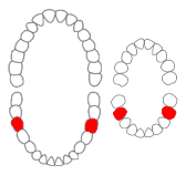
A removable partial denture (RPD) is a denture for a partially edentulous patient who desires to have replacement teeth for functional or aesthetic reasons and who cannot have a bridge for any reason, such as a lack of required teeth to serve as support for a bridge or financial limitations.

The maxillary central incisor is a human tooth in the front upper jaw, or maxilla, and is usually the most visible of all teeth in the mouth. It is located mesial to the maxillary lateral incisor. As with all incisors, their function is for shearing or cutting food during mastication (chewing). There is typically a single cusp on each tooth, called an incisal ridge or incisal edge. Formation of these teeth begins at 14 weeks in utero for the deciduous (baby) set and 3–4 months of age for the permanent set.

The maxillary lateral incisors are a pair of upper (maxillary) teeth that are located laterally from both maxillary central incisors of the mouth and medially from both maxillary canines. As with all incisors, their function is for shearing or cutting food during mastication, commonly known as chewing. There are generally no cusps on the teeth, but the rare condition known as talon cusps are most prevalent on the maxillary lateral incisors. The surface area of the tooth used in eating is called an incisal ridge or incisal edge. Though relatively the same, there are some minor differences between the deciduous (baby) maxillary lateral incisor and that of the permanent maxillary lateral incisor. The maxillary lateral incisors occlude in opposition to the mandibular lateral incisors.

The maxillary first molar is the human tooth located laterally from both the maxillary second premolars of the mouth but mesial from both maxillary second molars.

The maxillary second molar is the tooth located distally from both the maxillary first molars of the mouth but mesial from both maxillary third molars. This is true only in permanent teeth. In deciduous (baby) teeth, the maxillary second molar is the last tooth in the mouth and does not have a third molar behind it. The function of this molar is similar to that of all molars in regard to grinding being the principal action during mastication, commonly known as chewing. There are usually four cusps on maxillary molars, two on the buccal and two palatal.

The mandibular central incisor is the tooth located on the jaw, adjacent to the midline of the face. It is mesial from both mandibular lateral incisors. As with all incisors, its function includes shearing or cutting food during mastication, commonly known as chewing. There are no cusps on the tooth. Instead, the surface area of the tooth used in eating is called an incisal ridge or incisal edge. Though the two are similar, there are some minor differences between the deciduous (baby) mandibular central incisor and that of the permanent mandibular central incisor. The mandibular central incisors are usually the first teeth to appear in the mouth, typically around the age of 6–8 months.

The mandibular first premolar is the tooth located laterally from both the mandibular canines of the mouth but mesial from both mandibular second premolars. The function of this premolar is similar to that of canines in regard to tearing being the principal action during mastication, commonly known as chewing. Mandibular first premolars have two cusps. The one large and sharp is located on the buccal side of the tooth. Since the lingual cusp is small and nonfunctional, the mandibular first premolar resembles a small canine. There are no deciduous (baby) mandibular premolars. Instead, the teeth that precede the permanent mandibular premolars are the deciduous mandibular molars.
The mandibular first molar or six-year molar is the tooth located distally from both the mandibular second premolars of the mouth but mesial from both mandibular second molars. It is located on the mandibular (lower) arch of the mouth, and generally opposes the maxillary (upper) first molars and the maxillary 2nd premolar in normal class I occlusion. The function of this molar is similar to that of all molars in regard to grinding being the principal action during mastication, commonly known as chewing. There are usually five well-developed cusps on mandibular first molars: two on the buccal, two lingual, and one distal. The shape of the developmental and supplementary grooves, on the occlusal surface, are described as being M-shaped. There are great differences between the deciduous (baby) mandibular molars and those of the permanent mandibular molars, even though their function are similar. The permanent mandibular molars are not considered to have any teeth that precede it. Despite being named molars, the deciduous molars are followed by permanent premolars.

The mandibular second molar is the tooth located distally from both the mandibular first molars of the mouth but mesial from both mandibular third molars. This is true only in permanent teeth. The function of this molar is similar to that of all molars in regard to grinding being the principal action during mastication, commonly known as chewing. Though there is more variation between individuals than that of the first mandibular molar, there are usually four cusps on mandibular second molars: two on the buccal and two lingual. There are great differences between the deciduous (baby) mandibular molars and those of the permanent mandibular molars, even though their function is similar. The permanent mandibular molars are not considered to have any teeth that precede them. Despite being named molars, the deciduous molars are followed by permanent premolars.
Dental anatomy is a field of anatomy dedicated to the study of human tooth structures. The development, appearance, and classification of teeth fall within its purview. Tooth formation begins before birth, and the teeth's eventual morphology is dictated during this time. Dental anatomy is also a taxonomical science: it is concerned with the naming of teeth and the structures of which they are made, this information serving a practical purpose in dental treatment.

A cusp is a pointed, projecting, or elevated feature. In animals, it is usually used to refer to raised points on the crowns of teeth. The concept is also used with regard to the leaflets of the four heart valves. The mitral valve, which has two cusps, is also known as the bicuspid valve, and the tricuspid valve has three cusps.
This is a list of definitions of commonly used terms of location and direction in dentistry. This set of terms provides orientation within the oral cavity, much as anatomical terms of location provide orientation throughout the body.

In dentistry, a furcation defect is bone loss, usually a result of periodontal disease, affecting the base of the root trunk of a tooth where two or more roots meet. The extent and configuration of the defect are factors in both diagnosis and treatment planning.
Height of curvature in the tooth can be defined as the line encircling a tooth at its greatest bulge to a selected path of insertion. The height of curvature is the same as the height of contour.

The Lewis offset is a term for the portion of the central groove on a permanent mandibular first molar which lies between the two central pits. It was named for long time dental anatomy instructor Dr. Christopher S. Lewis, a Mercer Island, WA dentist.
Serial extraction is the planned extraction of certain deciduous teeth and specific permanent teeth in an orderly sequence and predetermined pattern to guide the erupting permanent teeth into a more favorable position.
Occlusion according to The Glossary of Prosthodontic Terms Ninth Edition is defined as "the static relationship between the incising or masticating surfaces of the maxillary or mandibular teeth or tooth analogues".

Lonchocyon is an extinct genus of arctoid carnivorans, with possible affinities to amphicyonids or hemicyonine bears. It contains a single species, Lonchocyon qiui, known from a single left mandible discovered at the fossil-bearing locality Haerhada at the base of the Baron Sog Formation, which is located in Inner Mongolia, China, and dates to the late Eocene. This taxon is notable for its large size in comparison to other arctoid carnivorans of the Eocene epoch, and for its hypercarnivorous adaptions, most notably its large canine and strongly reduced premolars. The genus name is a combination of Greek lonch, meaning spear and referencing the spear-like paraconid on its lower carnassial, and cyon, meaning dog. The specific name honours Professor Zhan-Xiang Qiu.
