Gingival and periodontal pocket

Last updated
A) Crown of the tooth, covered by enamel
B) Root of the tooth, covered by cementum
C) Alveolar bone
D) Subepithelial connective tissue
E) Oral epithelium
F) Free gingival margin
G) Gingival sulcus (extensions of which are the gingival and periodontal pockets)
H) Principal gingival fibers
I) Alveolar crest fibers of the periodontal ligament (PDL)
J) Horizontal fibers of the PDL
K) Oblique fibers of the PDL Periodontium.svg
A) Crown of the tooth, covered by enamel
B) Root of the tooth, covered by cementum
C) Alveolar bone
D) Subepithelial connective tissue
E) Oral epithelium
F) Free gingival margin
G) Gingival sulcus (extensions of which are the gingival and periodontal pockets)
H) Principal gingival fibers
I) Alveolar crest fibers of the periodontal ligament (PDL)
J) Horizontal fibers of the PDL
K) Oblique fibers of the PDL


In dental anatomy, the gingival and periodontal pockets (also informally referred to as gum pockets [1] ) are dental terms indicating the presence of an abnormal depth of the gingival sulcus near the point at which the gingival (gum) tissue contacts the tooth.

Contents

Tooth gingival interface

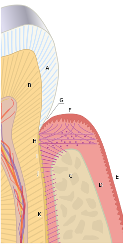
The interface between a tooth and the surrounding gingival tissue is a dynamic structure. [2] The gingival tissue forms a crevice surrounding the tooth, similar to a miniature, fluid-filled moat, wherein food debris, endogenous and exogenous cells, and chemicals float. The depth of this crevice, known as a sulcus, is in a constant state of flux due to microbial invasion and subsequent immune response. Located at the depth of the sulcus is the epithelial attachment, consisting of approximately 1 mm of junctional epithelium and another 1 mm of gingival fiber attachment, comprising the 2 mm of biologic width naturally found in the oral cavity. The sulcus is literally the area of separation between the surrounding epithelium and the surface of the encompassed tooth.

Gingival pocket

A gingival pocket presents when the marginal gingiva experiences an edematous reaction, whether due to localized irritation and subsequent inflammation, systemic issues, or drug induced gingival hyperplasia. Regardless of the etiology, when gingival hyperplasia occurs, greater than normal (the measurement in a pre-pathological state) periodontal probing measurements can be read, creating the illusion that periodontal pockets have developed. This phenomenon is also referred to as a false pocket or pseudopocket. The epithelial attachment does not migrate, it simply remains at the same attachment level found in pre-pathological health. The only anatomical landmark experiencing migration is the gingival margin in a coronal direction.

In a gingival pocket, no destruction of the connective tissue fibers (gingival fibers) or alveolar bone occurs. This early sign of disease in the mouth is completely reversible when the etiology of the edematous reaction is eliminated and frequently occurs without dental surgical therapy. However, in certain situations, a gingivectomy is necessary to reduce the gingival pocket depths to a healthy 1–3 mm.

Periodontal pocket

1: Total loss of attachment (clinical attachment loss, CAL) is the sum of 2: Gingival recession, and 3: Probing depth Periodontal terms diagram gingival recession.png
1: Total loss of attachment (clinical attachment loss, CAL) is the sum of 2: Gingival recession, and 3: Probing depth

As the original sulcular depth increases and the apical migration of the junctional epithelium has simultaneously occurred, the pocket is now lined by pocket epithelium (PE) instead of junctional epithelium (JE). [3] To have a true periodontal pocket, a probing measurement of 4 mm or more must be clinically evidenced. In this state, much of the gingival fibers that initially attached the gingival tissue to the tooth have been irreversibly destroyed. The depth of the periodontal pockets must be recorded in the patient record for proper monitoring of periodontal disease. Unlike in clinically healthy situations, parts of the sulcular epithelium can sometimes be seen in periodontally involved gingival tissue if air is blown into the periodontal pocket, exposing the newly denuded roots of the tooth. A periodontal pocket can become an infected space and may result in an abscess formation with a papule on the gingival surface. Incision and drainage of the abscess may be necessary, as well as systemic antibiotics; placement of local antimicrobial delivery systems within the periodontal pocket to reduce localized infections may also be considered. It is classified as supra bony and infra bony based on its depth in relation to alveolar bone. [4]

Mucogingival defect

If the destruction continues unabated apically and reaches the junction of the attached gingiva and alveolar mucosa, the pocket would thus be in violation of the mucogingival junction and would be termed a mucogingival defect. [5]

Pocket formation

For the periodontal pocket to form, several elements need to be present. It all starts with the dental plaque [ tone ]. The invasion of the bacteria from the plaque eventually triggers inflammatory response. This in turn results in the gradual destruction of the tissues surrounding the teeth, known as the periodontium. [6] Plaque that has been present long enough to harden and calcify will welcome additional bacteria to the pocket and make it virtually impossible to clean by means of a traditional toothbrush. [7] Continuous destruction of surrounding tissues due to inflammation will lead to degradation of attachment and bone, eventually causing tooth loss. Certain circumstances can worsen the condition and are known as risk factors. These can either be systemic (like diabetes or smoking) or local (like overhanging dental restorative materials causing food trap). [8] It is, therefore, important to manage plaque levels by appropriate oral hygiene measures. The importance of using interdental brushes along with standard or electric toothbrushing should be stressed early on. Early detection of high plaque levels at routine dental visits are found to be beneficial to avoid progression of the pocket formation. [9]

Related Research Articles

<span class="mw-page-title-main">Periodontal disease</span> Disease of the tissues surrounding the teeth (periodontium)

Periodontal disease, also known as gum disease, is a set of inflammatory conditions affecting the tissues surrounding the teeth. In its early stage, called gingivitis, the gums become swollen and red and may bleed. It is considered the main cause of tooth loss for adults worldwide. In its more serious form, called periodontitis, the gums can pull away from the tooth, bone can be lost, and the teeth may loosen or fall out. Halitosis may also occur.

<span class="mw-page-title-main">Gums</span> Soft tissue surrounding the roots of the teeth

The gums or gingiva consist of the mucosal tissue that lies over the mandible and maxilla inside the mouth. Gum health and disease can have an effect on general health.

Periodontology or periodontics is the specialty of dentistry that studies supporting structures of teeth, as well as diseases and conditions that affect them. The supporting tissues are known as the periodontium, which includes the gingiva (gums), alveolar bone, cementum, and the periodontal ligament. A periodontist is a dentist that specializes in the prevention, diagnosis and treatment of periodontal disease and in the placement of dental implants.

<span class="mw-page-title-main">Periodontal probe</span> Dental instrument for measuring depths of gum pockets around a tooth

In dentistry, a periodontal probe is a dental instrument which is usually long, thin, and blunted at the end. Its main function is to evaluate the depth of the pockets surrounding a tooth in order to determine the periodontium's overall health. For accuracy and readability, the instrument's head has markings written on it.

<span class="mw-page-title-main">Gingival sulcus</span> Space between a tooth and gums

In dental anatomy, the gingival sulcus is an area of potential space between a tooth and the surrounding gingival tissue and is lined by sulcular epithelium. The depth of the sulcus is bounded by two entities: apically by the gingival fibers of the connective tissue attachment and coronally by the free gingival margin. A healthy sulcular depth is three millimeters or less, which is readily self-cleansable with a properly used toothbrush or the supplemental use of other oral hygiene aids.

<span class="mw-page-title-main">Scaling and root planing</span> Dental procedure

Scaling and root planing, also known as conventional periodontal therapy, non-surgical periodontal therapy or deep cleaning, is a procedure involving removal of dental plaque and calculus and then smoothing, or planing, of the (exposed) surfaces of the roots, removing cementum or dentine that is impregnated with calculus, toxins, or microorganisms, the agents that cause inflammation. It is a part of non-surgical periodontal therapy. This helps to establish a periodontium that is in remission of periodontal disease. Periodontal scalers and periodontal curettes are some of the tools involved.

In dental anatomy, the junctional epithelium (JE) is that epithelium which lies at, and in health also defines, the base of the gingival sulcus. The probing depth of the gingival sulcus is measured by a calibrated periodontal probe. In a healthy-case scenario, the probe is gently inserted, slides by the sulcular epithelium (SE), and is stopped by the epithelial attachment (EA). However, the probing depth of the gingival sulcus may be considerably different from the true histological gingival sulcus depth.

<span class="mw-page-title-main">Gingival margin</span> Region of exposed gums bordering each tooth

In dental anatomy, the free gingival margin is the interface between the sulcular epithelium and the epithelium of the oral cavity. This interface exists at the most coronal point of the gingiva, otherwise known as the crest of the marginal gingiva.

A mucogingival junction is an anatomical feature found on the intraoral mucosa. The mucosa of the cheeks and floor of the mouth are freely moveable and fragile, whereas the mucosa around the teeth and on the palate are firm and keratinized. Where the two tissue types meet is known as a mucogingival junction.

In dental anatomy, the gingival fibers are the connective tissue fibers that inhabit the gingival tissue (gums) adjacent to teeth and help hold the tissue firmly against the teeth. They are primarily composed of type I collagen, although type III fibers are also involved.

Gingivectomy is a dental procedure in which a dentist or oral surgeon cuts away part of the gums in the mouth.

Dental pertains to the teeth, including dentistry. Topics related to the dentistry, the human mouth and teeth include:

Guided bone regeneration (GBR) and guided tissue regeneration (GTR) are dental surgical procedures that use barrier membranes to direct the growth of new bone and gingival tissue at sites with insufficient volumes or dimensions of bone or gingiva for proper function, esthetics or prosthetic restoration. Guided bone regeneration typically refers to ridge augmentation or bone regenerative procedures; guided tissue regeneration typically refers to regeneration of periodontal attachment.

<span class="mw-page-title-main">Debridement (dental)</span> Removal of plaque and calculus from teeth

In dentistry, debridement refers to the removal by dental cleaning of accumulations of plaque and calculus (tartar) in order to maintain dental health. Debridement may be performed using ultrasonic instruments, which fracture the calculus, thereby facilitating its removal, as well as hand tools, including periodontal scaler and curettes, or through the use of chemicals such as hydrogen peroxide.

Chronic periodontitis is one of the seven categories of periodontitis as defined by the American Academy of Periodontology 1999 classification system. Chronic periodontitis is a common disease of the oral cavity consisting of chronic inflammation of the periodontal tissues that is caused by the accumulation of profuse amounts of dental plaque. Periodontitis initially begins as gingivitis and can progress onto chronic and subsequent aggressive periodontitis according to the 1999 classification.

In dentistry, numerous types of classification schemes have been developed to describe the teeth and gum tissue in a way that categorizes various defects. All of these classification schemes combine to provide the periodontal diagnosis of the aforementioned tissues in their various states of health and disease.

Clinical attachment loss (CAL) is the predominant clinical manifestation and determinant of periodontal disease.

<span class="mw-page-title-main">Periodontal charting</span>

Periodontal charting refers to a chart utilized by a dental care professional to write and record gingival and overall oral conditions relating to oral and periodontal health or disease.

Periodontal surgery is a form of dental surgery that prevents or corrects anatomical, traumatic, developmental, or plaque-induced defects in the bone, gingiva, or alveolar mucosa. The objectives of this surgery include accessibility of instruments to the root surface, elimination of inflammation, creation of an oral environment for plaque control, periodontal disease control, oral hygiene maintenance, maintaining proper embrasure space, addressing gingiva–alveolar mucosa problems, and esthetic improvement. Surgical procedures include crown lengthening, frenectomy, and mucogingival flap surgery.

References

  1. "What do your Gum Pocket Measurements really mean?" (Staff Blog). Lorne Park Dental Associates. 3 May 2017. Retrieved 4 December 2018.
  2. Fermin A. Carranza. CARRANZA'S CLINICAL PERIODONTOLOGY, 9th edition, 2002. page 101
  3. Antonio Nanci, Ten Cate's Oral Histology, Elsevier, 2007, page 383
  4. Illustrated Dental Embryology, Histology, and Anatomy, Bath-Balogh and Fehrenbach, Elsevier, 2011, page 129
  5. Carranza's Clinical Periodontology, Newman, et al, Elsevier, 2011
  6. Lamont, Thomas; Worthington, Helen V; Clarkson, Janet E; Beirne, Paul V (2018-12-27). Cochrane Oral Health Group (ed.). "Routine scale and polish for periodontal health in adults". Cochrane Database of Systematic Reviews. 12 (4): CD004625. doi:10.1002/14651858.CD004625.pub5. PMC   6516960 . PMID   30590875.
  7. Worthington, Helen V; MacDonald, Laura; Poklepovic Pericic, Tina; Sambunjak, Dario; Johnson, Trevor M; Imai, Pauline; Clarkson, Janet E (2019-04-10). Cochrane Oral Health Group (ed.). "Home use of interdental cleaning devices, in addition to toothbrushing, for preventing and controlling periodontal diseases and dental caries". Cochrane Database of Systematic Reviews. 2020 (4): CD012018. doi:10.1002/14651858.CD012018.pub2. PMC   6953268 . PMID   30968949.
  8. Manresa, Carolina; Sanz-Miralles, Elena C.; Twigg, Joshua; Bravo, Manuel (1 January 2018). "Supportive periodontal therapy (SPT) for maintaining the dentition in adults treated for periodontitis". The Cochrane Database of Systematic Reviews. 1 (1): CD009376. doi:10.1002/14651858.CD009376.pub2. ISSN   1469-493X. PMC   6491071 . PMID   29291254.
  9. "CKS is only available in the UK". NICE. Retrieved 2020-02-19.