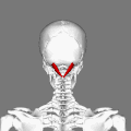
| Rectus capitis posterior major muscle | |
|---|---|
 Deep muscles of the back. (Rect. post. major visible at upper left.) | |
| Details | |
| Origin | Spinous process of the axis (C2) |
| Insertion | Inferior nuchal line of the occipital bone |
| Artery | Occipital artery |
| Nerve | Dorsal ramus of C1 (suboccipital nerve), sub-occipital nerve |
| Actions | Ipsilateral rotation of head and extension |
| Identifiers | |
| Latin | musculus rectus capitis posterior major |
| TA98 | A04.2.02.004 |
| TA2 | 2249 |
| FMA | 32525 |
| Anatomical terms of muscle | |
The rectus capitis posterior major (or rectus capitis posticus major[ citation needed ]) is a muscle in the upper back part of the neck. It is one of the suboccipital muscles. Its inferior attachment is at the spinous process of the axis (Second cervical vertebra); its superior attachment is onto the outer surface of the occipital bone on and around the side part of the inferior nuchal line. The muscle is innervated by the suboccipital nerve (the posterior ramus of cervical spinal nerve C1). The muscle acts to extend the head and rotate the head to its side.