Anal sinuses

Last updated
Anal sinuses
Gray1078.png
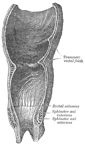
Coronal section of rectum and anal canal
Gray1080.png
The interior of the anal canal and lower part of the rectum, showing the anal columns and the anal valves between their lower ends (the columns were more numerous in the specimen than usual)
Details
Identifiers
Latin Sinus anales
TA98 A05.7.05.006
TA2 3013
FMA 76584
Anatomical terminology

An anal sinus (rectal sinus) is a furrow formed between any two adjacent anal columns of the anal canal. An anal sinus is limited inferiorly by an anal valve (which unites the inferior ends of a pair of adjacent anal columns). The anal glands open into the anal sinuses. [1]

Related Research Articles

<span class="mw-page-title-main">Levator ani</span> Broad, thin muscle group, situated on either side of the pelvis

The levator ani is a broad, thin muscle group, situated on either side of the pelvis. It is formed from three muscle components: the pubococcygeus, the iliococcygeus, and the puborectalis.

<span class="mw-page-title-main">Fistula</span> Abnormal connection between two epithelialized surfaces, often organs

A fistula in anatomy is an abnormal connection between two hollow spaces, such as blood vessels, intestines, or other hollow organs. Types of fistula can be described by their location. Anal fistulas connect the anal canal and the perianal skin. Anovaginal or rectovaginal fistulas occur when a hole develops between the anus or rectum and the vagina. Colovaginal fistulas occur between the colon and the vagina. Urinary tract fistulas are abnormal openings within the urinary tract or an abnormal connection between the urinary tract and another organ such as between the bladder and the uterus in a vesicouterine fistula, between the bladder and the vagina in a vesicovaginal fistula, and between the urethra and the vagina in urethrovaginal fistula. When occurring between two parts of the intestine, it is known as an enteroenteral fistula, between the small intestine and the skin as an enterocutaneous fistula, and between the colon and the skin as a colocutaneous fistula.

<span class="mw-page-title-main">Sigmoid colon</span> Section of the large intestine closest to the rectum and anus

The sigmoid colon is the part of the large intestine that is closest to the rectum and anus. It forms a loop that averages about 35–40 centimetres (14–16 in) in length. The loop is typically shaped like a Greek letter sigma (ς) or Latin letter S. This part of the colon normally lies within the pelvis, but due to its freedom of movement it is liable to be displaced into the abdominal cavity.

<span class="mw-page-title-main">Anal canal</span> Functional segment of the large intestine

The anal canal is the part that connects the rectum to the anus, located below the level of the pelvic diaphragm. It is located within the anal triangle of the perineum, between the right and left ischioanal fossa. As the final functional segment of the bowel, it functions to regulate release of excrement by two muscular sphincter complexes. The anus is the aperture at the terminal portion of the anal canal.

<span class="mw-page-title-main">External anal sphincter</span> Flat plane of skeletal muscle fibers

The external anal sphincter is an oval tube skeletal muscle fibers. Distally, it is adherent to the skin surrounding the margin of the anus. The sphincter exhibits a resting state of tonical contraction.

<span class="mw-page-title-main">Internal anal sphincter</span> Ring of smooth muscle that surrounds part of the anal canal

The internal anal sphincter, IAS, is a ring of smooth muscle that surrounds about 2.5–4.0 cm of the anal canal. It is about 5 mm thick, and is formed by an aggregation of the smooth (involuntary) circular muscle fibers of the rectum. it terminates distally about 6 mm from the anal orifice.

<span class="mw-page-title-main">Middle rectal artery</span>

The middle rectal artery is an artery in the pelvis that supplies blood to the rectum.

<span class="mw-page-title-main">Pelvic cavity</span> Body cavity bounded by the bones of the pelvis

The pelvic cavity is a body cavity that is bounded by the bones of the pelvis. Its oblique roof is the pelvic inlet. Its lower boundary is the pelvic floor.

<span class="mw-page-title-main">Rectal venous plexus</span>

The rectal venous plexus is the venous plexus surrounding the rectum. It consists of an internal and an external rectal plexus. It is drained by the superior, middle, and inferior rectal veins. It forms a portosystemic (portocaval) anastomosis. This allows rectally administered medications to bypassing first pass metabolism.

<span class="mw-page-title-main">Superior rectal artery</span>

The superior rectal artery is an artery that descends into the pelvis to supply blood to the rectum.

<span class="mw-page-title-main">Inferior hypogastric plexus</span>

The inferior hypogastric plexus is a network of nerves that supplies the organs of the pelvic cavity. The inferior hypogastric plexus gives rise to the prostatic plexus in males and the uterovaginal plexus in females.

<span class="mw-page-title-main">Pelvic fascia</span>

The pelvic fasciae are the fascia of the pelvis and can be divided into:

<span class="mw-page-title-main">Anal columns</span>

Anal columns are a number of vertical folds, produced by an infolding of the mucous membrane and some of the muscular tissue in the upper half of the lumen of the anal canal. They are named after Giovanni Battista Morgagni, who has several other eponyms named after him.

<span class="mw-page-title-main">Anal valves</span>

An anal valve is a small horizontal fold uniting the inferior ends of any two adjacent anal columns of the anal canal. An anal valve forms the inferior boundary of an anal sinus. The anal valves demarcate the level of the pectinate line.

<span class="mw-page-title-main">Pararectal fossa</span>

The pararectal fossa is an inferior-ward extension of the peritoneum on either side of the rectum. It is formed by a (sacrogenital) fold of peritoneum extending inferior-ward from the posterolateral pelvic wall. It represents a lateral extension of the rectouterine pouch in the female, and of rectovesical pouch in the male. It varies in size with the distension of the rectum.

<span class="mw-page-title-main">Pectinate line</span> Line dividing the upper two-thirds and the lower third of the anal canal

The pectinate line is a line which divides the upper two-thirds and lower third of the anal canal. Developmentally, this line represents the hindgut-proctodeum junction.

<span class="mw-page-title-main">Outline of human anatomy</span> Overview of and topical guide to human anatomy

The following outline is provided as an overview of and topical guide to human anatomy:

<span class="mw-page-title-main">Rectum</span> Final portion of the large intestine

The rectum is the final straight portion of the large intestine in humans and some other mammals, and the gut in others. The adult human rectum is about 12 centimetres (4.7 in) long, and begins at the rectosigmoid junction at the level of the third sacral vertebra or the sacral promontory depending upon what definition is used. Its diameter is similar to that of the sigmoid colon at its commencement, but it is dilated near its termination, forming the rectal ampulla. It terminates at the level of the anorectal ring or the dentate line, again depending upon which definition is used. In humans, the rectum is followed by the anal canal which is about 4 centimetres (1.6 in) long, before the gastrointestinal tract terminates at the anal verge. The word rectum comes from the Latin rectumintestinum, meaning straight intestine.

<span class="mw-page-title-main">Pelvis</span> Lower part of the trunk of the human body between the abdomen and the thighs

The pelvis is the lower part of the trunk, between the abdomen and the thighs, together with its embedded skeleton.

<span class="mw-page-title-main">Rectococcygeal muscle</span>

The rectococcygeal muscles are two bands of smooth muscle tissue arising from the 2nd and 3rd coccygeal vertebrae, and passing downward and forward to blend with the rectal longitudinal smooth muscle fibers on the posterior wall of the anal canal.

References

  1. Sinnatamby, Chummy S. (2011). Last's Anatomy (12th ed.). p. 315. ISBN   978-0-7295-3752-0.