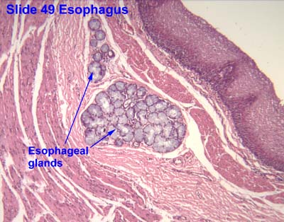
Esophageal gland

Last updated
Esophageal glands
Illu esophageal layers.jpg
Layers of Esophageal Wall:
1. Mucosa
2. Submucosa
3. Muscularis
4. Adventitia
5. Striated muscle
6. Striated and smooth
7. Smooth muscle
8. Lamina muscularis mucosae
9. Esophageal glands
Gray1033.png
Section of the human esophagus. Moderately magnified. The section is transverse and from near the middle of the gullet.
a. Fibrous covering.
b. Divided fibers of longitudinal muscular coat.
c. Transverse muscular fibers.
d. Submucous or areolar layer.
e. Muscularis mucosae.
f. Mucous membrane, with vessels and part of a lymphoid nodule.
g. Stratified epithelial lining.
h. Mucous gland.
i. Gland duct.
m’. Striated muscular fibers cut across.
Details
Identifiers
Latin glandulae oesophageae
TA98 A05.4.01.017
TA2 2893
FMA 71619
Anatomical terminology

The esophageal glands are glands that are part of the digestive system of various animals, including humans.

Contents

In humans

In humans the glands are known as the esophageal submucosal glands and are a part of the human digestive system. [1] They are a small compound racemose exocrine glands of the mucous type.[ citation needed ]

There are two types:

Each opens upon the surface by a long excretory duct.[ citation needed ]

In monoplacophorans

The esophageal gland is enlarged in large monoplacophoran species. [3]

In gastropods

The esophageal gland or oesophageal pouch is a part of the digestive system of some gastropods. The esophageal gland or pouch is a common feature in so-called basal gastropod clades, including Patelloidea, Vetigastropoda, Cocculiniformia, Neritimorpha and Neomphalina. [4]

The size of the esophageal gland of the scaly-foot gastropod Chrysomallon squamiferum (family Peltospiridae within Neomphalina) is about two orders of magnitude over the usual size. [4] The scaly-foot gastropod houses endosymbiotic bacteria in the esophageal gland. [4] Chrysomallon squamiferum was thought to be the only species of Peltospiridae, that has an enlarged esophageal gland, but later it was shown that both species Gigantopelta the gland also enlarged. [5] In other peltospirids, the posterior portion of the oesophagus forms a pair of blind mid-oesophageal pouches or gutters extending only to the anterior end of the foot ( Rhynchopelta , Peltospira , Nodopelta , Echinopelta , Pachydermia ). [4] The same situation is in Melanodrymia within the family Melanodrymiidae. [4] Bathyphytophilidae and Lepetellidae are also known to have enlarged esophageal pouches, however, though not to the extent of Chrysomallon. [4] Both are known to house endosymbiotic bacteria, in the case of bathyphytophilids most likely also in the esophageal glands but in the lepetellids the endosymbionts are spread in the hemocoel. [4]

Related Research Articles

<span class="mw-page-title-main">Exocrine gland</span> Gland that secretes substances onto an epithelial surface by way of a duct

Exocrine glands are glands that secrete substances onto an epithelial surface by way of a duct. Examples of exocrine glands include sweat, salivary, mammary, ceruminous, lacrimal, sebaceous, prostate and mucous. Exocrine glands are one of two types of glands in the human body, the other being endocrine glands, which secrete their products directly into the bloodstream. The liver and pancreas are both exocrine and endocrine glands; they are exocrine glands because they secrete products—bile and pancreatic juice—into the gastrointestinal tract through a series of ducts, and endocrine because they secrete other substances directly into the bloodstream. Exocrine sweat glands are part of the integumentary system; they have eccrine and apocrine types.

<span class="mw-page-title-main">Ileum</span> Final section of the small intestine

The ileum is the final section of the small intestine in most higher vertebrates, including mammals, reptiles, and birds. In fish, the divisions of the small intestine are not as clear and the terms posterior intestine or distal intestine may be used instead of ileum. Its main function is to absorb vitamin B12, bile salts, and whatever products of digestion that were not absorbed by the jejunum.

<span class="mw-page-title-main">Esophagus</span> Vertebrate organ through which food passes to the stomach

The esophagus or oesophagus, colloquially known also as the food pipe, food tube, or gullet, is an organ in vertebrates through which food passes, aided by peristaltic contractions, from the pharynx to the stomach. The esophagus is a fibromuscular tube, about 25 cm (10 in) long in adults, that travels behind the trachea and heart, passes through the diaphragm, and empties into the uppermost region of the stomach. During swallowing, the epiglottis tilts backwards to prevent food from going down the larynx and lungs. The word oesophagus is from Ancient Greek οἰσοφάγος (oisophágos), from οἴσω (oísō), future form of φέρω + ἔφαγον.

<span class="mw-page-title-main">Barrett's esophagus</span> Premalignant condition affecting the esophagus

Barrett's esophagus is a condition in which there is an abnormal (metaplastic) change in the mucosal cells lining the lower portion of the esophagus, from stratified squamous epithelium to simple columnar epithelium with interspersed goblet cells that are normally present only in the small intestine and large intestine. This change is considered to be a premalignant condition because of its potential to further transition to esophageal adenocarcinoma, an often-deadly cancer.

<span class="mw-page-title-main">Esophageal cancer</span> Gastrointestinal system cancer that is located in the esophagus

Esophageal cancer is cancer arising from the esophagus—the food pipe that runs between the throat and the stomach. Symptoms often include difficulty in swallowing and weight loss. Other symptoms may include pain when swallowing, a hoarse voice, enlarged lymph nodes ("glands") around the collarbone, a dry cough, and possibly coughing up or vomiting blood.

<span class="mw-page-title-main">Lamina propria</span> Thin connective layer forming part of the mucous membranes

The lamina propria is a thin layer of connective tissue that forms part of the moist linings known as mucous membranes or mucosae, which line various tubes in the body, such as the respiratory tract, the gastrointestinal tract, and the urogenital tract.

<span class="mw-page-title-main">Brunner's glands</span> Duodenal submucosal cells secreting bicarbonate-rich mucus

Brunner's glands are compound tubuloalveolar submucosal glands found in that portion of the duodenum proximal to the hepatopancreatic sphincter.

Gut-associated lymphoid tissue (GALT) is a component of the mucosa-associated lymphoid tissue (MALT) which works in the immune system to protect the body from invasion in the gut.

<span class="mw-page-title-main">Gastric glands</span> Glands in lining of the human stomach

The gastric glands are glands in the lining of the stomach that play an essential role in the process of digestion. All of the glands have mucus-secreting foveolar cells. Mucus lines the entire stomach, and protects the stomach lining from the effects of hydrochloric acid released from other cells in the glands.

<span class="mw-page-title-main">Muscularis mucosae</span> Thin layer of muscle of the gastrointestinal tract

The muscularis mucosae is a thin layer of muscle of the gastrointestinal tract, located outside the lamina propria, and separating it from the submucosa. It is present in a continuous fashion from the esophagus to the upper rectum. A discontinuous muscularis mucosae–like muscle layer is present in the urinary tract, from the renal pelvis to the bladder; as it is discontinuous, it should not be regarded as a true muscularis mucosae.

<span class="mw-page-title-main">Olfactory glands</span> Protein- and mucus-secreting glands in the olfactory mucosa

Olfactory glands, also known as Bowman's glands, are a type of nasal gland situated in the part of the olfactory mucosa beneath the olfactory epithelium, that is the lamina propria, a connective tissue also containing fibroblasts, blood vessels and bundles of fine axons from the olfactory neurons.

<span class="mw-page-title-main">Submucosa</span> Thin layer of tissue in various organs

The submucosa is a thin layer of tissue in various organs of the gastrointestinal, respiratory, and genitourinary tracts. It is the layer of dense irregular connective tissue that supports the mucosa and joins it to the muscular layer, the bulk of overlying smooth muscle.

<span class="mw-page-title-main">Neomphaloidea</span> Superfamily of gastropods

Neomphaloidea is a superfamily of deep-sea snails or limpets, marine gastropod mollusks. Neomphaloidea is the only superfamily in the order Neomphalida.

<span class="mw-page-title-main">Gastric pits</span> Indentations in the stomach

Gastric pits are indentations in the stomach which denote entrances to 3-5 tubular shaped gastric glands. They are deeper in the pylorus than they are in the other parts of the stomach. The human stomach has several million of these pits which dot the surface of the lining epithelium. Surface mucous cells line the pits themselves but give way to a series of other types of cells which then line the glands themselves.

<span class="mw-page-title-main">Gastric mucosa</span> Mucous membrane layer of the stomach

The gastric mucosa is the mucous membrane layer of the stomach, which contains the gastric pits, to which the gastric glands empty. In humans, it is about one mm thick, and its surface is smooth, soft, and velvety. It consists of simple secretory columnar epithelium, an underlying supportive layer of loose connective tissue called the lamina propria, and the muscularis mucosae, a thin layer of muscle that separates the mucosa from the underlying submucosa.

<span class="mw-page-title-main">Scaly-foot gastropod</span> Deep-sea gastropod

Chrysomallon squamiferum, commonly known as the scaly-foot gastropod, scaly-foot snail, sea pangolin, or volcano snail is a species of deep-sea hydrothermal-vent snail, a marine gastropod mollusc in the family Peltospiridae. This vent-endemic gastropod is known only from deep-sea hydrothermal vents in the Indian Ocean, where it has been found at depths of about 2,400–2,900 m (1.5–1.8 mi). C. squamiferum differs greatly from other deep-sea gastropods, even the closely related neomphalines. In 2019, it was declared endangered on the IUCN Red List, the first species to be listed as such due to risks from deep-sea mining of its vent habitat.

<span class="mw-page-title-main">Peltospiridae</span> Family of gastropods

Peltospiridae is a small family of gastropods that used to belong to the clade Vetigastropoda, but is now included in the clade Neomphalina

<span class="mw-page-title-main">Hyperplastic polyp</span> Type of colorectal polyp

A hyperplastic polyp is a type of gastric polyp or colorectal polyp.

<i>Gigantopelta chessoia</i> Species of gastropod

Gigantopelta chessoia is a species of deep sea snail from hydrothermal vents, a marine gastropod mollusk in the family Peltospiridae.

<span class="mw-page-title-main">Gastrointestinal wall</span> Digestive system structure

The gastrointestinal wall of the gastrointestinal tract is made up of four layers of specialised tissue. From the inner cavity of the gut outwards, these are the mucosa, the submucosa, the muscular layer and the serosa or adventitia.

References

PD-icon.svgThis article incorporates text in the public domain from page 1146 of the 20th edition of Gray's Anatomy (1918).
This article incorporates Creative Commons (CC-BY-4.0) text from the reference [4]

  1. Abdulnour-Nakhoul, Solange; Nakhoul, Nazih L.; Wheeler, Scott A.; Wang, Paul; Swenson, Eric R.; Orlando, Roy C. (April 2005). "HCO 3 − secretion in the esophageal submucosal glands". American Journal of Physiology-Gastrointestinal and Liver Physiology. 288 (4): G736–G744. doi:10.1152/ajpgi.00055.2004.
  2. 1 2 3 Nemeskeri, Agnes. Human Histology. Budapest: Apathy Istvan Foundation, Semmelweis University Budapest, Department of Human Morphology and Developmental Biology. p. 16.
  3. Chen, Chong; Uematsu, Katsuyuki; Linse, Katrin; Sigwart, Julia D. (2017). "By more ways than one: Rapid convergence at hydrothermal vents shown by 3D anatomical reconstruction of Gigantopelta (Mollusca: Neomphalina)". BMC Evolutionary Biology. 17 (1): 62. doi: 10.1186/s12862-017-0917-z . ISSN   1471-2148. PMC   5333402 . PMID   28249568.
  4. 1 2 3 4 5 6 7 8 Chen, C.; Copley, J.; Linse, K.; Rogers, A.; Sigwart, J. (2015). "The heart of a dragon: 3D anatomical reconstruction of the 'scaly-foot gastropod' (Mollusca: Gastropoda: Neomphalina) reveals its extraordinary circulatory system" (PDF). Frontiers in Zoology. 12: 13. doi: 10.1186/s12983-015-0105-1 . PMC   4470333 . PMID   26085836.
  5. Chen, C.; Linse, K.; Roterman, C. N.; Copley, J. T.; Rogers, A. D. (2015). "A new genus of large hydrothermal vent‐endemic gastropod (Neomphalina: Peltospiridae)". Zoological Journal of the Linnean Society . 175 (2): 319–335. doi: 10.1111/zoj.12279 .