Esophagogastric junction outflow obstruction

Last updated
Esophagogastric junction outflow obstruction
Other namesEGJOO
Specialty Gastroenterology
Symptoms Asymptomatic, dysphagia, chest pain
Usual onset56-57 years
TypesMechanical, functional, medication, artifact [1]
Risk factors Female gender, overweight
Diagnostic method High resolution manometry (esophageal manometry)
Differential diagnosis Achalasia
Treatment Pneumatic dilation, Per-oral endoscopic myotomy (POEM), botulinum toxin injection
Prognosis Depends on etiology
FrequencyUnknown

Esophagogastric junction outflow obstruction (EGJOO) is an esophageal motility disorder characterized by increased pressure where the esophagus connects to the stomach at the lower esophageal sphincter. EGJOO is diagnosed by esophageal manometry. However, EGJOO has a variety of etiologies; evaluating the cause of obstruction with additional testing, such as upper endoscopy, computed tomography (CT imaging), or endoscopic ultrasound may be necessary. [2] When possible, treatment of EGJOO should be directed at the cause of obstruction. When no cause for obstruction is found (functional EGJOO), observation alone may be considered if symptoms are minimal. Functional EGJOO with significant or refractory symptoms may be treated with pneumatic dilation, per-oral endoscopic myotomy (POEM), or botulinum toxin injection.

Contents

Types

EGJOO may be broadly categorized into 4 subgroups: mechanical, functional, medication-related, and artifact. [1]

Signs and symptoms

In some cases, EGJOO may cause no symptoms, and the manometry findings are identified during an evaluation prior to anti-reflux surgery. In other cases, EGJOO may be identified after an evaluation for esophageal symptoms. The most common symptoms are dysphagia (50-75%, typical reflux (29-67%), atypical reflux (21-36%), chest pain (16-46%), abdominal pain (43%) or regurgitation (38%). [1] One study suggested about 5% of individuals have no symptoms. [1]

Causes

Several causes for EGJOO exist. [2] Etiologies include early achalasia, mechanical processes (eosinophilic esophagitis, hiatal hernia, strictures, etc.), esophageal wall thickness (fibrosis, cancer, etc.), compression by nearby blood vessels (external vascular compression), obesity, opioid medication effect, or anatomic abnormalities. [2] The findings associated with EGJOO may be falsely abnormal due to measurement errors. [2]

Diagnosis

Normal in (A). EGJOO in (B).
Pressure waves in blue. Cross-sectional areas CSA) in fucsia. Esofago Distencion Contraccion EGJOO.png
Normal in (A). EGJOO in (B).
Pressure waves in blue. Cross-sectional areas CSA) in fucsia.

EGJOO is diagnosed using esophageal manometry. [2] High resolution esophageal manometry will show elevated pressure at the LES with normal peristalsis. [2] The LES pressure is evaluated immediately following a swallow, when the sphincter should relax. [3] The overall LES pressure after a swallow is represented by the integrated relaxation pressure (IRP). [3] If the IRP is abnormally elevated (>15 mmHg), this indicates an obstruction is present. Normal peristalsis with an obstruction at the esophagogastric junction (elevated IRP) is consistent with EGJOO. [3]

Upper endoscopy is used to evaluate for mechanical causes of obstruction. [2] Endoscopic findings may include a hiatal hernia, esophagitis, strictures, tumors, or masses. [2] Increased pressure at the LES over time may result in an epiphrenic diverticulum. [2] Further evaluation for mechanical causes of obstruction may include CT scans, MRI, or endoscopic ultrasound. [2]

Several additional tests may be used to further evaluate EGJOO. [2] Further evaluation of esophageal motor function may be accomplished with functional lumen imaging probe (FLIP). [2] Although not widely available, FLIP may help assess esophageal wall stiffness and compliance. [2] FLIP may help identify individuals with EGJOO who are likely to benefit from therapeutic procedures. [4]

Timed barium esophagram can help distinguish EGJOO from untreated achalasia. [2]

Treatment

Treatment primarily consists of addressing the underlying cause of EGJOO. [2] For example, gastroesophageal reflux disease (GERD) with reflux esophagitis is treated with proton pump inhibitors. Esophageal rings or strictures may be treated with esophageal dilation.

Simple observation may be considered, [5] especially if symptoms are minimal or absent. If symptoms are severe or persistent, peroral endoscopic myotomy (POEM) may be offered. [2]

Pneumatic dilation may be used for persistent symptoms in the absence of identified causes of mechanical obstruction. [2] Botulinum toxin may be considered, [5] especially for individuals who are unlikely to tolerate surgery. [2]

Prognosis

The prognosis for EGJOO depends on the etiology of obstruction. In the absence of anatomic or mechanical causes, such as cancer, outcomes are generally favorable. Individuals with minimal or no symptoms often experience resolution of the EGJOO, even without treatment. [2]

Epidemiology

The overall prevalence of EGJOO is unclear. [1] The prevalence of EGJOO among all patients undergoing high resolution manometry was up to 10 percent. [3] The diagnostic criteria were later adjusted to distinguish relevant (symptomatic) EGJOO from isolated manometric findings of EGJOO without symptoms. [3]

Individuals diagnosed with EGJOO based on Chicago 3.0 classification have an average age of 56–57 years. [1] EGJOO more commonly affects women (51-88%). [1] The average BMI is between 25 and 30. [1]

Related Research Articles

<span class="mw-page-title-main">Esophagus</span> Vertebrate organ through which food passes to the stomach

The esophagus or oesophagus, colloquially known also as the food pipe or gullet, is an organ in vertebrates through which food passes, aided by peristaltic contractions, from the pharynx to the stomach. The esophagus is a fibromuscular tube, about 25 cm (10 in) long in adults, that travels behind the trachea and heart, passes through the diaphragm, and empties into the uppermost region of the stomach. During swallowing, the epiglottis tilts backwards to prevent food from going down the larynx and lungs. The word oesophagus is from Ancient Greek οἰσοφάγος (oisophágos), from οἴσω (oísō), future form of φέρω + ἔφαγον.

<span class="mw-page-title-main">Esophageal achalasia</span> Rare, incurable, progressive motility disorder due to failure of esophogeal motor neurons

Esophageal achalasia, often referred to simply as achalasia, is a failure of smooth muscle fibers to relax, which can cause the lower esophageal sphincter to remain closed. Without a modifier, "achalasia" usually refers to achalasia of the esophagus. Achalasia can happen at various points along the gastrointestinal tract; achalasia of the rectum, for instance, may occur in Hirschsprung's disease. The lower esophageal sphincter is a muscle between the esophagus and stomach that opens when food comes in. It closes to avoid stomach acids from coming back up. A fully understood cause to the disease is unknown, as are factors that increase the risk of its appearance. Suggestions of a genetically transmittable form of achalasia exist, but this is neither fully understood, nor agreed upon.

Heartburn, also known as pyrosis, cardialgia or acid indigestion, is a burning sensation in the central chest or upper central abdomen. Heartburn is usually due to regurgitation of gastric acid into the esophagus. It is the major symptom of gastroesophageal reflux disease (GERD).

An esophageal motility disorder (EMD) is any medical disorder causing difficulty in swallowing, regurgitation of food and a spasm-type pain which can be brought on by an allergic reaction to certain foods. The most prominent one is dysphagia.
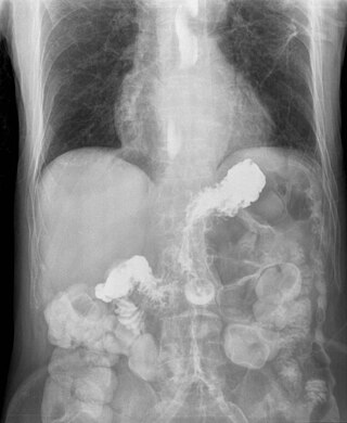
<span class="mw-page-title-main">Gastroesophageal reflux disease</span> Medical condition

Gastroesophageal reflux disease (GERD) or gastro-oesophageal reflux disease (GORD) is one of the upper gastrointestinal chronic diseases in which stomach content persistently and regularly flows up into the esophagus, resulting in symptoms and/or complications. Symptoms include dental corrosion, dysphagia, heartburn, odynophagia, regurgitation, non-cardiac chest pain, extraesophageal symptoms such as chronic cough, hoarseness, reflux-induced laryngitis, or asthma. In the long term, and when not treated, complications such as esophagitis, esophageal stricture, and Barrett's esophagus may arise.
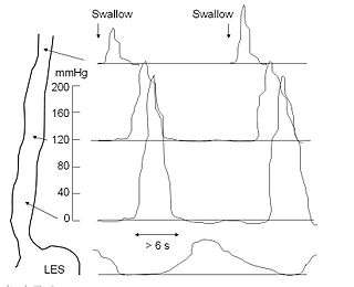
<span class="mw-page-title-main">Esophageal motility study</span> Medical test

An esophageal motility study (EMS) or esophageal manometry is a test to assess motor function of the upper esophageal sphincter (UES), esophageal body and lower esophageal sphincter (LES).
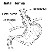
<span class="mw-page-title-main">Hiatal hernia</span> Type of hernia

A hiatal hernia or hiatus hernia is a type of hernia in which abdominal organs slip through the diaphragm into the middle compartment of the chest. This may result in gastroesophageal reflux disease (GERD) or laryngopharyngeal reflux (LPR) with symptoms such as a taste of acid in the back of the mouth or heartburn. Other symptoms may include trouble swallowing and chest pains. Complications may include iron deficiency anemia, volvulus, or bowel obstruction.
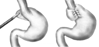
<span class="mw-page-title-main">Nissen fundoplication</span> Surgical procedure to treat gastric reflux and hiatal hernia

A Nissen fundoplication, or laparoscopic Nissen fundoplication when performed via laparoscopic surgery, is a surgical procedure to treat gastroesophageal reflux disease (GERD) and hiatal hernia. In GERD, it is usually performed when medical therapy has failed; but, with a Type II (paraesophageal) hiatus hernia, it is the first-line procedure. The Nissen fundoplication is total (360°), but partial fundoplications known as Thal, Belsey, Dor, Lind, and Toupet fundoplications are alternative procedures with somewhat different indications and outcomes.

<span class="mw-page-title-main">Eosinophilic esophagitis</span> Allergic inflammatory condition of the esophagus

Eosinophilic esophagitis (EoE) is an allergic inflammatory condition of the esophagus that involves eosinophils, a type of white blood cell. In healthy individuals, the esophagus is typically devoid of eosinophils. In EoE, eosinophils migrate to the esophagus in large numbers. When a trigger food is eaten, the eosinophils contribute to tissue damage and inflammation. Symptoms include swallowing difficulty, food impaction, vomiting, and heartburn.

Esophageal dysphagia is a form of dysphagia where the underlying cause arises from the body of the esophagus, lower esophageal sphincter, or cardia of the stomach, usually due to mechanical causes or motility problems.

Esophageal dilatation is a therapeutic endoscopic procedure that enlarges the lumen of the esophagus.

Esophageal spasm is a disorder of motility of the esophagus.
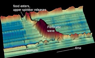
<span class="mw-page-title-main">Nutcracker esophagus</span> Medical condition

Nutcracker esophagus, jackhammer esophagus, or hypercontractile peristalsis, is a disorder of the movement of the esophagus characterized by contractions in the smooth muscle of the esophagus in a normal sequence but at an excessive amplitude or duration. Nutcracker esophagus is one of several motility disorders of the esophagus, including achalasia and diffuse esophageal spasm. It causes difficulty swallowing, or dysphagia, with both solid and liquid foods, and can cause significant chest pain; it may also be asymptomatic. Nutcracker esophagus can affect people of any age but is more common in the sixth and seventh decades of life.

<span class="mw-page-title-main">Esophageal stricture</span> Medical condition

A benign esophageal stricture, or peptic stricture, is a narrowing or tightening of the esophagus that causes swallowing difficulties.

<span class="mw-page-title-main">Esophageal food bolus obstruction</span> Medical condition

An esophageal food bolus obstruction is a medical emergency caused by the obstruction of the esophagus by an ingested foreign body.
<span class="mw-page-title-main">Esophageal pH monitoring</span>

Esophageal pH monitoring is the current gold standard for diagnosis of gastroesophageal reflux disease (GERD). It provides direct physiologic measurement of acid in the esophagus and is the most objective method to document reflux disease, assess the severity of the disease and monitor the response of the disease to medical or surgical treatment. It can also be used in diagnosing laryngopharyngeal reflux.

<span class="mw-page-title-main">Esophageal intramural pseudodiverticulosis</span> Medical condition

Esophageal intramucosal pseudodiverticulosis (EIPD) is a rare condition wherein the wall of the esophagus develops numerous small outpouchings (pseudodiverticulae). Individuals with the condition typically develop difficulty swallowing. The outpouchings represent the ducts of submucosal glands of the esophagus. It typically affects individuals in their sixth and seventh decades of life. While it is associated with certain chronic conditions, particularly alcoholism, diabetes and gastroesophageal reflux disease, the cause of the condition is unknown. Treatment involves medications to treat concomitant conditions such as reflux and esophageal spasm, and dilation of strictures in the esophagus.

The per-oral endoscopic myotomy, or POEM, is a minimally invasive surgical procedure for the treatment of achalasia wherein the inner circular muscle layer of the lower esophageal sphincter is divided through a submucosal tunnel. This enables food and liquids to pass into the stomach, a process that is impaired in achalasia. The tunnel is created, and the myotomy performed, using a flexible endoscope, meaning the entire procedure can be done without external incisions.

<span class="mw-page-title-main">Lymphocytic esophagitis</span> Medical condition

Lymphocytic esophagitis is a rare and poorly understood medical disorder involving inflammation in the esophagus. The disease is named from the primary inflammatory process, wherein lymphocytes are seen within the esophageal mucosa. Symptoms of the condition include difficulty swallowing, heartburn and food bolus obstruction. The condition was first described in 2006 by Rubio and colleagues. Initial reports questioned whether this was a true medical disorder, or whether the inflammation was secondary to another condition, such as gastroesophageal reflux disease.

Functional Lumen Imaging Probe (FLIP) is a test used to evaluate the function of the esophagus, by measuring the dimensions of the esophageal lumen using impedance planimetry. Typically performed with sedation during upper endoscopy, FLIP is used to evaluate for esophageal motility disorders, such as achalasia, diffuse esophageal spasm, etc.

References

  1. 1 2 3 4 5 6 7 8 Zikos, TA; Triadafilopoulos, G; Clarke, JO (2020-02-05). "Esophagogastric Junction Outflow Obstruction: Current Approach to Diagnosis and Management". Current Gastroenterology Reports. 22 (2): 9. doi:10.1007/s11894-020-0743-0. PMID   32020310. S2CID   211034929.
  2. 1 2 3 4 5 6 7 8 9 10 11 12 13 14 15 16 17 18 19 Samo, S; Qayed, E (2019-01-28). "Esophagogastric junction outflow obstruction: Where are we now in diagnosis and management?". World Journal of Gastroenterology. 25 (4): 411–417. doi: 10.3748/wjg.v25.i4.411 . PMC   6350167 . PMID   30700938.
  3. 1 2 3 4 5 Yadlapati, R; Kahrilas, PJ; Fox, MR; Bredenoord, AJ; Prakash Gyawali, C; Roman, S; Babaei, A; Mittal, RK; Rommel, N; Savarino, E; Sifrim, D; Smout, A; Vaezi, MF; Zerbib, F; Akiyama, J; Bhatia, S; Bor, S; Carlson, DA; Chen, JW; Cisternas, D; Cock, C; Coss-Adame, E; de Bortoli, N; Defilippi, C; Fass, R; Ghoshal, UC; Gonlachanvit, S; Hani, A; Hebbard, GS; Wook Jung, K; Katz, P; Katzka, DA; Khan, A; Kohn, GP; Lazarescu, A; Lengliner, J; Mittal, SK; Omari, T; Park, MI; Penagini, R; Pohl, D; Richter, JE; Serra, J; Sweis, R; Tack, J; Tatum, RP; Tutuian, R; Vela, MF; Wong, RK; Wu, JC; Xiao, Y; Pandolfino, JE (January 2021). "Esophageal motility disorders on high-resolution manometry: Chicago classification version 4.0©". Neurogastroenterology and Motility. 33 (1): e14058. doi:10.1111/nmo.14058. PMC   8034247 . PMID   33373111.
  4. Savarino, E; di Pietro, M; Bredenoord, AJ; Carlson, DA; Clarke, JO; Khan, A; Vela, MF; Yadlapati, R; Pohl, D; Pandolfino, JE; Roman, S; Gyawali, CP (November 2020). "Use of the Functional Lumen Imaging Probe in Clinical Esophagology". The American Journal of Gastroenterology. 115 (11): 1786–1796. doi: 10.14309/ajg.0000000000000773 . PMC   9380028 . PMID   33156096.
  5. 1 2 Garbarino, S; von Isenburg, M; Fisher, DA; Leiman, DA (January 2020). "Management of Functional Esophagogastric Junction Outflow Obstruction: A Systematic Review". Journal of Clinical Gastroenterology. 54 (1): 35–42. doi:10.1097/MCG.0000000000001156. PMID   30575636. S2CID   58589325.