Congenital diaphragmatic hernia

Last updated
Congenital diaphragmatic hernia
Other namesCDH
Hernia de Morgani.JPG
Morgagni hernia seen on a chest radiograph.
Specialty Medical genetics, pediatrics   OOjs UI icon edit-ltr-progressive.svg

Congenital diaphragmatic hernia (CDH) is a birth defect of the diaphragm. The most common type of CDH is a Bochdalek hernia ; other types include Morgagni hernia, diaphragm eventration and central tendon defects of the diaphragm. Malformation of the diaphragm allows the abdominal organs to push into the chest cavity, hindering proper lung formation.[ citation needed ]

Contents

CDH is a life-threatening pathology in infants and a major cause of death due to two complications: pulmonary hypoplasia and pulmonary hypertension. [1] Experts disagree on the relative importance of these two conditions, with some focusing on hypoplasia, others on hypertension. [2] Newborns with CDH often have severe respiratory distress which can be life-threatening unless treated appropriately.

Classification

Bochdalek hernia

The Bochdalek hernia, also known as a postero-lateral diaphragmatic hernia, is the most common manifestation of CDH, accounting for more than 95% of cases. In this instance the diaphragm abnormality is characterized by a hole in the postero-lateral corner of the diaphragm which allows passage of the abdominal viscera into the chest cavity. The majority of Bochdalek hernias (80–85%) occur on the left side of the diaphragm, a large proportion of the remaining cases occur on the right side. To date, it carries a high mortality [3] and is an active area of clinical research.[ citation needed ]

Morgagni hernia

CT of the chest demonstrates a Morgagni hernia (red arrow) Morgagni Hernia.PNG
CT of the chest demonstrates a Morgagni hernia (red arrow)

This rare anterior defect of the diaphragm is variably referred to as a Morgagni, retrosternal, or parasternal hernia. Accounting for approximately 2% of all CDH cases, it is characterized by herniation through the foramina of Morgagni which are located immediately adjacent and posterior to the xiphoid process of the sternum. [4]

Diaphragm eventration

The diagnosis of congenital diaphragmatic eventration is used when there is abnormal displacement (i.e. elevation) of part or all of an otherwise intact diaphragm into the chest cavity. This rare type of CDH occurs because in the region of eventration the diaphragm is thinner, allowing the abdominal viscera to protrude upwards.[ citation needed ]

Pathophysiology

There are genetic causes of CDH [5] including aneuploidies, chromosome copy number variants, and single gene mutations. Research implicates a few gene mutations including LONP1 [6] and MYRF. [7] It involves three major defects:[ citation needed ]

Diagnosis

Congenital diaphragmatic hernia: coronal obstetric ultrasound (head to right of image, thorax centre, abdomen left) shows the stomach and heart both within the thorax. Cdh0002.jpg
Congenital diaphragmatic hernia: coronal obstetric ultrasound (head to right of image, thorax centre, abdomen left) shows the stomach and heart both within the thorax.

This condition can often be diagnosed before birth and fetal intervention can sometimes help, depending on the severity of the condition. [8] Infants born with diaphragmatic hernia experience respiratory failure due to both pulmonary hypertension and pulmonary hypoplasia. The first condition is a restriction of blood flow through the lungs thought to be caused by defects in the lung. Pulmonary hypoplasia or decreased lung volume is directly related to the abdominal organs presence in the chest cavity which causes the lungs to be severely undersized, especially on the side of the hernia.[ citation needed ]

Survival rates for infants with this condition vary, but have generally been increasing through advances in neonatal medicine. Work has been done to correlate survival rates to ultrasound measurements of the lung volume as compared to the baby's head circumference. This figure known as the lung-to-head ratio (LHR). Still, LHR remains an inconsistent measure of survival. Outcomes of CDH are largely dependent on the severity of the defect and the appropriate timing of treatment.

A small percentage of cases go unrecognized into adulthood. [9]

Treatment

The first step in management is orogastric tube placement and securing the airway (intubation). Ideally, the baby will never take a breath, to avoid air going into the intestines and compressing the lungs and heart. The baby will then be immediately placed on a ventilator. Extracorporeal membrane oxygenation (ECMO) has been used as part of the treatment strategy at some hospitals. [10] [11] ECMO acts as a heart-lung bypass.

Diaphragm eventration is typically repaired thoracoscopically, by a technique called plication of the diaphragm. [12] Plication basically involves a folding of the eventrated diaphragm which is then sutured in order to “take up the slack” of the excess diaphragm tissue.[ citation needed ]

Prognosis

Congenital diaphragmatic hernia has a mortality rate of 40–62%, [13] with outcomes being more favorable in the absence of other congenital abnormalities. Individual rates vary greatly dependent upon multiple factors: size of hernia, organs involved, additional birth defects and/or genetic problems, amount of lung growth, age and size at birth, type of treatments, timing of treatments, complications (such as infections) and lack of lung function.[ citation needed ]

See also

Related Research Articles

<span class="mw-page-title-main">Hernia</span> Abnormal exit of tissues or organs from the cavity they usually reside in

A hernia is the abnormal exit of tissue or an organ, such as the bowel, through the wall of the cavity in which it normally resides. The term is also used for the normal development of the intestinal tract, referring to the retraction of the intestine from the extra-embryonal navel coelom into the abdomen in the healthy embryo at about 7½ weeks. Various types of hernias can occur, most commonly involving the abdomen, and specifically the groin. Groin hernias are most commonly inguinal hernias but may also be femoral hernias. Other types of hernias include hiatus, incisional, and umbilical hernias. Symptoms are present in about 66% of people with groin hernias. This may include pain or discomfort in the lower abdomen, especially with coughing, exercise, or urinating or defecating. Often, it gets worse throughout the day and improves when lying down. A bulge may appear at the site of hernia, that becomes larger when bending down. Groin hernias occur more often on the right than left side. The main concern is bowel strangulation, where the blood supply to part of the bowel is blocked. This usually produces severe pain and tenderness in the area. Hiatus, or hiatal hernias often result in heartburn but may also cause chest pain or pain while eating.

<span class="mw-page-title-main">Thoracic diaphragm</span> Sheet of internal skeletal muscle

The thoracic diaphragm, or simply the diaphragm, is a sheet of internal skeletal muscle in humans and other mammals that extends across the bottom of the thoracic cavity. The diaphragm is the most important muscle of respiration, and separates the thoracic cavity, containing the heart and lungs, from the abdominal cavity: as the diaphragm contracts, the volume of the thoracic cavity increases, creating a negative pressure there, which draws air into the lungs. Its high oxygen consumption is noted by the many mitochondria and capillaries present; more than in any other skeletal muscle.

<span class="mw-page-title-main">Omphalocele</span> Rare abdominal wall defect in which internal organs remain outside of the abdomen in a sac

Omphalocele or omphalocoele also called exomphalos, is a rare abdominal wall defect. Beginning at the 6th week of development, rapid elongation of the gut and increased liver size reduces intra abdominal space, which pushes intestinal loops out of the abdominal cavity. Around 10th week, the intestine returns to the abdominal cavity and the process is completed by the 12th week. Persistence of intestine or the presence of other abdominal viscera in the umbilical cord results in an omphalocele.

<span class="mw-page-title-main">Polyhydramnios</span> Excess of amniotic fluid in the amniotic sac

Polyhydramnios is a medical condition describing an excess of amniotic fluid in the amniotic sac. It is seen in about 1% of pregnancies. It is typically diagnosed when the amniotic fluid index (AFI) is greater than 24 cm. There are two clinical varieties of polyhydramnios: chronic polyhydramnios where excess amniotic fluid accumulates gradually, and acute polyhydramnios where excess amniotic fluid collects rapidly.

<span class="mw-page-title-main">Pulmonary sequestration</span> Medical condition

A pulmonary sequestration is a medical condition wherein a piece of tissue that ultimately develops into lung tissue is not attached to the pulmonary arterial blood supply, as is the case in normally developing lung. This sequestered tissue is therefore not connected to the normal bronchial airway architecture, and fails to function in, and contribute to, respiration of the organism.

<span class="mw-page-title-main">Vincent Bochdalek</span> Czech anatomist

Vincent Alexander Bochdalek was a Bohemian anatomist and pathologist. His first name has also been given as Vincenc and Vincenz. Bochdalek was elected as member of the German Academy of Sciences Leopoldina.

<span class="mw-page-title-main">Scimitar syndrome</span> Medical condition

Scimitar syndrome, or congenital pulmonary venolobar syndrome, is a rare congenital heart defect characterized by anomalous venous return from the right lung. This anomalous pulmonary venous return can be either partial (PAPVR) or total (TAPVR). The syndrome associated with PAPVR is more commonly known as Scimitar syndrome after the curvilinear pattern created on a chest radiograph by the pulmonary veins that drain to the inferior vena cava. This radiographic density often has the shape of a scimitar, a type of curved sword. The syndrome was first described by Catherine Neill in 1960.

<span class="mw-page-title-main">Diaphragmatic hernia</span> Medical condition

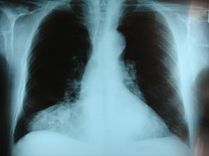
Diaphragmatic hernia is a defect or hole in the diaphragm that allows the abdominal contents to move into the chest cavity. Treatment is usually surgical.

<span class="mw-page-title-main">Catamenial pneumothorax</span> Medical condition

Catamenial pneumothorax is a spontaneous pneumothorax that recurs during menstruation, within 72 hours before or after the onset of a cycle. It usually involves the right side of the chest and right lung, and is associated with thoracic endometriosis. A third to a half of patients have pelvic endometriosis as well. Despite this association, CP is still considered to be misunderstood as is endometriosis considered to be underdiagnosed. The lack of a clear cause means that diagnosis and treatment is difficult. The disease is believed to be largely undiagnosed or misdiagnosed, leaving the true frequency unknown in the general population.

<span class="mw-page-title-main">Pentalogy of Cantrell</span> Medical condition

Pentalogy of Cantrell is an extremely rare congenital syndrome that causes defects involving the diaphragm, abdominal wall, pericardium, heart and lower sternum.

<span class="mw-page-title-main">Bochdalek hernia</span> Medical condition

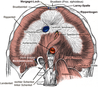
Bochdalek hernia is one of two forms of a congenital diaphragmatic hernia, the other form being Morgagni hernia. A Bochdalek hernia is a congenital abnormality in which an opening exists in the infant's diaphragm, allowing normally intra-abdominal organs to enter into the thoracic cavity. In the majority of people, the affected lung will be deformed, and the resulting lung compression can be life-threatening. Bochdalek hernias occur more commonly on the posterior left side.

<span class="mw-page-title-main">ZFPM2</span> Protein-coding gene in the species Homo sapiens

Zinc finger protein ZFPM2, i.e. zinc finger protein, FOG family member 2, but also termed Friend of GATA2, Friend of GATA-2, FOG2, or FOG-2, is a protein that in humans is encoded by the ZFPM2 and in mice by the Zfpm2 gene.

<span class="mw-page-title-main">Diaphragmatic rupture</span> Tear in the thoracic diaphragm, usually caused by physical trauma

Diaphragmatic rupture is a tear of the diaphragm, the muscle across the bottom of the ribcage that plays a crucial role in breathing. Most commonly, acquired diaphragmatic tears result from physical trauma. Diaphragmatic rupture can result from blunt or penetrating trauma and occurs in about 0.5% of all people with trauma.

<span class="mw-page-title-main">Pulmonary hypoplasia</span> Congenital disorder of respiratory system

Pulmonary hypoplasia is incomplete development of the lungs, resulting in an abnormally low number or small size of bronchopulmonary segments or alveoli. A congenital malformation, it most often occurs secondary to other fetal abnormalities that interfere with normal development of the lungs. Primary (idiopathic) pulmonary hypoplasia is rare and usually not associated with other maternal or fetal abnormalities.

<span class="mw-page-title-main">Fryns syndrome</span> Medical condition

Fryns syndrome is an autosomal recessive multiple congenital anomaly syndrome that is usually lethal in the neonatal period. Fryns (1987) reviewed the syndrome.

<span class="mw-page-title-main">Donnai–Barrow syndrome</span> Medical condition

Donnai–Barrow syndrome is a genetic disorder first described by Dian Donnai and Margaret Barrow in 1993. It is associated with LRP2. It is an inherited (genetic) disorder that affects many parts of the body.

Abdominal wall defects are a type of congenital defect that allows the stomach, the intestines, or other organs to protrude through an unusual opening that forms on the abdomen.

Lung hernia is a protrusion of lung outside of thoracic wall. In 20% patients with lung hernia, the incidence is congenital. In 80% of the cases, the hernia is noted after chest trauma, thoracic surgery or certain pulmonary diseases. Congenital hernia occurs because of the weakness of the suprapleural membrane or neck muscles. In pulmonary diseases such as asthma, frequent coughing can lead to high intra thoracic pressure, causing the lung to herniate out. Lung hernia may occur near the neck (cervical), between the ribs (intercostal), near the vertebrae (paravertebral) or near the sternum (parasternal).

<span class="mw-page-title-main">Pulmonary agenesis</span> Medical condition

Pulmonary agenesis is an inborn lung underdevelopment that is rare and potentially lethal. The disorder is caused by a complete developmental arrest of the primitive lung during embryonic life, and it is often associated with other developmental defects. Bilateral and unilateral pulmonary agenesis are classified, depending on whether one side of the lung or both sides are affected. Bilateral pulmonary agenesis is lethal, while the mortality rate of unilateral pulmonary agenesis is higher than 50%. Depending on the severity, the symptom ranges from none to various respiratory complaints. It is detectable prenatally, however, its nonspecific clinical features act as the obstacle for diagnosing. The exact cause of pulmonary agenesis is still obscure. However, theories have been raised regarding the vascular, iatrogenic, viral and genetic causes of pulmonary agenesis in an attempt to explain the pathogenesis of the disorder. In most cases of pulmonary agenesis, surgical resection is performed to remove the malformed lobe or the entire defected lung of the patient depending on the severity of the respiratory impairment.

Meacham syndrome is a rare genetic disorder which is characterized by lung, diaphragmatic and genitourinary anomalies.

References

  1. Gaxiola A, Varon J, Valladolid G (April 2009). "Congenital diaphragmatic hernia: an overview of the etiology and current management". Acta Paediatrica. 98 (4): 621–7. doi: 10.1111/j.1651-2227.2008.01212.x . PMID   19154527.
  2. Migliazza L, Bellan C, Alberti D, Auriemma A, Burgio G, Locatelli G, Colombo A (September 2007). "Retrospective study of 111 cases of congenital diaphragmatic hernia treated with early high-frequency oscillatory ventilation and presurgical stabilization". Journal of Pediatric Surgery. 42 (9): 1526–32. doi:10.1016/j.jpedsurg.2007.04.015. PMID   17848243.
  3. Herling, Anique; Makhdom, Fahd; Al-Shehri, Abdullah; Mulder, David S. (August 2014). "Bochdalek Hernia in a Symptomatic Adult". The Annals of Thoracic Surgery. 98 (2): 701–704. doi: 10.1016/j.athoracsur.2013.09.074 . PMID   25087794.
  4. Arráez-Aybar LA, González-Gómez CC, Torres-García AJ (2009). "Morgagni-Larrey parasternal diaphragmatic hernia in the adult". Rev Esp Enferm Dig. 101 (5): 357–66. doi: 10.4321/S1130-01082009000500009 . PMID   19527083.
  5. Wynn, Julia; Lan, Yu; Chung, Wendy (2014). "Genetic causes of congenital diaphragmatic hernia". Seminars in Fetal and Neonatal Medicine. 19 (6): 324–330. doi:10.1016/j.siny.2014.09.003. PMC   4259843 . PMID   25447988.
  6. Qiao, Lu; et al. (2021). "Rare and de novo variants in 827 congenital diaphragmatic hernia probands implicate LONP1 as candidate risk gene". American Journal of Human Genetics. 108 (10): 1964–1980. doi:10.1016/j.ajhg.2021.08.011. PMC   8546037 . PMID   34547244.
  7. Qi, Hongjian; Yu, Lan; Zhou, Xueya; Wynn, Julia; Zhao, Haoquan; Guo, Yicheng; Zhu, Na; Kitaygorodsky, Alexander; Hernan, Rebecca; Aspelund, Gudrun; Lim, Foong-Yen; Crombleholme, Timothy; Cusick, Robert; Azarow, Kenneth; Danko, Melissa E.; Chung, Dai; Warner, Brad W.; Mychaliska, George B.; Potoka, Douglas; Wagner, Amy J.; Elfiky, Mahmoud; Wilson, Jay M.; Nickerson, Debbie; Bamshad, Michael; High, Frances A.; Longoni, Mauro; Donahoe, Patricia K.; Chung, Wendy K.; Shen, Yufeng (2018). "De novo variants in congenital diaphragmatic hernia identify MYRF as a new syndrome and reveal genetic overlaps with other developmental disorders". PLOS Genetics. 14 (12): e1007822. doi: 10.1371/journal.pgen.1007822 . PMC   6301721 . PMID   30532227. S2CID   54480742.
  8. "Deadly hernia corrected in womb – Surgeons have developed an operation to repair a potentially fatal abnormality in babies before they are born". BBC News. 2004-07-26. Retrieved 2006-07-14. – report of new operation, pioneered at London's King's College Hospital which reduced death rates in the most at risk by 50%
  9. Swain F, Klaus A, Achem S, Hinder R (2001). "Congenital Diaphragmatic Hernia in Adults". Surgical Innovation. 8 (4): 246–255. doi:10.1177/155335060100800404. PMID   11813142. S2CID   30028643.
  10. Tiruvoipati R, Vinogradova Y, Faulkner G, Sosnowski AW, Firmin RK, Peek GJ (2007). "Predictors of outcome in patients with congenital diaphragmatic hernia requiring extracorporeal membrane oxygenation". J. Pediatr. Surg. 42 (8): 1345–50. doi:10.1016/j.jpedsurg.2007.03.031. PMID   17706494.
  11. Logan JW, Rice HE, Goldberg RN, Cotten CM (2007). "Congenital diaphragmatic hernia: a systematic review and summary of best-evidence practice strategies". Journal of Perinatology. 27 (9): 535–49. doi:10.1038/sj.jp.7211794. PMID   17637787. S2CID   15451125.
  12. Becmeur F, Talon I, Schaarschmidt K, et al. (2005). "Thoracoscopic diaphragmatic eventration repair in children: about 10 cases". J. Pediatr. Surg. 40 (11): 1712–5. doi:10.1016/j.jpedsurg.2005.07.008. PMID   16291157.
  13. Pediatric Congenital Diaphragmatic Hernia at eMedicine