Entamoeba is a genus of Amoebozoa found as internal parasites or commensals of animals.

Entamoeba histolytica is an anaerobic parasitic amoebozoan, part of the genus Entamoeba. Predominantly infecting humans and other primates causing amoebiasis, E. histolytica is estimated to infect about 35-50 million people worldwide. E. histolytica infection is estimated to kill more than 55,000 people each year. Previously, it was thought that 10% of the world population was infected, but these figures predate the recognition that at least 90% of these infections were due to a second species, E. dispar. Mammals such as dogs and cats can become infected transiently, but are not thought to contribute significantly to transmission.
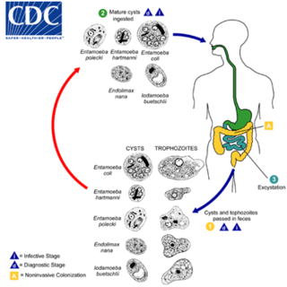
Entamoeba coli is a non-pathogenic species of Entamoeba that frequently exists as a commensal parasite in the human gastrointestinal tract. E. coli is important in medicine because it can be confused during microscopic examination of stained stool specimens with the pathogenic Entamoeba histolytica. This amoeba does not move much by the use of its pseudopod, and creates a "sur place (non-progressive) movement" inside the large intestine. Usually, the amoeba is immobile, and keeps its round shape. This amoeba, in its trophozoite stage, is only visible in fresh, unfixed stool specimens. Sometimes the Entamoeba coli have parasites as well. One is the fungus Sphaerita spp. This fungus lives in the cytoplasm of the E. coli. While this differentiation is typically done by visual examination of the parasitic cysts via light microscopy, new methods using molecular biology techniques have been developed. The scientific name of the amoeba, E. coli, is often mistaken for the bacterium, Escherichia coli. Unlike the bacterium, the amoeba is mostly harmless, and does not cause as many intestinal problems as some strains of the E. coli bacterium. To make the naming of these organisms less confusing, "alternate contractions" are used to name the species for the purpose making the naming easier; for example, using Esch. coli and Ent. coli for the bacterium and amoeba, instead of using E. coli for both.
Amoebozoa is a major taxonomic group containing about 2,400 described species of amoeboid protists, often possessing blunt, fingerlike, lobose pseudopods and tubular mitochondrial cristae. In traditional and currently no longer supported classification schemes, Amoebozoa is ranked as a phylum within either the kingdom Protista or the kingdom Protozoa. In the classification favored by the International Society of Protistologists, it is retained as an unranked "supergroup" within Eukaryota. Molecular genetic analysis supports Amoebozoa as a monophyletic clade. Modern studies of eukaryotic phylogenetic trees identify it as the sister group to Opisthokonta, another major clade which contains both fungi and animals as well as several other clades comprising some 300 species of unicellular eukaryotes. Amoebozoa and Opisthokonta are sometimes grouped together in a high-level taxon, variously named Unikonta, Amorphea or Opimoda.

Abdominal pain, also known as a stomach ache, is a symptom associated with both cancer and serious medical issues.

Gastrointestinal diseases refer to diseases involving the gastrointestinal tract, namely the esophagus, stomach, small intestine, large intestine and rectum, and the accessory organs of digestion, the liver, gallbladder, and pancreas.

Echinococcus multilocularis is a small cyclophyllid tapeworm found extensively in the northern hemisphere. E. multilocularis, along with other members of the Echinococcus genus, produce diseases known as echinococcosis. Unlike E. granulosus,E. multilocularis produces many small cysts that spread throughout the internal organs of the infected animal. The resultant disease is called Alveolar echinococcosis, and is caused by ingesting the eggs of E. multilocularis.

Balantidiasis is a protozoan infection caused by infection with Balantidium coli.

Ischemic colitis is a medical condition in which inflammation and injury of the large intestine result from inadequate blood supply. Although uncommon in the general population, ischemic colitis occurs with greater frequency in the elderly, and is the most common form of bowel ischemia. Causes of the reduced blood flow can include changes in the systemic circulation or local factors such as constriction of blood vessels or a blood clot. In most cases, no specific cause can be identified.

In the anatomy of humans and homologous primates, the descending colon is the part of the colon extending from the left colic flexure to the level of the iliac crest. The function of the descending colon in the digestive system is to store the remains of digested food that will be emptied into the rectum.

Balamuthia mandrillaris is a free-living amoeba that causes the rare but deadly neurological condition granulomatous amoebic encephalitis (GAE). B. mandrillaris is a soil-dwelling amoeba and was first discovered in 1986 in the brain of a mandrill that died in the San Diego Wild Animal Park.

A liver abscess is a mass filled with pus inside the liver. Common causes are abdominal conditions such as appendicitis or diverticulitis due to haematogenous spread through the portal vein. It can also develop as a complication of a liver injury.
Protozoan infections are parasitic diseases caused by organisms formerly classified in the kingdom Protozoa. They are usually contracted by either an insect vector or by contact with an infected substance or surface and include organisms that are now classified in the supergroups Excavata, Amoebozoa, SAR, and Archaeplastida.

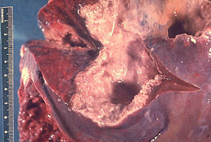
Amoebiasis, or amoebic dysentery, is an infection of the intestines caused by a parasitic amoeba Entamoeba histolytica. Amoebiasis can be present with no, mild, or severe symptoms. Symptoms may include lethargy, loss of weight, colonic ulcerations, abdominal pain, diarrhea, or bloody diarrhea. Complications can include inflammation and ulceration of the colon with tissue death or perforation, which may result in peritonitis. Anemia may develop due to prolonged gastric bleeding.

A pyogenic liver abscess is a type of liver abscess caused by bacteria.

Amoebic brain abscess is an affliction caused by the anaerobic parasitic protist Entamoeba histolytica. It is extremely rare; the first case being reported in 1849. Brain abscesses resulting from Entamoeba histolytica are difficult to diagnose and very few case reports suggest complete recovery even after the administration of appropriate treatment regimen.
Entamoeba moshkovskii is part of the genus Entamoeba. It is found in areas with polluted water sources, and is prevalent in places such as Malaysia, India, and Bangladesh, but more recently has made its way to Turkey, Australia, and North America. This amoeba is said to rarely infect humans, but recently this has changed. It is in question as to whether it is pathogenic or not. Despite some sources stating this is a free living amoeba, various studies worldwide have shown it contains the ability to infect humans, with some cases of pathogenic potential being reported. Some of the symptoms that often occur are diarrhea, weight loss, bloody stool, and abdominal pain. The first known human infection also known as the "Laredo strain" of Entamoebic mushkovskii was in Laredo, Texas in 1991, although it was first described by a man named Tshalaia in 1941 in Moscow, Russia. It is known to affect people of all ages and genders.
Entamoeba invadens is an amoebozoa parasite of reptiles, within the genus Entamoeba. It is closely related to the human parasite Entamoeba histolytica, causing similar invasive disease in reptiles, in addition to a similar morphology and lifecycle.

Naegleria fowleri, colloquially known as a "brain-eating amoeba", is a species of the genus Naegleria, belonging to the phylum Percolozoa, which is technically not classified as a true amoeba, but a shapeshifting amoeboflagellate excavate. It is a free-living, bacteria-eating microorganism that can be pathogenic, causing an extremely rare, sudden, severe, and usually fatal brain infection called naegleriasis or primary amoebic meningoencephalitis (PAM). This microorganism is typically found in bodies of warm freshwater, such as ponds, lakes, rivers, hot springs, warm water discharge from industrial or power plants, geothermal well water, poorly maintained or minimally chlorinated swimming pools, water heaters, soil, and pipes connected to tap water. It can be seen in either an amoeboid or temporary flagellate stage.
Checkpoint inhibitor induced colitis is an inflammatory condition affecting the colon (colitis), which is caused by cancer immunotherapy. Symptoms typically consist of diarrhea, abdominal pain and rectal bleeding. Less commonly, nausea and vomiting may occur, which may suggest the present of gastroenteritis. The severity of diarrhea and colitis are graded based on the frequency of bowel movements and symptoms of colitis, respectively.