Acanthamoeba keratitis

Last updated
Acanthamoeba keratitis
Parasite140120-fig1 Acanthamoeba keratitis Figure 1B.png
Fluorescein observation of an eye with Acanthamoeba keratitis
Specialty Ophthalmology, infectious diseases   OOjs UI icon edit-ltr-progressive.svg
Complications Visual impairment, blindness
Risk factors Contact lens wearer, contaminated water supply, low socioeconomic status
TreatmentTopical medications, surgical debridement, corneal transplantation
Frequency1.2–3 million people per year; 1 per 10,000 contact wearers [1]

Acanthamoeba keratitis (AK) is a rare disease in which amoebae of the genus Acanthamoeba invade the clear portion of the front (cornea) of the eye. It affects roughly 100 people in the United States each year. [2] Acanthamoeba are protozoa found nearly ubiquitously in soil and water and can cause infections of the skin, eyes, and central nervous system. [3]

Contents

Infection of the cornea by Acanthamoeba is difficult to treat with conventional medications, and AK may cause permanent visual impairment or blindness, due to damage to the cornea or through damage to other structures important to vision. [4] [5] Recently, AK has been recognized as an orphan disease and a funded project, orphan diseases Acanthamoeba keratitis (ODAK), has tested the effects of a diverse range drugs and biocides on AK. [6]

Pathogenesis

In the United States, Acanthamoeba keratitis is nearly always associated with soft contact lens use. [7] Acanthamoeba spp. is most commonly introduced to the eye by contact lenses that have been exposed to the organism through the use of contaminated lens solution, using homemade saline-based solution or tap water, or from wearing contact lenses while bathing or swimming. [8] [9] However, it may also be introduced to the eye by exposure to soil or vegetation, or by trauma. [2] In fact, the first case of Acanthamoeba keratitis described was due to ocular trauma. [2] Once on the contact lens, Acanthamoeba is able to survive in the space between the contact lens and the surface of the eye. [9] [10] [11] Soft contact lenses are more adherent to the corneal surface than hard lenses, which allows the Acanthamoeba organism to bind to mannosylated glycoproteins on the corneal surface. [12] Expression of these proteins on the corneal surface is increased by contact lens use. [11] This increase in glycoprotein content, along with microtrauma to the corneal epithelial surface due to contact lens use increases the risk for infection. [12] [13] Once the organism has gained access to the surface of the eye, it is able to invade through the epithelium and Bowman's layer. In some cases, the infection can then group around corneal nerves, producing radial deposits (radial keratoneuritis), and causing extreme pain. These are features also seen in viral and bacterial keratitis, and may be misleading. [14] [12] [11] The organism is also capable of invading deeper into the cornea; using metalloproteases it is able to penetrate deep into the stroma of the cornea. [12] As the disease progresses, it may penetrate through the cornea but very rarely causes infection inside the eye (endophthalmitis) due to a robust neutrophil response in the anterior chamber. [12] [11]

While the vast majority of cases of Acanthamoeba keratitis occur in contact lens wearers, there have been many cases of Acanthamoeba described in those who do not wear contact lenses, especially outside the United States. [15] [16] In non-contact lens users, the greatest risks for developing Acanthamoeba infection are trauma and exposure to contaminated water. [17] Further predisposing factors include contaminated home water supply, and low socioeconomic status. Infection is also more commonly seen in tropical or sub-tropical climates. [17]

Beyond the route of inoculation into the eye and external risk factors, host factors are also likely to play a significant role in the development of Acanthamoeba keratitis. In fact, studies of contact lens users in the United Kingdom, Japan, and New Zealand found that 400 to 800 per 10,000 asymptomatic contact lens users had lens storage cases contaminated with Acanthamoeba spp. [4] However, the rate of Acanthamoeba keratitis among these patients was only 0.01 to 1.49 per 10,000 contact lens users. [4] Although the exact host factors have not been fully described, it is likely that corneal epithelial defects, tear film composition, eye surface pH, and the level of anti-Acanthamoeba IgA antibodies in the tear film play a role in the development of Acanthamoeba keratitis. [7] [4]

Life cycle

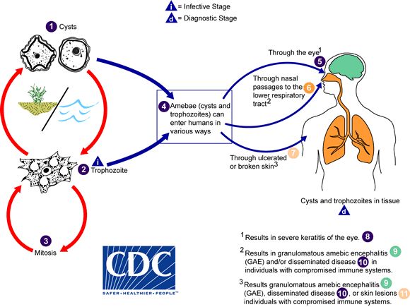
Species within the genus, Acanthamoeba, are generally free-living trophozoites. These trophozoites are relatively ubiquitous and can live in, but are not restricted to, tap water, freshwater lakes, rivers and soil. [18] In addition to the trophozoite stage, the organism can also form a double-walled cyst which may also be present in the environment, and can be very difficult to eradicate through medical treatment. Both of these stages are usually non-nucleated and reproduce by the means of binary fission. [19]

Acanthamoeba trophozoite. Scale bar: 10 mm Parasite140120-fig3 Acanthamoeba keratitis Figure 3B.png
Acanthamoeba trophozoite. Scale bar: 10 μm

Diagnosis

Due to the relative rarity of Acanthamoeba keratitis (AK) compared to other causes of keratitis (bacterial, viral, etc.), it is often misdiagnosed, especially in the early stages of the disease. [20] AK should be considered in all patients who use contact lenses, and following corneal abrasions or trauma. A thorough history should be obtained, especially relating to contact lens use and any recent changes in contact lens solution, exposure of the eyes to water or foreign objects, and symptoms that the patient is experiencing. The symptoms classically attributed to AK include decreased or blurred vision, sensitivity to light (photophobia), redness of the eye (conjunctival hyperemia), and pain out of proportion to physical exam findings. [15] [7] Another clinical feature that can distinguish Acanthamoeba from bacterial causes of keratitis is a lack of discharge from the eye. [12] [4]

On physical exam, findings will depend on the stage of the disease. Early manifestations in the cornea can be seen as punctate keratopathy, pseudodendrites, and epithelial or subepithelial corneal deposits. [11] These features can lead an examiner to confuse AK with a viral keratitis, such as that caused by varicella zoster virus or herpes simplex virus. [20] As the disease progresses and infiltrates the corneal stroma, a classic "ring infiltrate" may be present on examination (although this is only seen in about 50% of cases). [11] [12] Corneal ulceration, or in severe cases, perforation, can also occur and may be accompanied by hypopyon. [12] [21]

In cases of keratitis, diagnosis is typically achieved through evaluation of corneal scrapings. Scrapings are taking from the cornea, and plated on agar for culture, and also can be stained using Gram stain and Giemsa stain to differentiate between bacterial keratitis and AK. To culture Acanthamoeba, scrapings are placed on a non-nutrient agar saline plate seeded with a gram-negative bacteria such as E. coli . If Acanthamoeba are present, they will reproduce readily and become visible on the plate under 10–20 times objective on an inverted microscope. Polymerase chain reaction (PCR) can be used to confirm a diagnosis of Acanthamoeba keratitis, especially when contact lenses are not involved. [22] Confocal microscopy is a non-invasive technique that allows visualization of Acanthamoeba in vivo in cases in which corneal scraping, culture, and cytology do not yield a diagnosis. [23]

Treatment

Once Acanthamoeba keratitis is diagnosed, initiation of timely and appropriate treatment will have a significant impact on visual outcomes. Medical therapy aims to eradicate both trophozoite and cystic forms of Acanthamoeba and also control the inflammatory response.[ citation needed ]

Medical therapy

Multiple classes of drugs have been found to be effective in killing the trophozoite form of Acanthamoeba, including anti-bacterial, anti-fungal, anti-protozoal, and anti-neoplastic agents. However, no single therapy has been found to eliminate both trophozoite and cystic forms, and to eradicate corneal infection. [4] [15] [12]

One class of medications used in treatment is the biguanides, which include polyhexamethylene biguanide (PHMB) 0.02% to 0.06% drops, and chlorhexidine 0.02 to 0.2% drops. [12] [4] [21] These medications disrupt the cell wall of the trophozoite organism, leading to its death. However, these agents have shown limited efficacy against the cystic forms. [12] [24] Due to the efficacy of these drugs against the Acanthamoeba, as well as their low toxicity to the cornea, they are commonly used as the first line medications in the treatment of AK. [12] [21] Biguanides have also been found to act synergistically when used in combination with diamidines, with propamidine isethionate and hexamidine being the most commonly used. [25] A limitation of diamidine use is relative corneal toxicity with long term use. [12] A combined regimen of propamidine, miconazole nitrate, and neomycin has also been suggested. [26] [27] [28] Due to the potential for negative longterm visual outcomes with AK, therapy is usually started with a combination of a biguanide and a diamidine. Early use of high dose dual therapy helps to eliminate both trophozoite and cyst forms of the organism, while also preventing deep penetration of cysts into the corneal stroma. Cysts that are not eradicated from the cornea will cause recurrence. [4] [15] [12] The treatment is often initiated by instilling drops onto the surface of the eye every hour, 24 hours a day, for at least the first 48–72 hours. If an appropriate response to therapy, this may be reduced to hourly administrations during the day only, which is continued for several weeks to months. [4] [12]

Beyond anti-amoebic therapies, there is also a role for topical steroids of anti-inflammatory medications in the treatment of Acanthamoeba keratitis. During infection, severe inflammation in the cornea and anterior chamber can cause more severe symptoms including pain and visual disturbance. [12] Topical steroids may be used to reduce this inflammation and thereby alleviate symptoms. [12] [21] However, the role of steroids is typically very limited, because their dampening of the immune response may lead to worsening of the infection. [4] [21] Additionally, steroids can increase the number of trophozoites in the cornea by inducing excystation. [29] Therefore it is typically recommended that steroids be used briefly to aid in symptom resolution, and that anti-amoebic agents be used both during, and for several weeks after topical steroid use. [15]

Surgical treatment

Surgical debridement of an infected cornea can also be used to reduce organism load and excise devitalized tissue from the cornea. It may also improve the efficacy of medical therapy by promoting penetration of medication into deeper layers of the cornea. [4] [12] In cases of corneal ulceration or perforation, or if corneal scarring is severe, corneal transplant may be required. [24] [25] This typically involves full thickness transplantation of the cornea from a healthy donor eye. The size of the graft should be kept as small as possible, as larger grafts carry a great risk of host rejection, and due to the possibility of graft revision surgery. While surgery is capable of restoring vision by replacing a damaged cornea, it also carries risks of recurrent Acanthamoeba infection or graft failure. For this reason, anti-amoebic medications should be started prior to surgery, and continued for several weeks afterward. If there is suspicion or evidence of recurrent infection, cultures should be sent. If cultures are positive, anti-amoebic therapy should be continued for 6 months. [4] [12] [20]

Outcomes following surgery are typically much better for patients who receive surgery for vision improvement following infection resolution, and therefore all efforts should be made to maximize medical management before attempting surgery. [12]

Epidemiology

A study in Austria reported a total of 154 cases of Acanthamoeba keratitis over a 20-year period. The age of those with AK ranged from 8 to 82 years old and 58% of the people were female. The data showed that 89% of the infected patients were contact lens wearers, almost all cases occurred only in one eye, and 19% required a corneal transplant. [30]

Related Research Articles

<span class="mw-page-title-main">Contact lens</span> Lenses placed on the eyes surface

Contact lenses, or simply contacts, are thin lenses placed directly on the surface of the eyes. Contact lenses are ocular prosthetic devices used by over 150 million people worldwide, and they can be worn to correct vision or for cosmetic or therapeutic reasons. In 2010, the worldwide market for contact lenses was estimated at $6.1 billion, while the US soft lens market was estimated at $2.1 billion. Multiple analysts estimated that the global market for contact lenses would reach $11.7 billion by 2015. As of 2010, the average age of contact lens wearers globally was 31 years old, and two-thirds of wearers were female.

<span class="mw-page-title-main">Keratoconus</span> Medical condition involving the eye

Keratoconus (KC) is a disorder of the eye that results in progressive thinning of the cornea. This may result in blurry vision, double vision, nearsightedness, irregular astigmatism, and light sensitivity leading to poor quality-of-life. Usually both eyes are affected. In more severe cases a scarring or a circle may be seen within the cornea.

<i>Acanthamoeba</i> Genus of protozoans

Acanthamoeba is a genus of amoebae that are commonly recovered from soil, fresh water, and other habitats. The genus Acanthamoeba has two stages in its life cycle, the metabolically active trophozoite stage and a dormant, stress-resistant cyst stage. In nature, Acanthamoeba species are generally free-living bacterivores. However, they are also opportunistic pathogens able to cause serious and sometimes fatal infections in humans and other animals.

<span class="mw-page-title-main">Keratitis</span> Medical condition

Keratitis is a condition in which the eye's cornea, the clear dome on the front surface of the eye, becomes inflamed. The condition is often marked by moderate to intense pain and usually involves any of the following symptoms: pain, impaired eyesight, photophobia, red eye and a 'gritty' sensation. Diagnosis of infectious keratitis is usually made clinically based on the signs and symptoms as well as eye examination, but corneal scrapings may be obtained and evaluated using microbiological culture or other testing to identify the causative pathogen.

Free-living amoebae in the Amoebozoa group are important causes of disease in humans and animals.

<span class="mw-page-title-main">Dry eye syndrome</span> Medical condition

Dry eye syndrome, also known as keratoconjunctivitis sicca, is the condition of having dry eyes. Symptoms include dryness in the eye, irritation, redness, discharge, blurred vision, and easily fatigued eyes. Symptoms range from mild and occasional to severe and continuous. Dry eye syndrome can lead to blurred vision, instability of the tear film, increased risk of damage to the ocular surface such as scarring of the cornea, and changes in the eye including the neurosensory system.

<span class="mw-page-title-main">Thygeson's superficial punctate keratopathy</span> Medical condition

Thygeson's superficial punctate keratopathy (TSPK) is a disease of the eyes. The causes of TSPK are not currently known, but details of the disease were first published in the Journal of the American Medical Association in 1950 by renowned American ophthalmologist Phillips Thygeson (1903–2002), after whom it is named.

<span class="mw-page-title-main">Red eye (medicine)</span> Eye that appears red due to illness or injury

A red eye is an eye that appears red due to illness or injury. It is usually injection and prominence of the superficial blood vessels of the conjunctiva, which may be caused by disorders of these or adjacent structures. Conjunctivitis and subconjunctival hemorrhage are two of the less serious but more common causes.

<span class="mw-page-title-main">Corneal abrasion</span> Scratch to the surface of the cornea of the eye

Corneal abrasion is a scratch to the surface of the cornea of the eye. Symptoms include pain, redness, light sensitivity, and a feeling like a foreign body is in the eye. Most people recover completely within three days.

<span class="mw-page-title-main">Corneal ulcers in animals</span> Veterinary inflammatory condition of the cornea

A corneal ulcer, or ulcerative keratitis, is an inflammatory condition of the cornea involving loss of its outer layer. It is very common in dogs and is sometimes seen in cats. In veterinary medicine, the term corneal ulcer is a generic name for any condition involving the loss of the outer layer of the cornea, and as such is used to describe conditions with both inflammatory and traumatic causes.

Fungal keratitis is a fungal infection of the cornea, which can lead to blindness. It generally presents with a red, painful eye and blurred vision. There is also increased sensitivity to light, and excessive tears or discharge.

<span class="mw-page-title-main">Corneal neovascularization</span> Medical condition

Corneal neovascularization (CNV) is the in-growth of new blood vessels from the pericorneal plexus into avascular corneal tissue as a result of oxygen deprivation. Maintaining avascularity of the corneal stroma is an important aspect of healthy corneal physiology as it is required for corneal transparency and optimal vision. A decrease in corneal transparency causes visual acuity deterioration. Corneal tissue is avascular in nature and the presence of vascularization, which can be deep or superficial, is always pathologically related.

<span class="mw-page-title-main">Corneal ulcer</span> Medical condition of the eye

Corneal ulcer, also called keratitis, is an inflammatory or, more seriously, infective condition of the cornea involving disruption of its epithelial layer with involvement of the corneal stroma. It is a common condition in humans particularly in the tropics and in farming. In developing countries, children afflicted by vitamin A deficiency are at high risk for corneal ulcer and may become blind in both eyes persisting throughout life. In ophthalmology, a corneal ulcer usually refers to having an infection, while the term corneal abrasion refers more to a scratch injury.

<span class="mw-page-title-main">Fungal contamination of contact lenses</span>

Microbial corneal infection is the most serious and "most common vision threatening" complication of contact lens wear, which is believed to be strongly associated with contact lens cases. Such infections "are being increasingly recognized as an important cause of morbidity and blindness" and "may even be life-threatening." While the cornea is believed to be the most common site for fungal eye infections, other parts of the eye such as the orbit, sclera, eyelids, and more may also be involved. Contact lens cases are recognized as a "potential source of pathogens associated with corneal ulcers" and according to Moorfields Eye Hospital, contact lens wear is “the most prevalent risk factor for new cases of corneal ulcers.” Contaminants "isolated from contact lens associated corneal ulcers have often been shown to be" the same as found in the patient's contact lens case, thus providing evidence contaminated contact lens cases may be a "replenishable source of pathogenic microbes."

<span class="mw-page-title-main">Herpes simplex keratitis</span> Medical condition

Herpetic simplex keratitis is a form of keratitis caused by recurrent herpes simplex virus (HSV) infection in the cornea.

<span class="mw-page-title-main">Herpes zoster ophthalmicus</span> Shingles in the human eye

Herpes zoster ophthalmicus (HZO), also known as ophthalmic zoster, is shingles involving the eye or the surrounding area. Common signs include a rash of the forehead with swelling of the eyelid. There may also be eye pain and redness, inflammation of the conjunctiva, cornea or uvea, and sensitivity to light. Fever and tingling of the skin and allodynia near the eye may precede the rash. Complications may include visual impairment, increased pressure within the eye, chronic pain, and stroke.

Neurotrophic keratitis (NK) is a degenerative disease of the cornea caused by damage of the trigeminal nerve, which results in impairment of corneal sensitivity, spontaneous corneal epithelium breakdown, poor corneal healing and development of corneal ulceration, melting and perforation. This is because, in addition to the primary sensory role, the nerve also plays a role maintaining the integrity of the cornea by supplying it with trophic factors and regulating tissue metabolism.

Herbert Edward Kaufman is an American ophthalmologist who discovered idoxuridine, the first clinically useful antiviral agent; co-developed with William Bourne the clinical specular microscope to view the live corneal endothelium, co-developed timolol with Thomas Zimmerman, a new class of medications to treat glaucoma; corneal storage media for eye banks; natamycin, the first commercially available medication to treat fungal infections of the eye; co-developed with Tony Gasset the use of bandage contact lenses; and was involved in the first laser vision photorefractive keratectomy of the eye with Marguarite McDonald.

Exposure keratopathy is medical condition affecting the cornea of eyes. It can lead to corneal ulceration and permanent loss of vision due to corneal opacity.

Peripheral Ulcerative Keratitis (PUK) is a group of destructive inflammatory diseases involving the peripheral cornea in human eyes. The symptoms of PUK include pain, redness of the eyeball, photophobia, and decreased vision accompanied by distinctive signs of crescent-shaped damage of the cornea. The causes of this disease are broad, ranging from injuries, contamination of contact lenses, to association with other systemic conditions. PUK is associated with different ocular and systemic diseases. Mooren's ulcer is a common form of PUK. The majority of PUK is mediated by local or systemic immunological processes, which can lead to inflammation and eventually tissue damage. Standard PUK diagnostic test involves reviewing the medical history and a completing physical examinations. Two major treatments are the use of medications such as corticosteroids or other immunosuppressive agents and surgical resection of the conjunctiva. The prognosis of PUK is unclear with one study providing potential complications. PUK is a rare condition with an estimated incidence of 3 per million annually.

References

  1. "Acanthamoeba Infection: Background, Pathophysiology, Epidemiology". Medscape Reference. 2020-03-26. Retrieved 2020-05-30.
  2. 1 2 3 Scruggs BA, Quist TS, Salinas JL, Greiner MA, et al. (2019). Notes from the Field: Acanthamoeba Keratitis Cases — Iowa, 2002–2017. Morbidity and Mortality Weekly Report (MMWR).
  3. "CDC - Acanthamoeba Infection". www.cdc.gov. 2017-06-21. Retrieved 2018-11-06.
  4. 1 2 3 4 5 6 7 8 9 10 11 12 Lorenzo-Morales J, Khan NA, Walochnik J (2015). "An update on Acanthamoeba keratitis: diagnosis, pathogenesis and treatment". Parasite. 22: 10. doi:10.1051/parasite/2015010. PMC   4330640 . PMID   25687209. Open Access logo PLoS transparent.svg
  5. "CDC - Acanthamoeba Infection - General Information - Acanthamoeba Keratitis FAQs". Cdc.gov. Retrieved 2013-08-02.
  6. Baig AM (2019). "Drug targeting in Acanthamoeba keratitis: rational of using drugs that are already approved for ocular use in non-keratitis indications". Eye. 33 (3): 509–518. doi:10.1038/s41433-018-0245-6. PMC   6460712 . PMID   30356128.
  7. 1 2 3 Auran JD, Starr MB, Jakobiec FA (1987). "Acanthamoeba keratitis. A review of the literature". Cornea. 6 (1): 2–26. doi:10.1097/00003226-198706010-00002. PMID   3556011. S2CID   220579859.
  8. Ahmad SS (2018-07-01). "Water related ocular diseases". Saudi Journal of Ophthalmology. 32 (3): 227–233. doi:10.1016/j.sjopt.2017.10.009. PMC   6137694 . PMID   30224888.
  9. 1 2 JOHN D.T. (1993) Opportunistically pathogenic free-living amebae. In: J.P. Kreier and J.R. Baker (Eds.), Parasitic Protozoa. Vol. 3. Academic Press, New York, pp. 143–246.
  10. Badenoch PR, Adams M, Coster DJ (February 1995). "Corneal virulence, cytopathic effect on human keratocytes and genetic characterization of Acanthamoeba". International Journal for Parasitology. 25 (2): 229–39. doi:10.1016/0020-7519(94)00075-Y. PMID   7622330.
  11. 1 2 3 4 5 6 Niederkorn JY, Alizadeh H, Leher H, McCulley JP (May 1999). "The pathogenesis of Acanthamoeba keratitis". Microbes and Infection. 1 (6): 437–43. doi: 10.1016/S1286-4579(99)80047-1 . PMID   10602676.
  12. 1 2 3 4 5 6 7 8 9 10 11 12 13 14 15 16 17 18 19 20 pubmeddev. "Update on Acanthamoeba Keratitis: Diagnosis, Treatment, and Outcomes - PubMed - NCBI". www.ncbi.nlm.nih.gov. Retrieved 2018-11-12.
  13. Anger C, Lally JM (September 2008). "Acanthamoeba: a review of its potential to cause keratitis, current lens care solution disinfection standards and methodologies, and strategies to reduce patient risk". Eye & Contact Lens. 34 (5): 247–53. doi:10.1097/ICL.0b013e31817e7d83. PMID   18779663. S2CID   43868038.
  14. Alfawaz A (July 2011). "Radial keratoneuritis as a presenting sign in acanthamoeba keratitis". Middle East African Journal of Ophthalmology. 18 (3): 252–5. doi: 10.4103/0974-9233.84062 . PMC   3162742 . PMID   21887085.
  15. 1 2 3 4 5 Sharma S, Garg P, Rao GN (October 2000). "Patient characteristics, diagnosis, and treatment of non-contact lens related Acanthamoeba keratitis". The British Journal of Ophthalmology. 84 (10): 1103–8. doi:10.1136/bjo.84.10.1103. PMC   1723254 . PMID   11004092.
  16. Bharathi JM, Srinivasan M, Ramakrishnan R, Meenakshi R, Padmavathy S, Lalitha PN (2007). "A study of the spectrum of Acanthamoeba keratitis: a three-year study at a tertiary eye care referral center in South India". Indian Journal of Ophthalmology. 55 (1): 37–42. doi: 10.4103/0301-4738.29493 . PMID   17189885.
  17. 1 2 Garg P, Kalra P, Joseph J (November 2017). "Acanthamoeba keratitis". Indian Journal of Ophthalmology. 65 (11): 1079–1086. doi: 10.4103/ijo.IJO_826_17 . PMC   5700572 . PMID   29133630.
  18. "Acanthamoeba-General Information-Acanthamoeba keratitis". CDC. 2019-01-04.
  19. Neelam S, Niederkorn JY (June 2017). "Acanthamoeba Keratitis: Insights from Animal Models". The Yale Journal of Biology and Medicine. 90 (2): 261–268. PMC   5482302 . PMID   28656012.
  20. 1 2 3 Lorenzo-Morales J, Khan NA, Walochnik J (2015). "An update on Acanthamoeba keratitis: diagnosis, pathogenesis and treatment". Parasite. 22: 10. doi:10.1051/parasite/2015010. PMC   4330640 . PMID   25687209.
  21. 1 2 3 4 5 Hammersmith KM (August 2006). "Diagnosis and management of Acanthamoeba keratitis". Current Opinion in Ophthalmology. 17 (4): 327–31. doi:10.1097/01.icu.0000233949.56229.7d. PMID   16900022. S2CID   19843745.
  22. Pasricha G, Sharma S, Garg P, Aggarwal RK (July 2003). "Use of 18S rRNA gene-based PCR assay for diagnosis of acanthamoeba keratitis in non-contact lens wearers in India". Journal of Clinical Microbiology. 41 (7): 3206–11. doi:10.1128/JCM.41.7.3206-3211.2003. PMC   165372 . PMID   12843065.
  23. Nakano E, Oliveira M, Portellinha W, de Freitas D, Nakano K (September 2004). "Confocal microscopy in early diagnosis of Acanthamoeba keratitis". Journal of Refractive Surgery. 20 (5 Suppl): S737–40. doi:10.3928/1081-597X-20040903-23. PMID   15521280.
  24. 1 2 Alkharashi M, Lindsley K, Law HA, Sikder S (February 2015). "Medical interventions for acanthamoeba keratitis". The Cochrane Database of Systematic Reviews. 2 (2): CD010792. doi:10.1002/14651858.CD010792.pub2. PMC   4730543 . PMID   25710134.
  25. 1 2 Lindsay RG, Watters G, Johnson R, Ormonde SE, Snibson GR (September 2007). "Acanthamoeba keratitis and contact lens wear". Clinical & Experimental Optometry. 90 (5): 351–60. doi:10.1111/j.1444-0938.2007.00172.x. PMID   17697181. S2CID   3244497.
  26. Baig AM (2019). "Drug targeting in Acanthamoeba keratitis: Rational of using drugs that are already approved for ocular use in non-keratitis indications". Eye. 33 (3): 509–518. doi:10.1038/s41433-018-0245-6. PMC   6460712 . PMID   30356128.
  27. "Acanthamoeba: Treatment & Medication - eMedicine Infectious Diseases" . Retrieved 2009-02-27.
  28. Singh S, Sachdeva MP (July 1994). "Acanthamoeba keratitis". BMJ. 309 (6949): 273. doi:10.1136/bmj.309.6949.273. PMC   2540756 . PMID   7802782.
  29. McClellan K, Howard K, Niederkorn JY, Alizadeh H (2001-11-01). "Effect of Steroids on Acanthamoeba Cysts and Trophozoites". Investigative Ophthalmology & Visual Science. 42 (12): 2885–2893. ISSN   1552-5783. PMID   11687533.
  30. Walochnik J, Scheikl U, Haller-Schober EM (2014). "Twenty years of acanthamoeba diagnostics in Austria". The Journal of Eukaryotic Microbiology. 62 (1): 3–11. doi:10.1111/jeu.12149. PMC   4342769 . PMID   25047131.