Persistent tunica vasculosa lentis

Last updated
Persistent tunica vasculosa lentis
Schematic diagram of the human eye en.svg
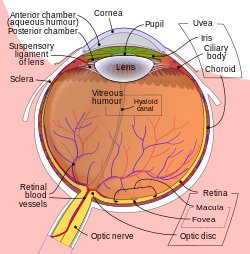
Anatomy human eye (vitreous humor)
Specialty Ophthalmology

Persistent tunica vasculosa lentis is a congenital ocular anomaly. It is a form of persistent fetal vasculature (PFV).

It is a developmental disorder of the vitreous. It is usually unilateral and first noticed in the neonatal period. It may be associated with microphthalmos, cataracts, and increased intraocular pressure. Elongated ciliary processes are visible through the dilated pupil. A USG B-scan confirms diagnosis in the presence of a cataract.

See also

Related Research Articles

<span class="mw-page-title-main">Superior vena cava</span> One of two veinous trunks bringing deoxygenated blood back to the heart

The superior vena cava (SVC) is the superior of the two venae cavae, the great venous trunks that return deoxygenated blood from the systemic circulation to the right atrium of the heart. It is a large-diameter (24 mm) short length vein that receives venous return from the upper half of the body, above the diaphragm. Venous return from the lower half, below the diaphragm, flows through the inferior vena cava. The SVC is located in the anterior right superior mediastinum. It is the typical site of central venous access via a central venous catheter or a peripherally inserted central catheter. Mentions of "the cava" without further specification usually refer to the SVC.

<span class="mw-page-title-main">Lens (vertebrate anatomy)</span> Eye structure

The lens, or crystalline lens, is a transparent biconvex structure in most land vertebrate eyes. Along with the cornea, aqueous and vitreous humours it refracts light, focusing it onto the retina. In many land animals the shape of the lens can be altered, effectively changing the focal length of the eye, enabling them to focus on objects at various distances. This adjustment of the lens is known as accommodation. In many fully aquatic vertebrates such as fish other methods of accommodation are used such as changing the lens's position relative to the retina rather than changing lens shape. Accommodation is analogous to the focusing of a photographic camera via changing its lenses. In land vertebrates the lens is flatter on its anterior side than on its posterior side, while in fish the lens is often close to spherical.

<span class="mw-page-title-main">Allantois</span> Embryonic structure

The allantois is a hollow sac-like structure filled with clear fluid that forms part of a developing amniote's conceptus. It helps the embryo exchange gases and handle liquid waste.

Tunica may refer to:

<span class="mw-page-title-main">Capsule of lens</span> Membrane surrounding the lens within the eyeball

The lens capsule is a component of the globe of the eye. It is a clear elastic basement membrane composed of collagen IV laminin etc. a quality that keeps it under constant tension. As a result, the lens naturally tends towards a rounder or more globular configuration, a shape it must assume for the eye to focus at a near distance. The lens capsule is the thickest basement membrane in the body.

<span class="mw-page-title-main">Schlemm's canal</span> Lymphatic-like vessel in the eye

Schlemm's canal is a circular lymphatic-like vessel in the eye. It collects aqueous humor from the anterior chamber and delivers it into the episcleral blood vessels. Canaloplasty may be used to widen it.

<span class="mw-page-title-main">Ectopia lentis</span> Medical condition

Ectopia lentis is a displacement or malposition of the eye's crystalline lens from its normal location. A partial dislocation of a lens is termed lens subluxation or subluxated lens; a complete dislocation of a lens is termed lens luxation or luxated lens.

<span class="mw-page-title-main">Leukocoria</span> Medical condition

Leukocoria is an abnormal white reflection from the retina of the eye. Leukocoria resembles eyeshine, but leukocoria can also occur in animals that lack eyeshine because their retina lacks a tapetum lucidum.

<span class="mw-page-title-main">Persistent pupillary membrane</span> Medical condition

Persistent pupillary membrane (PPM) is a condition of the eye involving remnants of a fetal membrane that persist as strands of tissue crossing the pupil. The pupillary membrane in mammals exists in the fetus as a source of blood supply for the lens. It normally atrophies from the time of birth to the age of four to eight weeks. PPM occurs when this atrophy is incomplete. It generally does not cause any symptoms. The strands can connect to the cornea or lens, but most commonly to other parts of the iris. Attachment to the cornea can cause small corneal opacities, while attachment to the lens can cause small cataracts. Using topical atropine to dilate the pupil may help break down PPMs.

<span class="mw-page-title-main">Tunica albuginea of testis</span> Tissue surrounding the testicle

The tunica albugine is a dense, blue-white layer of fibrous tissue surrounding the testis. It is the middle of three enveloppes forming the capsule of the testis; it is deep to the visceral layer of tunica vaginalis, and superficial to the tunica vasculosa testis.

The tunica vasculosa lentis is an extensive capillary network, spreading over the posterior and lateral surfaces of the lens of the eye. It disappears shortly after birth.

The tunica vasculosa testis is the inner-most of the three layers that form the capsule of the testis. It consists of a vascular plexus and loose connective tissue. It extends into the testis itself to line the surfaces of individual septa of testis.

Tunica vasculosa can refer to:

<span class="mw-page-title-main">Tunica albuginea (penis)</span> Anatomical structure of the penis

The tunica albuginea is the fibrous envelope that extends the length of the corpora cavernosa penis and corpus spongiosum penis. It is a bi-layered structure that includes an outer longitudinal layer and an inner circular layer.

<span class="mw-page-title-main">KLF2</span> Protein-coding gene in the species Homo sapiens

Krüppel-like Factor 2 (KLF2), also known as lung Krüppel-like Factor (LKLF), is a protein that in humans is encoded by the KLF2 gene on chromosome 19. It is in the Krüppel-like factor family of zinc finger transcription factors, and it has been implicated in a variety of biochemical processes in the human body, including lung development, embryonic erythropoiesis, epithelial integrity, T-cell viability, and adipogenesis.

<span class="mw-page-title-main">Persistent fetal vasculature</span> Medical condition

Persistent fetal vasculature(PFV), also known as persistent fetal vasculature syndrome (PFSV), and until 1997 known primarily as persistent hyperplastic primary vitreous (PHPV), is a rare congenital anomaly which occurs when blood vessels within the developing eye, known as the embryonic hyaloid vasculature network, fail to regress as they normally would in-utero after the eye is fully developed. Defects which arise from this lack of vascular regression are diverse; as a result, the presentation, symptoms, and prognosis of affected patients vary widely, ranging from clinical insignificance to irreversible blindness. The underlying structural causes of PFV are considered to be relatively common, and the vast majority of cases do not warrant additional intervention. When symptoms do manifest, however, they are often significant, causing detrimental and irreversible visual impairment. Persistent fetal vasculature heightens the lifelong risk of glaucoma, cataracts, intraocular hemorrhages, and Retinal detachments, accounting for the visual loss of nearly 5% of the blind community in the developed world. In diagnosed cases of PFV, approximately 90% of patients with a unilateral disease have associated poor vision in the affected eye.

Fuchs heterochromic iridocyclitis (FHI) is a chronic unilateral uveitis appearing with the triad of heterochromia, predisposition to cataract and glaucoma, and keratitic precipitates on the posterior corneal surface. Patients are often asymptomatic and the disease is often discovered through investigation of the cause of the heterochromia or cataract. Neovascularisation is possible and any eye surgery, such as cataract surgery, can cause bleeding from the fragile vessels in the atrophic iris causing accumulation of blood in the anterior chamber of the eye, also known as hyphema.

In biology, a tunica is a layer, coat, sheath, or similar covering. The word came to English from the Neo-Latin of science and medicine. Its literal sense is about the same as that of the word tunic, with which it is cognate. In biology one of its senses used to be the taxonomic name of a genus of plants, but the nomenclature has been revised and those plants are now included in the genus Petrorhagia.

PFV may refer to:

Arthur Brückner was a German-Swiss ophthalmologist, known for his research in sensory physiology and his studies involving the cytology of the eye.

References