| Chemosis | |
|---|---|
 | |
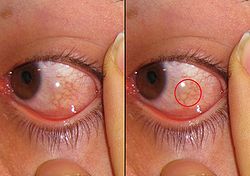
| Example of Chemosis | |
| Specialty | Immunology |
Chemosis is the swelling (or edema) of the conjunctiva. The term derives from the Greek words cheme and -osis, cheme meaning cockleshell due to the swollen conjunctiva resembling it, and -osis meaning condition. [1] The swelling is due to the oozing of exudate from abnormally permeable capillaries. In general, chemosis is a nonspecific sign of eye irritation. The outer surface covering appears to have fluid in it. The conjunctiva becomes swollen and gelatinous in appearance. Often, the eye area swells so much that the eyes become difficult or impossible to close fully. [2] Sometimes, it may also appear as if the iris has moved slightly backwards from the white part of the eye due to the fluid filled in the conjunctiva all over the eyes except the iris. The iris is not covered by this fluid and so it appears to be moved slightly inwards.