Problem with focusing light accurately on the retina due to the shape of the eye
Medical condition
Refractive error
Other names
Refraction error
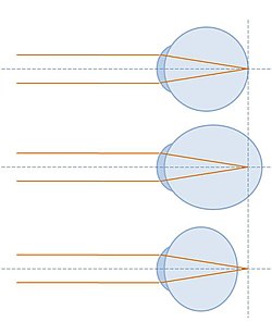
A correctly-focused eye (top), and two showing refractive error: In the middle image, the light is focused too far forward; in the bottom image, the focal point is behind the eye.
Refractive error is a problem with focusing light accurately on the retina due to the shape of the eye and/or cornea.[3] The most common types of refractive error are near-sightedness, far-sightedness, astigmatism, and presbyopia. Near-sightedness results in far away objects being blurry, far-sightedness and presbyopia result in close objects being blurry, and astigmatism causes objects to appear stretched out or blurry. Other symptoms may include double vision, headaches, and eye strain.[3]
Near-sightedness is due to the length of the eyeball being too long; far-sightedness the eyeball too short; astigmatism the cornea being the wrong shape, while presbyopia results from aging of the lens of the eye such that it cannot change shape sufficiently.[3] Some refractive errors occur more often among those whose parents are affected. Diagnosis is by eye examination.
Refractive errors are corrected with eyeglasses, contact lenses, or surgery.[3] Eyeglasses are the easiest and safest method of correction. Contact lenses can provide a wider field of vision; however they are associated with a risk of infection. Refractive surgery may consist of either permanently changing the shape of the cornea or, alternatively, implanting intraocular lenses.[3][4]
The number of people globally with refractive errors has been estimated at one to two billion.[5] Rates vary between regions of the world with about 25% of Europeans and 80% of Asians affected.[5] Near-sightedness is the most common disorder.[6] Rates among adults are between 15 and 49% while rates among children are between 1.2 and 42%.[7] Far-sightedness more commonly affects young children and the elderly.[8][9] Presbyopia affects most people over the age of 35.[3]
The number of people with refractive errors that have not been corrected was estimated at 660 million (10 per 100 people) in 2013.[10] Of these 9.5million were blind due to the refractive error.[10] It is one of the most common causes of vision loss along with cataracts, macular degeneration, and vitamin A deficiency.[11]
Classification
Top: farsighted corrected using convex lens. Bottom: nearsighted corrected using concave lens.
Refractive error – sometimes called "ametropia" – refers to a condition in which the refractive power of an eye does not match the length of the eye, so the image is focused away from the central retina, instead of directly on it.[12]
Myopia or nearsightedness: When the refractive power is too strong for the length of the eyeball, this is called myopia or nearsightedness. People with myopia typically have blurry vision when viewing distant objects because the eye is refracting more than necessary. Myopia can be corrected with a concave lens, which causes the divergence of light rays before they reach the cornea.[13]
Hyperopia or farsightedness: When the refractive power is too weak for the length of the eyeball, one has hyperopia or farsightedness. People with hyperopia have blurry vision when viewing near objects because the eye is unable to focus the light sufficiently. This can be corrected with convex lenses, which cause light rays to converge prior to hitting the cornea.[14]
Presbyopia: When the flexibility of the lens declines, typically due to age, the individual experiences difficulty in near vision, often relieved by reading glasses, bifocal, or progressive lenses.[15]
Astigmatism occurs when the refractive power of the eye is not uniform across the surface of the cornea because of asymmetry. In other words, the eye focuses light more strongly in one direction than another, leading to distortion of the image.[16]
Children are typically born hyperopic and shift toward emmetropia or myopia as their eyes lengthen through childhood.[17]
Refractive errors are typically measured using three numbers: sphere, cylinder, and axis.[20]
Sphere: This number denotes the strength of the lens needed to correct your vision. A "–" indicates nearsightedness while a "+" indicates farsightedness. Higher numbers indicate more power in either direction.
Cylinder: This number denotes the amount of astigmatism, if any.
Axis: This number notes the direction of the astigmatism and is written in degrees between 1 and 180.
An eye that has no refractive error when viewing distant objects is said to have emmetropia or be emmetropic meaning the eye is in a state in which it can focus parallel rays of light (light from distant objects) on the retina, without using any accommodation. A distant object, in this case, is defined as an object located beyond 6 meters, or 20 feet, from the eye, since the light from those objects arrives as essentially parallel rays when considering the limitations of human perception.[21]
There is evidence to suggest genetic predilection for refractive error. Individuals that have parents with certain refractive errors are more likely to have similar refractive errors.[3]
In studies of the genetic predisposition of refractive error, there is a correlation between environmental factors and the risk of developing myopia.[24] Myopia has been observed in individuals with visually intensive occupations.[23]Reading has also been found to be a predictor of myopia in children. It has been reported that children with myopia spent significantly more time reading than non-myopic children who spent more time playing outdoors.[23] Additionally, focusing on near objects for long periods of time - such as when reading, looking at close screens, or writing - has been associated with myopia.[25][26]Socioeconomic status and higher levels of education have also been reported to be a risk factor for myopia.[27]Blepharoptosis can also induce refractive errors.[28]
Normal refraction
In order to see a clear image, the eye must focus rays of light on to the light-sensing part of the eye – the retina, which is located in the back of the eye. This focusing – called refraction – is performed mainly by the cornea and the lens, which are located at the front of the eye, the anterior segment.[29]
When an eye focuses light correctly on to the retina when viewing distant objects, this is called emmetropia or being emmetropic. This means that the refractive power of the eye matches what is needed to focus parallel rays of light onto the retina. A distant object is defined as an object located beyond 6 meters (20 feet) from the eye.[citation needed]
When an object is located close to the eye, the rays of light from this object no longer approach the eye parallel to each other. Consequently, the eye must increase its refractive power to bring those rays of light together on the retina. This is called accommodation, and is accomplished by the eye thickening the lens.[29]
Diagnosis
A doctor uses a trial frame and trial lenses to measure the person's refractive error.
Blurry vision may result from any number of conditions not necessarily related to refractive errors. The diagnosis of a refractive error is usually confirmed by an eye care professional during an eye examination using a large number of lenses of different optical powers, and often a retinoscope (a procedure entitled retinoscopy). The eye care professional instructs the patient to view a distant spot while the clinician changes the lenses held before the person's eye and watches the pattern of reflection of a small light shone on the eye. Once the clinician arrives at an estimate of the patient's "objective" refractive error, the clinician typically shows the patient lenses of progressively higher or weaker powers in a process known as subjective refraction. Cycloplegic agents are frequently used to more accurately determine the amount of refractive error, particularly in children.[24]
Vision defects caused by refractive error can be distinguished from other problems using a pinhole occluder, which will improve vision only in the case of refractive error.[32]
Screening
When refractive errors in children are not treated, the child may be at risk of developing ambylopia, where vision may remain permanently blurry.[33] Because young children typically do not complain of blurry vision, the American Academy of Pediatrics recommends that children have yearly vision screening starting at three years old so that unknown refractive errors or other ophthalmic conditions can be found and treated if deemed necessary by healthcare professionals.[33][34]
Management
The management of refractive error is done post-diagnosis of the condition by either optometrists, ophthalmologists, refractionists, or ophthalmic medical practitioners.[35]
How refractive errors are treated or managed depends upon the amount and severity of the condition. Those who possess mild amounts of refractive error may elect to leave the condition uncorrected, particularly if the person is asymptomatic. For those who are symptomatic, glasses, contact lenses, refractive surgery, or a combination are typically used.[29][20][22]
Glasses
These are the most effective ways of correcting the refractive error. However, the availability and affordability of eyeglasses can present a difficulty for people in many low income settings of the world. Glasses also pose a challenge to children to whom they are prescribed to, due to children's tendency to not wear them as consistently as recommended.[36]
As mentioned earlier refractive errors are because of the improper focusing of the light in the retina. Eyeglasses work as an added lens of the eye serving to bend the light to bring it to focus on the retina. Depending on the eyeglasses, they serve many functions.[citation needed]
Reading glasses
These are general over-the-counter glasses which can be worn for easier reading, especially for defective vision due to aging called presbyopia.[citation needed]
Single vision prescription lenses
They can correct only one form of defective vision, either far-sightedness or near-sightedness.[citation needed]
Multifocal lenses
The multifocal lenses can correct defective vision in multiple focus, for example: near-vision as well as far-vision. This is particularly beneficial for presbyobia.[37]
Contact lenses
Alternatively, many people choose to wear contact lenses. One style is hard contact lenses, which can distort the shape of the cornea to a desired shape. Another style, soft contact lenses, are made of silicone or hydrogel. Depending on the duration they are designed for, they may be worn daily or may be worn for an extended period of time, such as for weeks.[35]
There are a number of complication associated with contact lenses. Typically the ones that are used daily.[citation needed][clarification needed]
Caused in response to the allergen present in the material from which the contact lens is made from. There is often discomfort in the eye after wearing and vision may be affected. Choosing the right lens material and changing it regularly might prevent conjunctivitis.
Caused by a foreign body, dust, sand, or grit trapped under the lens.
Corneal edema
Caused by decreased oxygen delivery to the tissue compressed by the lens. Usually resolved after the removal of the lenses. Discomfort upon lens removal may be seen.
New blood vessels may form in the iris region and the limbus. This may impair vision.
Infections
Various viral, bacterial, and fungal infection may be seen in the eye post-contact-lens wear, if proper lens hygiene is not maintained. Acanthamoeba are the most common infections in the people using contact lenses.
If redness, itching, and difficulty in vision develops, the use of the lenses should be stopped immediately and the consultation of ophthalmologists may be sought.
Surgery
Laser in situ keratomileusis (LASIK) and photorefractive keratectomy (PRK) are popular procedures; while use of laser epithelial keratomileusis (LASEK) is increasing. Other surgical treatments for severe myopia include insertion of implants after clear lens extraction (refractive lens exchange). Full thickness corneal graft may be a final option for patients with advanced keratoconus although currently there is interest in new techniques that involve collagen crosslinking. As with any surgical procedure, complications may arise post-operatively. Post-operative monitoring is normally undertaken by the specialist ophthalmic surgical clinic and optometry services. Any patient reporting pain and redness after surgery should be referred urgently to their ophthalmic surgeon.[38][39]
Medical treatment
Atropine is believed to slow the progression of near-sightedness when administered in combination with multifocal lenses. This, however, needs further research.[40][41]
Prevention
Strategies being studied to slow worsening include adjusting working conditions, increasing the time children spend outdoors,[23] and special types of contact lenses.[42] In children special contact lenses appear to slow worsening of nearsightedness.[42][43]
A number of questionnaires exist to determine quality of life impact of refractive errors and their correction.[44][45]
Epidemiology
DALYs per 100,000 people due to refractive errors in 2004.
No data
Less than 100
100–170
170–240
240–310
310–380
380–450
450–520
520–590
590–660
660–730
730–800
More than 800
It is estimated that at least 2 billion people in the world have refractive errors.[5] The number of people globally with refractive errors that have not been corrected was estimated at 660 million (10 per 100 people) in 2013.[10]
Refractive errors are the first common cause of visual impairment and second most common cause of visual loss.[47] The assessment of refractive error is now done in DALY (disability-adjusted life years) which showed an 8% increase from 1990 to 2019.[48]
The number of people globally with significant refractive errors has been estimated at one to two billion.[5] Rates vary between regions of the world with about 25% of Europeans and 80% of Asians affected.[5] Near-sightedness is one of the most prevalent disorders of the eye.[6] Rates among adults are between 15 and 49% while rates among children are between 1.2 and 42%.[7] Far-sightedness more commonly affects young children, whose eyes have yet to grow to their full length, and the elderly, who have lost the ability to compensate with their accommodation system.[8][9] Presbyopia affects most people over the age of 35, and nearly 100% of people by the ages of 55–65.[3] Uncorrected refractive error is responsible for visual impairment and disability for many people worldwide.[10] It is one of the most common causes of vision loss along with cataracts, macular degeneration, and vitamin A deficiency.[11]
Cost
The yearly cost of correcting refractive errors is estimated at 3.9 to 7.2billion dollars in the United States.[49]
↑ Lawrenson, John G.; Huntjens, Byki; Virgili, Gianni; Ng, Sueko; Dhakal, Rohit; Downie, Laura E.; Verkicharla, Pavan K.; Kernohan, Ashleigh; Li, Tianjing; Walline, Jeffrey J. (13 February 2025). "Interventions for myopia control in children: a living systematic review and network meta-analysis". The Cochrane Database of Systematic Reviews. 2025 (2) CD014758. doi:10.1002/14651858.CD014758.pub3. ISSN1469-493X. PMC11822883. PMID39945354.
↑ Lee, Jong-Jer; Fang, Po-Chiung; Yang, I-Hui; Chen, Chih-Hsin; Lin, Pei-Wen; Lin, Sue-Ann; Kuo, Hsi-Kung; Wu, Pei-Chang (February 2006). "Prevention of Myopia Progression with 0.05% Atropine Solution". Journal of Ocular Pharmacology and Therapeutics. 22 (1): 41–46. doi:10.1089/jop.2006.22.41. ISSN1080-7683. PMID16503774.
1 2 Li, X; Friedman, IB; Medow, NB; Zhang, C (1 May 2017). "Update on Orthokeratology in Managing Progressive Myopia in Children: Efficacy, Mechanisms, and Concerns". Journal of Pediatric Ophthalmology and Strabismus. 54 (3): 142–148. doi:10.3928/01913913-20170106-01. PMID28092397.
↑ Kandel H, Khadka J, Goggin M, Pesudovs K (2017). "Patient-reported outcomes for assessment of quality of life in refractive error: a systematic review". Optometry and Vision Science. 94 (12): 1102–1119. doi:10.1097/OPX.0000000000001143. PMID29095758. S2CID21512136.
↑ Kandel H, Khadka J, Lundström M, Goggin M, Pesudovs K (2017). "Questionnaires for measuring refractive surgery outcomes". Journal of Refractive Surgery. 33 (6): 416–424. doi:10.3928/1081597X-20170310-01. PMID28586503.
This page is based on this Wikipedia article Text is available under the CC BY-SA 4.0 license; additional terms may apply. Images, videos and audio are available under their respective licenses.