Ophthalmology is a clinical and surgical specialty within medicine that deals with the diagnosis and treatment of eye disorders. A former term is oculism.

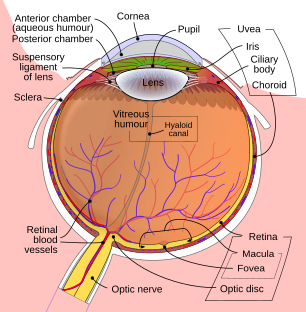
The sclera, also known as the white of the eye or, in older literature, as the tunica albuginea oculi, is the opaque, fibrous, protective outer layer of the eye containing mainly collagen and some crucial elastic fiber.

The cornea is the transparent front part of the eye that covers the iris, pupil, and anterior chamber. Along with the anterior chamber and lens, the cornea refracts light, accounting for approximately two-thirds of the eye's total optical power. In humans, the refractive power of the cornea is approximately 43 dioptres. The cornea can be reshaped by surgical procedures such as LASIK.

The choroid, also known as the choroidea or choroid coat, is a part of the uvea, the vascular layer of the eye. It contains connective tissues, and lies between the retina and the sclera. The human choroid is thickest at the far extreme rear of the eye, while in the outlying areas it narrows to 0.1 mm. The choroid provides oxygen and nourishment to the outer layers of the retina. Along with the ciliary body and iris, the choroid forms the uveal tract.

Eye surgery, also known as ophthalmic surgery or ocular surgery, is surgery performed on the eye or its adnexa. Eye surgery is part of ophthalmology and is performed by an ophthalmologist or eye surgeon. The eye is a fragile organ, and requires due care before, during, and after a surgical procedure to minimize or prevent further damage. An eye surgeon is responsible for selecting the appropriate surgical procedure for the patient, and for taking the necessary safety precautions. Mentions of eye surgery can be found in several ancient texts dating back as early as 1800 BC, with cataract treatment starting in the fifth century BC. It continues to be a widely practiced class of surgery, with various techniques having been developed for treating eye problems.

The aqueous humour is a transparent water-like fluid similar to blood plasma, but containing low protein concentrations. It is secreted from the ciliary body, a structure supporting the lens of the eyeball. It fills both the anterior and the posterior chambers of the eye, and is not to be confused with the vitreous humour, which is located in the space between the lens and the retina, also known as the posterior cavity or vitreous chamber. Blood cannot normally enter the eyeball.
This is a partial list of human eye diseases and disorders.

The uvea, also called the uveal layer, uveal coat, uveal tract, vascular tunic or vascular layer, is the pigmented middle layer of the three concentric layers that make up an eye, precisely between the inner retina and the outer fibrous layer composed of the sclera and cornea.

The human eye is a sensory organ in the visual system that reacts to visible light allowing eyesight. Other functions include maintaining the circadian rhythm, and keeping balance.

An eye examination, commonly known as an eye test, is a series of tests performed to assess vision and ability to focus on and discern objects. It also includes other tests and examinations of the eyes. Eye examinations are primarily performed by an optometrist, ophthalmologist, or an orthoptist. Health care professionals often recommend that all people should have periodic and thorough eye examinations as part of routine primary care, especially since many eye diseases are asymptomatic. Typically, a healthy individual who otherwise has no concerns with their eyes receives an eye exam once in their 20s and twice in their 30s.

The accommodation reflex is a reflex action of the eye, in response to focusing on a near object, then looking at a distant object, comprising coordinated changes in vergence, lens shape (accommodation) and pupil size. It is dependent on cranial nerve II, superior centers (interneuron) and cranial nerve III. The change in the shape of the lens is controlled by ciliary muscles inside the eye. Changes in contraction of the ciliary muscles alter the focal distance of the eye, causing nearer or farther images to come into focus on the retina; this process is known as accommodation. The reflex, controlled by the parasympathetic nervous system, involves three responses: pupil constriction, lens accommodation, and convergence.

The ophthalmic artery (OA) is an artery of the head. It is the first branch of the internal carotid artery distal to the cavernous sinus. Branches of the ophthalmic artery supply all the structures in the orbit around the eye, as well as some structures in the nose, face, and meninges. Occlusion of the ophthalmic artery or its branches can produce sight-threatening conditions.

The long ciliary nerves are 2-3 nerves that arise from the nasociliary nerve (itself a branch of the ophthalmic branch (CN V1) of the trigeminal nerve (CN V)). They enter the eyeball to provide sensory innervation to parts of the eye, and sympathetic visceral motor innervation to the dilator pupillae muscle.

The short posterior ciliary arteries are a number of branches of the ophthalmic artery. They pass forward with the optic nerve to reach the eyeball, piercing the sclera around the entry of the optic nerve into the eyeball.

Eye formation in the human embryo begins at approximately three weeks into embryonic development and continues through the tenth week. Cells from both the mesodermal and the ectodermal tissues contribute to the formation of the eye. Specifically, the eye is derived from the neuroepithelium, surface ectoderm, and the extracellular mesenchyme which consists of both the neural crest and mesoderm.
A staphyloma is an abnormal protrusion of the uveal tissue through a weak point in the eyeball. The protrusion is generally black in colour, due to the inner layers of the eye. It occurs due to weakening of outer layer of eye by an inflammatory or degenerative condition. It may be of five types, depending on the location on the eyeball.
Mammals normally have a pair of eyes. Although mammalian vision is not as excellent as bird vision, it is at least dichromatic for most of mammalian species, with certain families possessing a trichromatic color perception.

Diktyoma, or ciliary body medulloepithelioma, or teratoneuroma, is a rare tumor arising from primitive medullary epithelium in the ciliary body of the eye. Almost all diktyomas arise in the ciliary body, although, rarely, they may arise from the optic nerve head or retina.

The eagle eye is among the sharpest in the animal kingdom, with an eyesight estimated at 4 to 8 times stronger than that of the average human. Although an eagle may only weigh 4.5 kilograms (10 lb), its eyes are roughly the same size as those of a human. Eagle weight varies: a small eagle could weigh 700 grams (1.5 lb), while a larger one could weigh 6.5 kilograms (14 lb); an eagle of about 4.5 kilograms (9.9 lb) weight could have eyes as big as that of a human who weighs 91 kilograms (200 lb). Although the size of the eagle eye is about the same as that of a human being, the back side shape of the eagle eye is flatter. Their eyes are stated to be larger than their brain, by weight. Color vision with resolution and clarity are the most prominent features of eagles' eyes, hence sharp-sighted people are sometimes referred to as "eagle-eyed". Eagles can identify five distinctly colored squirrels and locate their prey even if hidden.
Open-globe injuries are full-thickness eye-wall wounds requiring urgent diagnosis and treatment.