Neuropil is any area in the nervous system composed of mostly unmyelinated axons, dendrites and glial cell processes that forms a synaptically dense region containing a relatively low number of cell bodies. The most prevalent anatomical region of neuropil is the brain which, although not completely composed of neuropil, does have the largest and highest synaptically-concentrated areas of neuropil in the body. For example, the neocortex and olfactory bulb both contain neuropil.

Rod cells are photoreceptor cells in the retina of the eye that can function in less intense light than the other type of visual photoreceptor, cone cells. Rods are usually found concentrated at the outer edges of the retina and are used in peripheral vision. On average, there are approximately 92 million rod cells in the human retina. Rod cells are more sensitive than cone cells and are almost entirely responsible for night vision. However, rods have little role in color vision, which is the main reason why colors are much less apparent in dim light, and not at all at night.

Cone cells, or cones, are photoreceptor cells in the retinas of vertebrate eyes. They respond differently to light of different wavelengths, and are thus responsible for color vision and function best in relatively bright light, as opposed to rod cells, which work better in dim light. Cone cells are densely packed in the fovea centralis, a 0.3 mm diameter rod-free area with very thin, densely packed cones which quickly reduce in number towards the periphery of the retina. There are about six to seven million cones in a human eye and are most concentrated towards the macula. The commonly cited figure of six million cone cells in the human eye was found by Osterberg in 1935. Oyster's textbook (1999) cites work by Curcio et al. (1990) indicating an average close to 4.5 million cone cells and 90 million rod cells in the human retina.
As a part of the retina, bipolar cells exist between photoreceptors and ganglion cells. They act, directly or indirectly, to transmit signals from the photoreceptors to the ganglion cells.

The tunica media, or media for short, is the middle tunica (layer) of an artery or vein. It lies between the tunica intima on the inside and the tunica externa on the outside.
A myelocyte is a young cell of the granulocytic series, occurring normally in bone marrow.
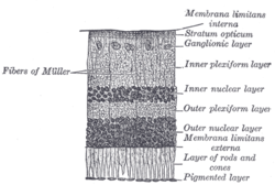
The inner plexiform layer is an area of the retina that is made up of a dense reticulum of fibrils formed by interlaced dendrites of retinal ganglion cells and cells of the inner nuclear layer. Within this reticulum a few branched spongioblasts are sometimes embedded.
The elements composing the Layer of Rods and Cones in the retina of the eye are of two kinds, rod cells and cone cells, the former being much more numerous than the latter except in the macula lutea.
The external limiting membrane is one of the ten distinct layers of the retina of the eye. It has a network-like structure and is situated at the bases of the rods and cones.
The outer plexiform layer is a layer of neuronal synapses in the retina of the eye. It consists of a dense network of synapses between dendrites of horizontal cells from the inner nuclear layer, and photoreceptor cell inner segments from the outer nuclear layer. It is much thinner than the inner plexiform layer, where amacrine cells synapse with retinal ganglion cells.
The retinal nerve fiber layer (RNFL) or nerve fiber layer, stratum opticum, is formed by the expansion of the fibers of the optic nerve; it is thickest near the optic disc, gradually diminishing toward the ora serrata.
The ganglion cell layer is a layer of the retina that consists of retinal ganglion cells and displaced amacrine cells.
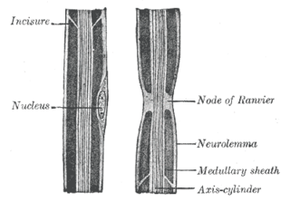
Myelin incisures, are small pockets of cytoplasm left behind during the Schwann cell myelination process. They are histological evidence of the small amount of cytoplasm that remains in the inner layer of the myelin sheath created by Schwann cells wrapping tightly around an axon.

The endoneurium is a layer of delicate connective tissue around the myelin sheath of each myelinated nerve fiber in the peripheral nervous system. Its component cells are called endoneurial cells. The endoneuria with their enclosed nerve fibers are bundled into groups called nerve fascicles, each fascicle within its own protective sheath called a perineurium. In sufficiently large nerves multiple fascicles, each with its blood supply and fatty tissue, may be bundled within yet another sheath, the epineurium.
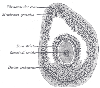
The larger ovarian follicles consist of an external fibrovascular coat, connected with the surrounding stroma of the ovary by a network of bloodvessels; and an internal coat, which consists of several layers of nucleated cells, called the membrana granulosa. It contains numerous granulosa cells.

The gastric mucosa is the mucous membrane layer of the stomach, which contains the glands and the gastric pits. In humans, it is about 1 mm thick, and its surface is smooth, soft, and velvety. It consists of simple columnar epithelium, lamina propria, and the muscularis mucosae.
The theca externa is the outer layer of the theca folliculi. It is derived from connective tissue, the cells resembling fibroblasts, and contains abundant collagen. During ovulation, the surge in luteinizing hormone increases cAMP which increases progesterone and PGF2α production. The PGF2α induces the contraction of the smooth muscle cells of the theca externa, increasing intrafollicular pressure. This aids in rupture of the mature oocyte, or immature oocyte at the germinal vesicle stage in the canine, along with plasmin and collagenase degradation of the follicle wall.

Mammals normally have a pair of eyes. Although mammalian vision is not so excellent as bird vision, it is at least dichromatic for most of mammalian species, with certain families possessing a trichromatic color perception.
The public domain consists of all the creative works to which no exclusive intellectual property rights apply. Those rights may have expired, been forfeited, expressly waived, or may be inapplicable.

Gray's Anatomy is an English language textbook of human anatomy originally written by Henry Gray and illustrated by Henry Vandyke Carter. Earlier editions were called Anatomy: Descriptive and Surgical, Anatomy of the Human Body and Gray's Anatomy: Descriptive and Applied, but the book's name is commonly shortened to, and later editions are titled, Gray's Anatomy. The book is widely regarded as an extremely influential work on the subject, and has continued to be revised and republished from its initial publication in 1858 to the present day. The latest edition of the book, the 41st, was published in September 2015.