Anterior segment of eyeball

Last updated
Anterior segment of eyeball
Cataract in human eye.png
Human eye Anterior Segment - Magnified view seen on examination with a slit lamp under diffuse illumination showing conjunctiva overlying the white sclera, transparent cornea, pharmacologically dilated pupil and cataract
Details
Identifiers
Latin segmentum anterius bulbi oculi
MeSH D000869
Anatomical terminology
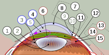
Diagram of anterior segment of a human eye (horizontal section of the right eye)
1. Lens, 2. Zonule of Zinn or ciliary zonule, 3. Posterior chamber and 4. Anterior chamber with 5. Aqueous humour flow; 6. Pupil, 7. Corneosclera with 8. Cornea, 9. Trabecular meshwork and Schlemm's canal. 10. Corneal limbus and 11. Sclera; 12. Conjunctiva, 13. Uvea with 14. Iris, 15. Ciliary body. Schematic diagram of anterior segment human eye.svg
Diagram of anterior segment of a human eye (horizontal section of the right eye)
1. Lens, 2. Zonule of Zinn or ciliary zonule, 3. Posterior chamber and 4. Anterior chamber with 5. Aqueous humour flow; 6. Pupil, 7. Corneosclera with 8. Cornea, 9. Trabecular meshwork and Schlemm's canal. 10. Corneal limbus and 11. Sclera; 12. Conjunctiva, 13. Uvea with 14. Iris, 15. Ciliary body.

The anterior segment or anterior cavity [1] is the front third of the eye that includes the structures in front of the vitreous humour: the cornea, iris, ciliary body, and lens. [2] [3]

Contents

Within the anterior segment are two fluid-filled spaces:

Aqueous humour fills these spaces within the anterior segment and provides nutrients to the surrounding structures.

Some ophthalmologists and optometrists specialize in the treatment and management of anterior segment disorders and diseases. [3]

See also

Related Research Articles

<span class="mw-page-title-main">Iris (anatomy)</span> Colored part of an eye

The iris is a thin, annular structure in the eye in most mammals and birds, responsible for controlling the diameter and size of the pupil, and thus the amount of light reaching the retina. In optical terms, the pupil is the eye's aperture, while the iris is the diaphragm. Eye color is defined by the iris.

<span class="mw-page-title-main">Floater</span> Deposits within the eyes vitreous humour

Floaters or eye floaters are sometimes visible deposits within the eye's vitreous humour, which is normally transparent, or between the vitreous and retina. They can become particularly noticeable when looking at a blank surface or an open monochromatic space, such as blue sky. Each floater can be measured by its size, shape, consistency, refractive index, and motility. They are also called muscae volitantes, or mouches volantes. The vitreous usually starts out transparent, but imperfections may gradually develop as one ages. The common type of floater, present in most people's eyes, is due to these degenerative changes of the vitreous. The perception of floaters, which may be annoying or problematic to some people, is known as myodesopsia, or, less commonly, as myodaeopsia, myiodeopsia, or myiodesopsia. It is not often treated, except in severe cases, where vitrectomy (surgery), laser vitreolysis, and medication may be effective.

<span class="mw-page-title-main">Vitreous body</span> Gel in eyeballs

The vitreous body is the clear gel that fills the space between the lens and the retina of the eyeball in humans and other vertebrates. It is often referred to as the vitreous humor, from Latin meaning liquid, or simply "the vitreous". Vitreous fluid or "liquid vitreous" is the liquid component of the vitreous gel, found after a vitreous detachment. It is not to be confused with the aqueous humor, the other fluid in the eye that is found between the cornea and lens.

<span class="mw-page-title-main">Eye surgery</span> Surgery performed on the eye or its adnexa

Eye surgery, also known as ophthalmic surgery or ocular surgery, is surgery performed on the eye or its adnexa. Eye surgery is part of ophthalmology and is performed by an ophthalmologist or eye surgeon. The eye is a fragile organ, and requires due care before, during, and after a surgical procedure to minimize or prevent further damage. An eye surgeon is responsible for selecting the appropriate surgical procedure for the patient, and for taking the necessary safety precautions. Mentions of eye surgery can be found in several ancient texts dating back as early as 1800 BC, with cataract treatment starting in the fifth century BC. It continues to be a widely practiced class of surgery, with various techniques having been developed for treating eye problems.

<span class="mw-page-title-main">Aqueous humour</span> Fluid in the anterior segment of the eye

The aqueous humour is a transparent water-like fluid similar to blood plasma, but containing low protein concentrations. It is secreted from the ciliary body, a structure supporting the lens of the eyeball. It fills both the anterior and the posterior chambers of the eye, and is not to be confused with the vitreous humour, which is located in the space between the lens and the retina, also known as the posterior cavity or vitreous chamber. Blood cannot normally enter the eyeball.

<span class="mw-page-title-main">Phacoemulsification</span> Method of cataract surgery

Phacoemulsification is a cataract surgery method in which the internal lens of the eye which has developed a cataract is emulsified with the tip of an ultrasonic handpiece and aspirated from the eye. Aspirated fluids are replaced with irrigation of balanced salt solution to maintain the volume of the anterior chamber during the procedure. This procedure minimises the incision size and reduces the recovery time and risk of surgery induced astigmatism.

<span class="mw-page-title-main">Synechia (eye)</span> Condition in which the iris adheres to the cornea or lens

Ocular synechia is an eye condition where the iris adheres to either the cornea or lens. Synechiae can be caused by ocular trauma, iritis or iridocyclitis and may lead to certain types of glaucoma. It is sometimes visible on careful examination but usually more easily through an ophthalmoscope or slit-lamp.

This is a partial list of human eye diseases and disorders.

<span class="mw-page-title-main">Cataract surgery</span> Removal of opacified lens from the eye

Cataract surgery, also called lens replacement surgery, is the removal of the natural lens of the eye that has developed a cataract, an opaque or cloudy area. The eye's natural lens is usually replaced with an artificial intraocular lens (IOL) implant.

<span class="mw-page-title-main">Hyphema</span> Hemorrhage in the front chamber of the eye

Hyphema is the medical condition of bleeding in the anterior chamber of the eye between the iris and the cornea. People usually first notice a loss or decrease in vision. The eye may also appear to have a reddish tinge, or it may appear as a small pool of blood at the bottom of the iris in the cornea. A traumatic hyphema is caused by a blow to the eye. A hyphema can also occur spontaneously.

<span class="mw-page-title-main">Anterior chamber of eyeball</span> Space in the eye

The anterior chamber (AC) is the aqueous humor-filled space inside the eye between the iris and the cornea's innermost surface, the endothelium. Hyphema, anterior uveitis and glaucoma are three main pathologies in this area. In hyphema, blood fills the anterior chamber as a result of a hemorrhage, most commonly after a blunt eye injury. Anterior uveitis is an inflammatory process affecting the iris and ciliary body, with resulting inflammatory signs in the anterior chamber. In glaucoma, blockage of the trabecular meshwork prevents the normal outflow of aqueous humour, resulting in increased intraocular pressure, progressive damage to the optic nerve head, and eventually blindness.

<span class="mw-page-title-main">Gonioscopy</span> Measurement of the angle between an eyes iris and cornea

In ophthalmology, gonioscopy is a routine procedure that measures the angle between the iris and the cornea, using a goniolens together with a slit lamp or operating microscope. Its use is important in diagnosing and monitoring various eye conditions associated with glaucoma.

<span class="mw-page-title-main">Ectopia lentis</span> Medical condition

Ectopia lentis is a displacement or malposition of the eye's crystalline lens from its normal location. A partial dislocation of a lens is termed lens subluxation or subluxated lens; a complete dislocation of a lens is termed lens luxation or luxated lens.

<span class="mw-page-title-main">Posterior chamber of eyeball</span> Region of the eyeball between the iris and lens

The posterior chamber is a narrow space behind the peripheral part of the iris, and in front of the suspensory ligament of the lens and the ciliary processes. The posterior chamber consists of small space directly posterior to the iris but anterior to the lens. The posterior chamber is part of the anterior segment and should not be confused with the vitreous chamber.

<span class="mw-page-title-main">Posterior segment of eyeball</span> Back two-thirds of the eye

The posterior segment or posterior cavity is the back two-thirds of the eye that includes the anterior hyaloid membrane and all of the optical structures behind it: the vitreous humor, retina, choroid, and optic nerve. The portion of the posterior segment visible during ophthalmoscopy is sometimes referred to as the posterior pole, or fundus. Some ophthalmologists specialize in the treatment and management of posterior segment disorders and diseases.

<span class="mw-page-title-main">Axenfeld–Rieger syndrome</span> Medical condition

Axenfeld–Rieger syndrome is a rare autosomal dominant disorder, which affects the development of the teeth, eyes, and abdominal region.

<span class="mw-page-title-main">Vitreous chamber</span> Space in the eye occupied by vitreous humor

The vitreous chamber is the largest of the three chambers in the eye and is located behind the lens and in front of the optic nerve. The vitreous chamber is located in the posterior cavity of the eye. This chamber is occupied with a thick, clear gel-like substance called the vitreous humor.

<span class="mw-page-title-main">Mammalian eye</span>

Mammals normally have a pair of eyes. Although mammalian vision is not so excellent as bird vision, it is at least dichromatic for most of mammalian species, with certain families possessing a trichromatic color perception.

Fuchs heterochromic iridocyclitis (FHI) is a chronic unilateral uveitis appearing with the triad of heterochromia, predisposition to cataract and glaucoma, and keratitic precipitates on the posterior corneal surface. Patients are often asymptomatic and the disease is often discovered through investigation of the cause of the heterochromia or cataract. Neovascularisation is possible and any eye surgery, such as cataract surgery, can cause bleeding from the fragile vessels in the atrophic iris causing accumulation of blood in the anterior chamber of the eye, also known as hyphema.

Open-globe injuries are full-thickness eye-wall wounds requiring urgent diagnosis and treatment.

References

  1. "WikiJournal of Medicine/Medical gallery of Blausen Medical 2014 - Wikiversity".
  2. 1 2 Cassin, B. and Solomon, S. Dictionary of Eye Terminology. Gainesville, Florida: Triad Publishing Company, 1990.
  3. 1 2 "Departments. Anterior segment." Archived September 27, 2006, at the Wayback Machine Cantabrian Institute of Ophthalmology.