Iris dilator muscle

Last updated
Iris dilator muscle
Gray878.png
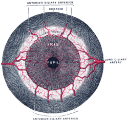
Iris, front view. (Muscle visible but not labeled.)
Gray883.png
The upper half of a sagittal section through the front of the eyeball. (Iris dilator muscle is NOT labeled and not to be confused with "Radiating fibers" labeled near center, which are part of the ciliary muscle.)
Details
Origin outer margins of iris [1]
Insertion inner margins of iris [1]
Nerve Long ciliary nerves (sympathetics)
Actions dilates pupil
Antagonist iris sphincter muscle
Identifiers
Latin musculus dilatator pupillae
TA98 A15.2.03.030
TA2 6763
FMA 49158
Anatomical terms of muscle

The iris dilator muscle (pupil dilator muscle, pupillary dilator, radial muscle of iris, radiating fibers), is a smooth muscle [2] of the eye, running radially in the iris and therefore fit as a dilator. The pupillary dilator consists of a spokelike arrangement of modified contractile cells called myoepithelial cells. These cells are stimulated by the sympathetic nervous system. [3] When stimulated, the cells contract, widening the pupil and allowing more light to enter the eye.

Contents

Structure

Innervation

It is innervated by the sympathetic system, which acts by releasing noradrenaline, which acts on α1-receptors. [4] Thus, when presented with a threatening stimulus that activates the fight-or-flight response, this innervation contracts the muscle and dilates the pupil, thus temporarily letting more light reach the retina.

The dilator muscle is innervated more specifically by postganglionic sympathetic nerves arising from the superior cervical ganglion as the sympathetic root of ciliary ganglion. From there, they travel via the internal carotid artery through the carotid canal to foramen lacerum. They then enter the middle cranial fossa above foramen lacerum, travel through the cavernous sinus in the middle cranial fossa and then travel with the ophthalmic artery in the optic canal or on the ophthalmic nerve through the superior orbital fissure. From there, they travel with the nasociliary nerve and then the long ciliary nerve. They then pierce the sclera, travel between sclera and choroid to reach the iris dilator muscle. They will also pass through ciliary ganglion and travel in short ciliary nerves to reach the iris dilator muscle.

Function

The pupil widens when the iris dilator muscle is stimulated and contracts. An extreme, though natural, case is shown Human eye in dim light.jpg
The pupil widens when the iris dilator muscle is stimulated and contracts. An extreme, though natural, case is shown

The pupillary dilator acts to increase the size of the pupil to allow more light to enter the eye. It works in opposition to the pupillary constrictor. [5] Pupil dilation occurs when there is insufficient light for the normal function of the eye, and during heightened sympathetic activity, for example in the "fight-or-flight reflex". [5] [6]

History

Etymology

The English name dilator pupillae muscle [7] as currently used in the list of English equivalents of the Terminologia Anatomica , the reference-work of the official anatomic nomenclature, [8] can be considered as a corruption [9] of the full Latin expression musculus dilatator pupillae. [10] The full Latin expression exhibits three words that each can be traced back to Roman antiquity. The Classical Latin name musculus is actually a diminutive of the Classical Latin name mus, [11] and can be translated as little mouse. [11] In the medical writings of Aulus Cornelius Celsus we can also find this specific name to refer to a muscle instead of its literal meaning. [11] Latin musculus can be explained by the fact that a muscle looks like a little mouse that moves under the skin. [12] In the writings of Greek philosopher Aristotle the Ancient Greek word for mouse, i.e. μῦς [13] is also used to refer to a muscle. [13]

Dilatator in the Latin expression musculus dilatator pupillae is derived from the classical Latin verb dilatare, [14] to dilate, to spread out. [11] Two possible explanations exist concerning the etymological derivation of this verb. The first explanation considers dilatare as frequentative of differere. [11] The Latin verb differe can mean, to carry different ways, to spread abroad, to scatter, [11] but also to delay. [11] The other explanation [12] considers dilatare as a compound from di- and latus, with the latter word meaning, broad or wide, [11] hence the German name Erweiterer for Latin dilatator. [14]

The expression dilator pupillae muscle, as used in the list of English equivalents of the Terminologia Anatomica, is actually partly Latin, i.e. dilator pupillae, with pupillae (=of the pupil [11] ), a noun in the genitive case modifying dilator, a noun in the nominative case, and partly English, i.e. muscle. In previous editions ( Nomina Anatomica ) this muscle was officially called the musculus dilator pupillae, [15] [16] [17] [18] [19] The Nomina Anatomica as authorized in 1895 in Basle [10] and in 1935 in Jena [20] [21] used the full Latin expression.

Additional images

See also

Related Research Articles

<span class="mw-page-title-main">Iris (anatomy)</span> Colored part of an eye

In humans and most mammals and birds, the iris is a thin, annular structure in the eye, responsible for controlling the diameter and size of the pupil, and thus the amount of light reaching the retina. Eye color is defined by the iris. In optical terms, the pupil is the eye's aperture, while the iris is the diaphragm.

<span class="mw-page-title-main">Mydriasis</span> Excessive dilation of the pupil

Mydriasis is the dilation of the pupil, usually having a non-physiological cause, or sometimes a physiological pupillary response. Non-physiological causes of mydriasis include disease, trauma, or the use of certain types of drug. It may also be of unknown cause.

<span class="mw-page-title-main">Oculomotor nerve</span> Cranial nerve III, for eye movements

The oculomotor nerve, also known as the third cranial nerve, cranial nerve III, or simply CN III, is a cranial nerve that enters the orbit through the superior orbital fissure and innervates extraocular muscles that enable most movements of the eye and that raise the eyelid. The nerve also contains fibers that innervate the intrinsic eye muscles that enable pupillary constriction and accommodation. The oculomotor nerve is derived from the basal plate of the embryonic midbrain. Cranial nerves IV and VI also participate in control of eye movement.

<span class="mw-page-title-main">Metacarpal bones</span> Bones of hand

In human anatomy, the metacarpal bones or metacarpus, also known as the "palm bones", are the appendicular bones that form the intermediate part of the hand between the phalanges (fingers) and the carpal bones, which articulate with the forearm. The metacarpal bones are homologous to the metatarsal bones in the foot.

<span class="mw-page-title-main">Internal carotid artery</span> Artery of the human brain

The internal carotid artery is an artery in the neck which supplies the anterior circulation of the brain.

<span class="mw-page-title-main">Pupillary light reflex</span> Eye reflex which alters the pupils size in response to light intensity

The pupillary light reflex (PLR) or photopupillary reflex is a reflex that controls the diameter of the pupil, in response to the intensity (luminance) of light that falls on the retinal ganglion cells of the retina in the back of the eye, thereby assisting in adaptation of vision to various levels of lightness/darkness. A greater intensity of light causes the pupil to constrict, whereas a lower intensity of light causes the pupil to dilate. Thus, the pupillary light reflex regulates the intensity of light entering the eye. Light shone into one eye will cause both pupils to constrict.

<span class="mw-page-title-main">Buccinator muscle</span> Muscle

The buccinator is a thin quadrilateral muscle occupying the interval between the maxilla and the mandible at the side of the face. It forms the anterior part of the cheek or the lateral wall of the oral cavity.

<span class="mw-page-title-main">Thyroid cartilage</span> Largest of nine cartilages that make up the laryngeal skeleton

The thyroid cartilage is the largest of the nine cartilages that make up the laryngeal skeleton, the cartilage structure in and around the trachea that contains the larynx. It does not completely encircle the larynx.

<span class="mw-page-title-main">Ciliary ganglion</span> Bundle of nerves, parasympathetic ganglion

The ciliary ganglion is a bundle of nerves, parasympathetic ganglion located just behind the eye in the posterior orbit. It is 1–2 mm in diameter and in humans contains approximately 2,500 neurons. The ganglion contains postganglionic parasympathetic neurons. These neurons supply the pupillary sphincter muscle, which constricts the pupil, and the ciliary muscle which contracts to make the lens more convex. Both of these muscles are involuntary since they are controlled by the parasympathetic division of the autonomic nervous system.

<span class="mw-page-title-main">Iris sphincter muscle</span> Muscle in the eye which constricts the pupil

The iris sphincter muscle is a muscle in the part of the eye called the iris. It encircles the pupil of the iris, appropriate to its function as a constrictor of the pupil.

<span class="mw-page-title-main">Anconeus muscle</span>

The anconeus muscle is a small muscle on the posterior aspect of the elbow joint.

<span class="mw-page-title-main">Ilium (bone)</span> Uppermost and largest part of the coxal bone

The ilium is the uppermost and largest part of the coxal bone, and appears in most vertebrates including mammals and birds, but not bony fish. All reptiles have an ilium except snakes, although some snake species have a tiny bone which is considered to be an ilium.

Nomina Anatomica (NA) was the international standard on human anatomic terminology from 1895 until it was replaced by Terminologia Anatomica in 1998.

<span class="mw-page-title-main">Internal carotid plexus</span>

The internal carotid plexus is a nerve plexus situated upon the lateral side of the internal carotid artery. It is composed of post-ganglionic sympathetic fibres which have synapsed at the superior cervical ganglion. The plexus gives rise to the deep petrosal nerve.

<span class="mw-page-title-main">Long ciliary nerves</span>

The long ciliary nerves are 2-3 nerves that arise from the nasociliary nerve (itself a branch of the ophthalmic branch (CN V1) of the trigeminal nerve (CN V)). They enter the eyeball to provide sensory innervation to parts of the eye, and sympathetic visceral motor innervation to the dilator pupillae muscle.

<span class="mw-page-title-main">Short ciliary nerves</span> Nerves of the orbit around the eye

The short ciliary nerves are nerves of the orbit around the eye. They are branches of the ciliary ganglion. They supply parasympathetic and sympathetic nerve fibers to the ciliary muscle, iris, and cornea. Damage to the short ciliary nerve may result in loss of the pupillary light reflex, or mydriasis.

The Terminologia Embryologica (TE) is a standardized list of words used in the description of human embryologic and fetal structures. It was produced by the Federative International Committee on Anatomical Terminology on behalf of the International Federation of Associations of Anatomists and posted on the Internet since 2010. It has been approved by the General Assembly of the IFAA during the seventeenth International Congress of Anatomy in Cape Town.

The Terminologia Histologica (TH) is the controlled vocabulary for use in cytology and histology. In April 2011, Terminologia Histologica was published online by the Federative International Programme on Anatomical Terminologies (FIPAT), the successor of FCAT.

The Federative International Programme for Anatomical Terminology (FIPAT) is a group of experts who review, analyze, and discuss the terms of the morphological structures of the human body. It was created by the International Federation of Associations of Anatomists (IFAA) and was previously known as the Federative Committee on Anatomical Terminology (FCAT) and the Federative International Committee on Anatomical Terminology (FICAT).

<span class="mw-page-title-main">Roots of the ciliary ganglion</span>

The ciliary ganglion is a parasympathetic ganglion located just behind the eye in the posterior orbit. Three types of axons enter the ciliary ganglion but only the preganglionic parasympathetic axons synapse there. The entering axons are arranged into three roots of the ciliary ganglion, which join enter the posterior surface of the ganglion.

References

  1. 1 2 Gest, Thomas R; Burkel, William E. (2000). "Anatomy Tables - Eye". Medical Gross Anatomy. University of Michigan Medical School. Archived from the original on 2010-05-26.{{cite web}}: CS1 maint: unfit URL (link)
  2. Pilar, G; Nuñez, R; McLennan, I. S.; Meriney, S. D. (1987). "Muscarinic and nicotinic synaptic activation of the developing chicken iris". The Journal of Neuroscience. 7 (12): 3813–26. doi: 10.1523/JNEUROSCI.07-12-03813.1987 . PMC   6569112 . PMID   2826718.
  3. Saladin, Kenneth (2012). Anatomy and Physiology. McGraw-Hill. pp. 616–7.
  4. Rang, H. P. (2003). Pharmacology. Edinburgh: Churchill Livingstone. ISBN   0-443-07145-4. page 163
  5. 1 2 "Reflexes of the Eye". Cleveland Clinic. Retrieved 3 August 2023.
  6. "What Happens to Your Body During the Fight-or-Flight Response?". Cleveland Clinic. 9 December 2019. Retrieved 3 August 2023.
  7. Federative Committee on Anatomical Terminology (FCAT) (1998). Terminologia Anatomica. Stuttgart: Thieme
  8. Kachlik, David; Baca, Vaclav; Bozdechova, Ivana; Cech, Pavel; Musil, Vladimir (2008). "Anatomical terminology and nomenclature: Past, present and highlights". Surgical and Radiologic Anatomy. 30 (6): 459–66. doi:10.1007/s00276-008-0357-y. PMID   18488135. S2CID   2273591.
  9. Marečková, Elena; Šimon, František; Červený, Ladislav (2001). "On the new anatomical nomenclature". Annals of Anatomy - Anatomischer Anzeiger. 183 (3): 201–7. doi:10.1016/S0940-9602(01)80215-6. PMC   2413596 . PMID   11396787.
  10. 1 2 His (1895). Die anatomische Nomenclatur. Nomina Anatomica. Der von der Anatomischen Gesellschaft auf ihrer IX. Versammlung in Basel angenommenen Namen. Leipzig: Verlag von Veit & Comp.[ page needed ]
  11. 1 2 3 4 5 6 7 8 9 Lewis, C.T. & Short, C. (1879). A Latin dictionary founded on Andrews' edition of Freund's Latin dictionary. Oxford: Clarendon Press.[ page needed ]
  12. 1 2 Kraus, L.A. (1844). Kritisch-etymologisches medicinisches Lexikon (Dritte Auflage). Göttingen: Verlag der Deuerlich- und Dieterichschen Buchhandlung.[ page needed ]
  13. 1 2 Liddell, H.G. & Scott, R. (1940). A Greek-English Lexicon. revised and augmented throughout by Sir Henry Stuart Jones. with the assistance of. Roderick McKenzie. Oxford: Clarendon Press.[ page needed ]
  14. 1 2 Foster, F.D. (1891-1893). An illustrated medical dictionary. Being a dictionary of the technical terms used by writers on medicine and the collateral sciences, in the Latin, English, French, and German languages. New York: D. Appleton and Company.[ page needed ]
  15. Donáth, T. & Crawford, G.C.N. (1969). Anatomical dictionary with nomenclature and explanatory notes. Oxford/London/Edinburgh/New York/Toronto/Syney/Paris/Braunschweig: Pergamon Press.[ page needed ]
  16. International Anatomical Nomenclature Committee (1966). Nomina Anatomica. Amsterdam: Excerpta Medica Foundation.[ page needed ]
  17. International Anatomical Nomenclature Committee (1977). Nomina Anatomica, together with Nomina Histologica and Nomina Embryologica. Amsterdam-Oxford: Excerpta Medica.[ page needed ]
  18. International Anatomical Nomenclature Committee (1983). Nomina Anatomica, together with Nomina Histologica and Nomina Embryologica. Baltimore/London: Williams & Wilkins[ page needed ]
  19. International Anatomical Nomenclature Committee (1989). Nomina Anatomica, together with Nomina Histologica and Nomina Embryologica. Edinburgh: Churchill Livingstone.[ page needed ]
  20. Kopsch, F. (1941). Die Nomina anatomica des Jahres 1895 (B.N.A.) nach der Buchstabenreihe geordnet und gegenübergestellt den Nomina anatomica des Jahres 1935 (I.N.A.) (3. Auflage). Leipzig: Georg Thieme Verlag.[ page needed ]
  21. Stieve, H. (1949). Nomina Anatomica. Zusammengestellt von der im Jahre 1923 gewählten Nomenklatur-Kommission, unter Berücksichtigung der Vorschläge der Mitglieder der Anatomischen Gesellschaft, der Anatomical Society of Great Britain and Ireland, sowie der American Association of Anatomists, überprüft und durch Beschluß der Anatomischen Gesellschaft auf der Tagung in Jena 1935 endgültig angenommen. (4th edition). Jena: Verlag Gustav Fischer.[ page needed ]