The retina is the innermost, light-sensitive layer of tissue of the eye of most vertebrates and some molluscs. The optics of the eye create a focused two-dimensional image of the visual world on the retina, which then processes that image within the retina and sends nerve impulses along the optic nerve to the visual cortex to create visual perception. The retina serves a function which is in many ways analogous to that of the film or image sensor in a camera.

The macula (/ˈmakjʊlə/) or macula lutea is an oval-shaped pigmented area in the center of the retina of the human eye and in other animals. The macula in humans has a diameter of around 5.5 mm (0.22 in) and is subdivided into the umbo, foveola, foveal avascular zone, fovea, parafovea, and perifovea areas.

Epithelium or epithelial tissue is a thin, continuous, protective layer of compactly packed cells with a little intercellular matrix. Epithelial tissues line the outer surfaces of organs and blood vessels throughout the body, as well as the inner surfaces of cavities in many internal organs. An example is the epidermis, the outermost layer of the skin. Epithelial tissue is one of the four basic types of animal tissue, along with connective tissue, muscle tissue and nervous tissue. These tissues also lack blood or lymph supply. The tissue is supplied by nerves.

A photoreceptor cell is a specialized type of neuroepithelial cell found in the retina that is capable of visual phototransduction. The great biological importance of photoreceptors is that they convert light into signals that can stimulate biological processes. To be more specific, photoreceptor proteins in the cell absorb photons, triggering a change in the cell's membrane potential.

Rod cells are photoreceptor cells in the retina of the eye that can function in lower light better than the other type of visual photoreceptor, cone cells. Rods are usually found concentrated at the outer edges of the retina and are used in peripheral vision. On average, there are approximately 92 million rod cells in the human retina. Rod cells are more sensitive than cone cells and are almost entirely responsible for night vision. However, rods have little role in color vision, which is the main reason why colors are much less apparent in dim light.
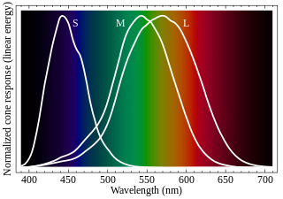
Cone cells, or cones, are photoreceptor cells in the retinas of vertebrates' eyes, including the human eye. They respond differently to light of different wavelengths, and the combination of their responses is responsible for color vision. Cones function best in relatively bright light, called the photopic region, as opposed to rod cells, which work better in dim light, or the scotopic region. Cone cells are densely packed in the fovea centralis, a 0.3 mm diameter rod-free area with very thin, densely packed cones which quickly reduce in number towards the periphery of the retina. Conversely, they are absent from the optic disc, contributing to the blind spot. There are about six to seven million cones in a human eye, with the highest concentration being towards the macula.

The choroid, also known as the choroidea or choroid coat, is a part of the uvea, the vascular layer of the eye. It contains connective tissues, and lies between the retina and the sclera. The human choroid is thickest at the far extreme rear of the eye, while in the outlying areas it narrows to 0.1 mm. The choroid provides oxygen and nourishment to the outer layers of the retina. Along with the ciliary body and iris, the choroid forms the uveal tract.

The fovea centralis is a small, central pit composed of closely packed cones in the eye. It is located in the center of the macula lutea of the retina.
Hemeralopia is the inability to see clearly in bright light and is the exact opposite of nyctalopia, the inability to see clearly in low light. Hemera was the Greek goddess of day, and Nyx was the goddess of night. However, it has been used in an opposite sense by many non-English-speaking doctors. It can be described as insufficient adaptation to bright light. It is also called "heliophobia" and "day blindness".

A cone dystrophy is an inherited ocular disorder characterized by the loss of cone cells, the photoreceptors responsible for both central and color vision.

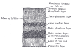
The pigmented layer of retina or retinal pigment epithelium (RPE) is the pigmented cell layer just outside the neurosensory retina that nourishes retinal visual cells, and is firmly attached to the underlying choroid and overlying retinal visual cells.
The inner nuclear layer or layer of inner granules, of the retina, is made up of a number of closely packed cells, of which there are three varieties, viz.: bipolar cells, horizontal cells, and amacrine cells.
The external limiting membrane is one of the ten distinct layers of the retina of the eye. It has a network-like structure and is situated at the bases of the rods and cones.
The outer plexiform layer is a layer of neuronal synapses in the retina of the eye. It consists of a dense network of synapses between dendrites of horizontal cells from the inner nuclear layer, and photoreceptor cell inner segments from the outer nuclear layer. It is much thinner than the inner plexiform layer, where amacrine cells synapse with retinal ganglion cells.
The outer nuclear layer, is one of the layers of the vertebrate retina, the light-detecting portion of the eye. Like the inner nuclear layer, the outer nuclear layer contains several strata of oval nuclear bodies; they are of two kinds, viz.: rod and cone granules, so named on account of their being respectively connected with the rods and cones of the next layer.

Epiretinal membrane or macular pucker is a disease of the eye in response to changes in the vitreous humor or more rarely, diabetes. Sometimes, as a result of immune system response to protect the retina, cells converge in the macular area as the vitreous ages and pulls away in posterior vitreous detachment (PVD).

Arthur Jacob (1790–1874) was an Irish ophthalmologist. He is known for founding several hospitals, a medical school, and a medical journal. He contributed to science and academia through his 41-year term as Professor of Anatomy at the Royal College of Surgeons in Ireland (RCSI) and as the first Irish ocular pathologist. He was elected President of RCSI in 1837 and 1864.
Mammals normally have a pair of eyes. Although mammalian vision is not so excellent as bird vision, it is at least dichromatic for most of mammalian species, with certain families possessing a trichromatic color perception.

The foveola is located within a region called the macula, a yellowish, cone photoreceptor filled portion of the human retina. Approximately 0.35 mm in diameter, the foveola lies in the center of the fovea and contains only cone cells and a cone-shaped zone of Müller cells. In this region the cone receptors are found to be longer, slimmer, and more densely packed than anywhere else in the retina, thus allowing that region to have the potential to have the highest visual acuity in the eye.
Proliferative vitreoretinopathy (PVR) is a disease that develops as a complication of rhegmatogenous retinal detachment. PVR occurs in about 8–10% of patients undergoing primary retinal detachment surgery and prevents the successful surgical repair of rhegmatogenous retinal detachment. PVR can be treated with surgery to reattach the detached retina but the visual outcome of the surgery is very poor. A number of studies have explored various possible adjunctive agents for the prevention and treatment of PVR, such as methotrexate, although none have yet been licensed for clinical use.